

Stages of ciliogenesis and regulation of ciliary length
- PMID: 22178116
- PMCID: PMC3269565
- DOI: 10.1016/j.diff.2011.11.015
Stages of ciliogenesis and regulation of ciliary length
Abstract
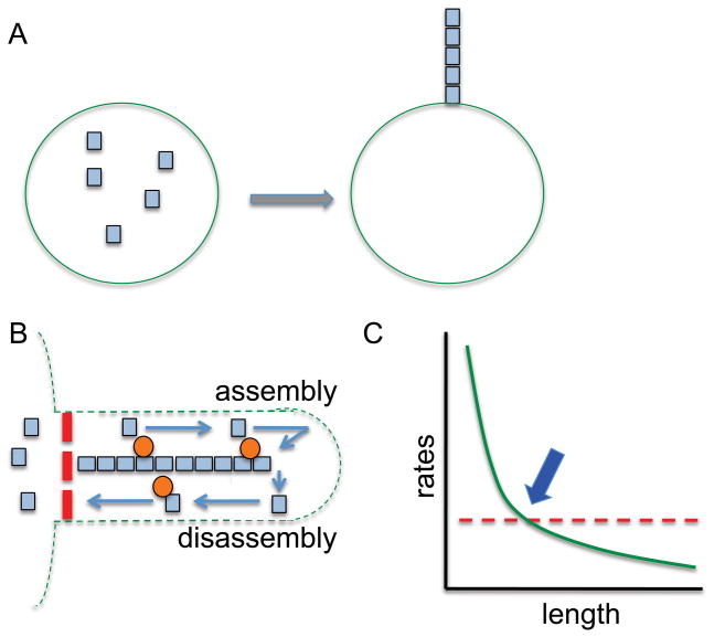
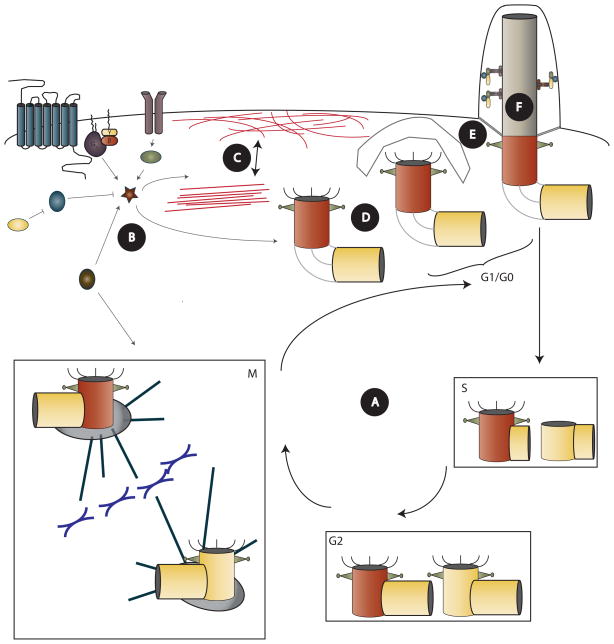
Cilia and flagella are highly conserved eukaryotic microtubule-based organelles that protrude from the surface of most mammalian cells. These structures require large protein complexes and motors for distal addition of tubulin and extension of the ciliary membrane. In order for ciliogenesis to occur, coordination of many processes must take place. An intricate concert of cell cycle regulation, vesicular trafficking, and ciliary extension must all play out with accurate timing to produce a cilium. Here, we review the stages of ciliogenesis as well as regulation of the length of the assembled cilium. Regulation of ciliogenesis during cell cycle progression centers on centrioles, from which cilia extend upon maturation into basal bodies. Centriole maturation involves a shift from roles in cell division to cilium nucleation via migration to the cell surface and docking at the plasma membrane. Docking is dependent on a variety of proteinaceous structures, termed distal appendages, acquired by the mother centriole. Ciliary elongation by the process of intraflagellar transport (IFT) ensues. Direct modification of ciliary structures, as well as modulation of signal transduction pathways, play a role in maintenance of the cilium. All of these stages are tightly regulated to produce a cilium of the right size at the right time. Finally, we discuss the implications of abnormal ciliogenesis and ciliary length control in human disease as well as some open questions.
Copyright © 2011 International Society of Differentiation. Published by Elsevier B.V. All rights reserved.
Figures
Similar articles
-
Cellular Mechanisms of Ciliary Length Control.Cells. 2016 Jan 29;5(1):6. doi: 10.3390/cells5010006. Cells. 2016. PMID: 26840332 Free PMC article. Review.
-
The ciliary cytoskeleton.Compr Physiol. 2012 Jan;2(1):779-803. doi: 10.1002/cphy.c110043. Compr Physiol. 2012. PMID: 23728985 Review.
-
Drosophila transition fibers are essential for IFT-dependent ciliary elongation but not basal body docking and ciliary budding.Curr Biol. 2023 Feb 27;33(4):727-736.e6. doi: 10.1016/j.cub.2022.12.046. Epub 2023 Jan 19. Curr Biol. 2023. PMID: 36669498
-
Intraflagellar transport proteins in ciliogenesis of photoreceptor cells.Biol Cell. 2011 Oct 1;103(10):449-66. doi: 10.1042/BC20110034. Biol Cell. 2011. PMID: 21732910
-
Intraflagellar transport (IFT) role in ciliary assembly, resorption and signalling.Curr Top Dev Biol. 2008;85:23-61. doi: 10.1016/S0070-2153(08)00802-8. Curr Top Dev Biol. 2008. PMID: 19147001 Review.
Cited by
-
Spatiotemporal dynamics of primary and motile cilia throughout lung development.bioRxiv [Preprint]. 2024 Oct 26:2024.10.25.620342. doi: 10.1101/2024.10.25.620342. bioRxiv. 2024. Update in: Dev Dyn. 2025 Mar 8. doi: 10.1002/dvdy.70008. PMID: 39484464 Free PMC article. Updated. Preprint.
-
ATP4a is required for development and function of the Xenopus mucociliary epidermis - a potential model to study proton pump inhibitor-associated pneumonia.Dev Biol. 2015 Dec 15;408(2):292-304. doi: 10.1016/j.ydbio.2015.03.013. Epub 2015 Apr 4. Dev Biol. 2015. PMID: 25848696 Free PMC article.
-
Nanotubes mediate niche-stem-cell signalling in the Drosophila testis.Nature. 2015 Jul 16;523(7560):329-32. doi: 10.1038/nature14602. Epub 2015 Jul 1. Nature. 2015. PMID: 26131929 Free PMC article.
-
Seeing cilia: imaging modalities for ciliary motion and clinical connections.Am J Physiol Lung Cell Mol Physiol. 2018 Jun 1;314(6):L909-L921. doi: 10.1152/ajplung.00556.2017. Epub 2018 Mar 1. Am J Physiol Lung Cell Mol Physiol. 2018. PMID: 29493257 Free PMC article. Review.
-
A Point Mutation in p190A RhoGAP Affects Ciliogenesis and Leads to Glomerulocystic Kidney Defects.PLoS Genet. 2016 Feb 9;12(2):e1005785. doi: 10.1371/journal.pgen.1005785. eCollection 2016 Feb. PLoS Genet. 2016. PMID: 26859289 Free PMC article.
References
-
- Afzelius BA. A human syndrome caused by immotile cilia. Science. 1976;193:317–319. - PubMed
-
- Arts HH, Bongers EM, Mans DA, van Beersum SE, Oud MM, Bolat E, Spruijt L, Cornelissen EA, Schuurs-Hoeijmakers JH, de Leeuw N, et al. C14ORF179 encoding IFT43 is mutated in Sensenbrenner syndrome. J Med Genet. 2011;48:390–395. - PubMed
Publication types
MeSH terms
Substances
Grants and funding
LinkOut - more resources
Full Text Sources
