Histopathological investigation in porcine infected with torque teno sus virus type 2 by inoculation
- PMID: 22171963
- PMCID: PMC3275549
- DOI: 10.1186/1743-422X-8-545
Histopathological investigation in porcine infected with torque teno sus virus type 2 by inoculation
Abstract
Background: Porcine torque teno sus virus (TTSuV) is a small icosahedral and non-enveloped virus which contains a single-stranded (ssDNA), circular and negative DNA genome and infects mainly vertebrates and is currently classified into the 'floating' genus Anellovirus of Circoviridae with two species. Viral DNA of both porcine TTSuV species has a high prevalence in both healthy and diseased pigs worldwide and multiple infections of TTSuV with distinct genotypes or subtypes of the same species has been documented in the United States, Europe and Asia. However, there exists no information about histopathological lesions caused by infection with porcine TTSuV2.
Methods: Porcine liver tissue homogenate with 1 ml of 6.91 × 10⁷ genomic copies viral loads of porcine TTSuV2 that had positive result for torque teno sus virus type 2 and negative result for torque teno sus virus type 1 and porcine pseudorabies virus type 2 were used to inoculate specific pathogen-free piglets by intramuscular route and humanely killed at 3,7,10,14,17,21 and 24 days post inoculation (dpi), the control pigs were injected intramuscularly with 1 ml of sterile DMEM and humanely killed the end of the study for histopathological examination routinely processed, respectively.
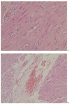
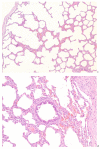
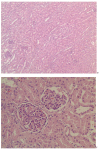
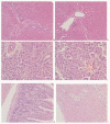
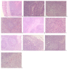
Results: All porcine TTSuV2 inoculated piglets were clinic asymptomatic but developed myocardial fibroklasts and endocardium, interstitial pneumonia, membranous glomerular nephropathy, and modest inflammatory cells infiltration in portal areas in the liver, foci of hemorrhage in some pancreas islet, a tiny amount red blood cells in venule of muscularis mucosae and outer longitudinal muscle, rarely red blood cells in the microvasculation and infiltration of inflammatory cells (lymphocytes and eosinophils) of tonsil and hilar lymph nodes, infiltration of inflammatory lymphocytes and necrosis or degeneration and focal gliosis of lymphocytes in the paracortical zone after inoculation with porcine TTSuV2-containing tissue homogenate.
Conclusions: Analysis of these presentations revealed that porcine TTSuV2 was readily transmitted to TTSuV-negative swine and that infection was associated with characteristic pathologic changes in specific pathogen-free piglets inoculated with porcine TTSuV2. Those results indicated no markedly histopathological changes happened in those parenchymatous organs, especially the digestive system and immune system when the specific pathogen-free pigs were infected with porcine TTSuV2, hence, to some extent, it was not remarkable pathological agent for domestic pigs at least. So, porcine TTSuV2 could be an unrecognized pathogenic viral infectious etiology of swine. This study indicated a directly related description of lesions responsible for TTSuV2 infection in swine.
Figures
Similar articles
-
The pathogenic role of torque teno sus virus 1 and 2 and their correlations with various viral pathogens and host immunocytes in wasting pigs.Vet Microbiol. 2015 Nov 18;180(3-4):186-95. doi: 10.1016/j.vetmic.2015.08.027. Epub 2015 Aug 29. Vet Microbiol. 2015. PMID: 26390821
-
Expression of the putative ORF1 capsid protein of Torque teno sus virus 2 (TTSuV2) and development of Western blot and ELISA serodiagnostic assays: correlation between TTSuV2 viral load and IgG antibody level in pigs.Virus Res. 2011 Jun;158(1-2):79-88. doi: 10.1016/j.virusres.2011.03.013. Epub 2011 Mar 23. Virus Res. 2011. PMID: 21439334
-
The prevalence of Torque teno sus virus (TTSuV) is common and increases with the age of growing pigs in the United States.J Virol Methods. 2012 Jul;183(1):40-4. doi: 10.1016/j.jviromet.2012.03.026. Epub 2012 Mar 31. J Virol Methods. 2012. PMID: 22484614
-
Torque teno sus virus in pigs: an emerging pathogen?Transbound Emerg Dis. 2012 Mar;59 Suppl 1:103-8. doi: 10.1111/j.1865-1682.2011.01289.x. Epub 2012 Jan 17. Transbound Emerg Dis. 2012. PMID: 22252126 Review.
-
Torque teno virus infection in the pig and its potential role as a model of human infection.Vet J. 2009 May;180(2):163-8. doi: 10.1016/j.tvjl.2007.12.005. Epub 2008 Mar 4. Vet J. 2009. PMID: 18296088 Review.
Cited by
-
The Synergic Role of Emerging and Endemic Swine Virus in the Porcine Respiratory Disease Complex: Pathological and Biomolecular Analysis.Vet Sci. 2023 Sep 27;10(10):595. doi: 10.3390/vetsci10100595. Vet Sci. 2023. PMID: 37888547 Free PMC article.
-
Analysis of TTSuV1b antibody in porcine serum and its correlation with four antibodies against common viral infectious diseases.Virol J. 2015 Aug 12;12:125. doi: 10.1186/s12985-015-0349-6. Virol J. 2015. PMID: 26260234 Free PMC article.
-
Identification of novel anelloviruses with broad diversity in UK rodents.J Gen Virol. 2014 Jul;95(Pt 7):1544-1553. doi: 10.1099/vir.0.065219-0. Epub 2014 Apr 17. J Gen Virol. 2014. PMID: 24744300 Free PMC article.
-
A Review on Pathological and Diagnostic Aspects of Emerging Viruses-Senecavirus A, Torque teno sus virus and Linda Virus-In Swine.Vet Sci. 2022 Sep 10;9(9):495. doi: 10.3390/vetsci9090495. Vet Sci. 2022. PMID: 36136710 Free PMC article. Review.
-
Molecular investigation of Torque teno sus virus in geographically distinct porcine breeding herds of Sichuan, China.Virol J. 2013 May 24;10:161. doi: 10.1186/1743-422X-10-161. Virol J. 2013. PMID: 23705989 Free PMC article.
References
-
- Mushahwar IK, Erker JC, Muerhoff AS, Leary TP, Simons JN, Birkenmeyer LG, Chalmers ML, Pilot-Matias TJ, Dexai SM. Molecular and biophysical characterization of TT virus: evidence for a new virus family infecting humans. Proc Natl Acad Sci USA. 1999;96:3177–3182. doi: 10.1073/pnas.96.6.3177. - DOI - PMC - PubMed
-
- Leary TP, Erker JC, Chalmers ML, Desai SM, Mushahwar IK. Improved detection systems for TT virus reveal high prevalence in humans, non-human primates and farm animals. J Gen Virol. 1999;80:2115. - PubMed
Publication types
MeSH terms
Substances
LinkOut - more resources
Full Text Sources

