. 2012 Apr 1;21(7):1457-69.
doi: 10.1093/hmg/ddr582. Epub 2011 Dec 13.

HSC20 interacts with frataxin and is involved in iron-sulfur cluster biogenesis and iron homeostasis

Affiliations

HSC20 interacts with frataxin and is involved in iron-sulfur cluster biogenesis and iron homeostasis

Yuxi Shan et al. Hum Mol Genet. .

Abstract

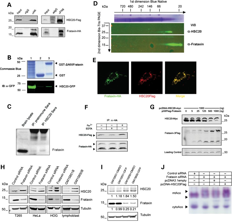
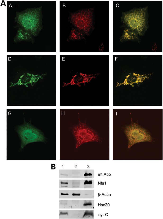
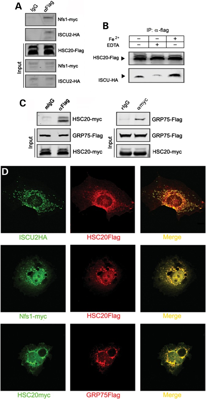
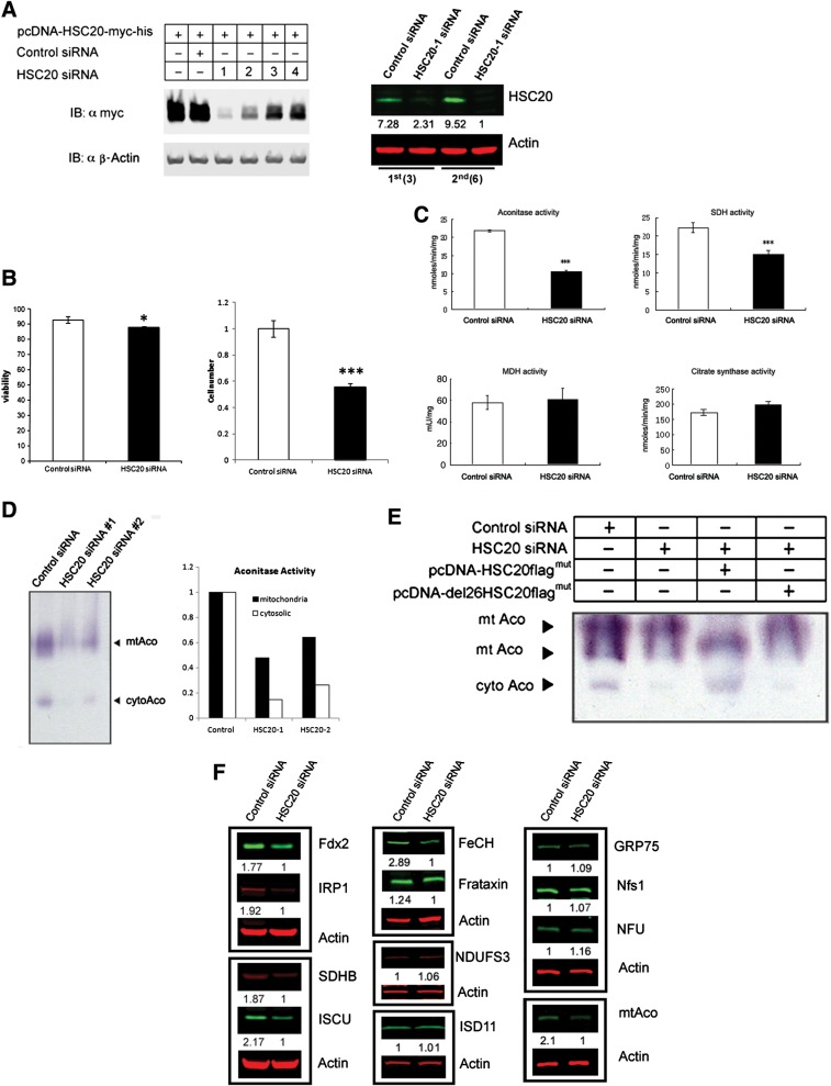
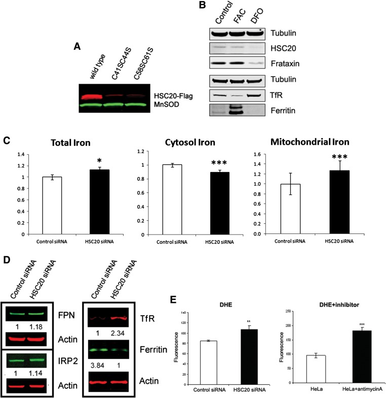
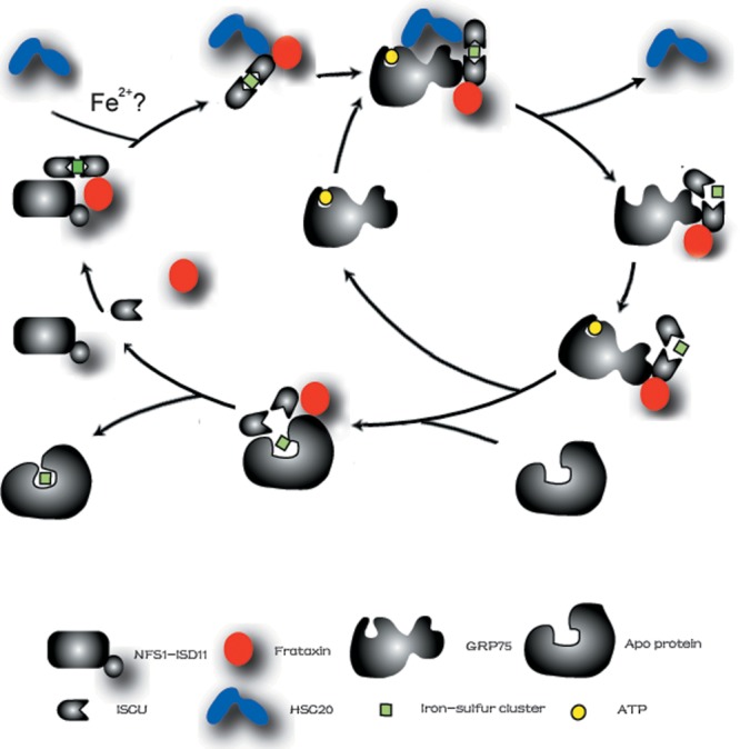
Friedreich's ataxia is a neurodegenerative disorder caused by mutations in the frataxin gene that produces a predominantly mitochondrial protein whose primary function appears to be mitochondrial iron-sulfur cluster (ISC) biosynthesis. Previously we demonstrated that frataxin interacts with multiple components of the mammalian ISC assembly machinery. Here we demonstrate that frataxin interacts with the mammalian mitochondrial chaperone HSC20. We show that this interaction is iron-dependent. We also show that like frataxin, HSC20 interacts with multiple proteins involved in ISC biogenesis including the ISCU/Nfs1 ISC biogenesis complex and the GRP75 ISC chaperone. Furthermore, knockdown of HSC20 caused functional defects in activity of mitochondrial ISC-containing enzymes and also defects in ISC protein expression. Alterations up or down of frataxin expression caused compensatory changes in HSC20 expression inversely, as expected of two cooperating proteins operating in the same pathway and suggesting a potential therapeutic strategy for the disease. Knockdown of HSC20 altered cytosolic and mitochondrial iron pools and increased the expression of transferrin receptor 1 and iron regulatory protein 2 consistent with decreased iron bioavailability. These results indicate that HSC20 interacts with frataxin structurally and functionally and is important for ISC biogenesis and iron homeostasis in mammals. Furthermore, they suggest that HSC20 may act late in the ISC pathway as a chaperone in ISC delivery to apoproteins and that HSC20 should be included in multi-protein complex studies of mammalian ISC biogenesis.


Figures

Figure 1.
Figure 1.
Homologs and functions of ISC proteins. Homologs of frataxin and frataxin-related iron–sulfur proteins are shown, and possible functions schematized. Inferred from Ref. (24).
Figure 2.
Figure 2.
Cloning and expression of HSC20. (A) Amino-acid sequence alignment of HSC20 from different species. The identical sequences are boxed in black and the similar are in gray. Four conserved cysteines were marked with an asterisk. (B) Motif analysis of HSC20 proteins from different species. (C) Phylogenetic analysis of evolutionary relationships among homologs of HSC20 proteins from different species. (D) HSC20 expression patterns in mitochondria of different mouse tissue and human cell line. (E) HSC20 protein overexpression in HEK293T cells. HEK293T cells were transfected with HSC20-EGFP, HSC20-Flag, HSC20-myc, empty EGFP, Frataxin-3Flag, Frataxin-myc. Left: cells were lysed and western blotting was performed with anti-myc (green) and anti-Flag (red) antibodies (upper panel), and then the same blot was stripped and probed with anti-HSC20 (green, bottom panel). Right: cell lysates were probed with anti-GFP (red) and HSC20 (green). (F) Mass spectrometry analysis was performed on immunoprecipitated HSC20. Two bands showed on the gel. P, precursor; M, mature form. The 25 kDa mature form band was sequenced and the position of obtained N-terminal residues (underlined) within the HSC20 sequence.
Figure 3.
Figure 3.
Frataxin interacts with HSC20. (A) Frataxin interacts with HSC20 in vivo. Interaction between HSC20 and frataxin was identified by co-immunoprecipitation. HEK293T cells were transiently transfected with pcDNA-Frataxin-HA and pcDNA-HSC20-Flag plasmids. After 48 h, cells were lysed and aliquoted and immunoprecipitated with indicated IgG or antibodies, followed by release of the immobile fraction and immunoblotting with anti-Flag (upper tier) and anti-HA antibodies (bottom tier). Input: 5% of the total cell lysates applied for the co-immunoprecipitation. (B) Frataxin interacts with HSC20 in vitro. GST-▵55 frataxin pulls down HSC20-GFP. HEK293T cells were transiently transfected with pEGFPN-HSC20. Expression of GFP-tagged HSC20 is shown (lane 1). GST alone (lane 2) or GST-tagged frataxin (lane 3) was bound to beads and incubated with lysates of HEK293T cells transfected with pEGFPN-HSC20. Bead-bound protein complexes were subjected to immunoblotting. The gel was stained with Coomassie blue (upper tier) and the blot was probed with polyclonal anti-GFP antibody (bottom tier). (C) Endogenous interaction between frataxin and HSC20. YG8 mouse brains were homogenized with CHAPS buffer and immunoprecipitated with preimmune sera or HSC20 sera, followed by immunoblotting with anti-frataxin antibody. (D) Frataxin co-isolates with HSC20. Blue-native and SDS–PAGE 2D was applied to separate mitochondrial proteins. The native fraction was separated on the gradient gel in the first dimension, then the gel strip was stacked over Bis–Tris gradient gel and separated, the blot was immunoblotted against the HSC20 and frataxin. (E) Frataxin co-localizes with HSC20. COS7 cells were transfected with Frataxin-HA and HSC20-Flag constructs. Then the cells were analyzed by immunofluorescence with monoclonal anti-Flag/goat anti-mouse rhodamine (red) and rabbit anti-HA/goat anti-rabbit fluorescein conjugate (green). The yellow figure is a merge of two figures. (F) Binding of HSC20 to frataxin is increased by iron. HEK293T cells were transfected with frataxin-HA and HSC20-Flag constructs and lysed/washed by HEPES buffer with corresponding components above the figure. Cell lysates were immunoprecipitated with anti-HA antibody and immunoblotted with anti-Flag antibody and HA antibody. (G) Frataxin inhibits the expression of HSC20. HEK293T cells were seeded in a 12-well plate and transfected with corresponding amount of Frataxin-Flag or HSC20myc plasmid. The cells were harvested after 48 h and probed by anti-Flag and anti-myc antibodies. (H) HSC20 expression increased in frataxin-deficient cells. Left three panels: T265 (Schwann's cell), HeLa and HOG cells were transfected with 50 nm scramble negative control siRNA or frataxin siRNA. Right panels: the lymphoblasts from the FRDA patient (GM15850) and his healthy brother (GM15851) were analyzed. (I) HSC20 was less up-modulated in the patients’ cells in the presence of a mitochondrion-permeable iron chelator DFP (25 µm). (J) Overexpression of HSC20 could overcome a loss of mitochondrial aconitase activity caused by frataxin knockdown. HeLa cells were transfected with corresponding oligos, and 100 µg of proteins were analyzed with Aconitase activity gel staining.
Figure 4.
Figure 4.
HSC20 is targeted to mitochondria. (A) HSC20 co-localizes with mitotracker in COS7 cells. COS7 cells were transfected with pcDNA-HSC20Flag (Top tier) or pcDNA-del26HSC20Flag vector (bottom tier), and analyzed by immunofluorescence with monoclonal anti-Flag/goat anti-mouse FITC (A and G, green) and Mitotrack (B and H, red). The middle tier: COS7 cells were analyzed by HSC20 poly-antibody/goat anti-rabbit FITC (D, green) and Mitotrack (E, red). The yellow pictures in the right column are the merge of the left two pictures, respectively. (B) HSC20 was located in mitochondria. Lane 1, whole-cell lysates of HeLa cells; lane 2, cytosolic lysates of HeLa cells; lane 3, mitochondrial lysates of HeLa cells. The blot was probed with anti-mitochondrial aconitase, anti-Nfs1, anti-β-actin, anti-HSC20 and anti-cytochrome-c antibodies, respectively.
Figure 5.
Figure 5.
HSC20 interacts with GRP75 and theNfs1/ISCU complex. (A) HSC20 interacts with Nfs1/ISCU complex. HEK293T cells were co-transfected with pcDNA-HSC20-Flag, pXS-Nfs1-myc and pSX-ISCU-HA. Cell lysates were immunoprecipitated with mouse IgG or Flag antibody. The precipitates were probed with myc, Flag and HA antibodies. (B) Binding of HSC20 to ISCU is increased by iron. HEK293T cells were transfected with ISCU-HA and HSC20-Flag constructs and lysed/washed by HEPES buffer with corresponding component above the figure. Cell lysates were immunoprecipitated with anti-Flag antibody and immunoblotted with both anti-Flag and HA antibody. (C) HSC20 interacts with GRP75. HEK293T cells were co-transfected with pcDNA-HSC20-myc and pcDNA-GRP75-Flag. Cell lysates were immuprecipitated with mouse IgG or Flag antibody (left), or with rabbit IgG or myc antibody. The precipitates were probed with Flag and HA antibodies. (D) HSC20 co-localizes with Nfs1/ISCU complex and GRP75. Top tier: COS7 cells were co-transfected with pcDNA-ISCU2HA and pcDNA-HSC20Flag; middle tier: COS7 cells were transfected with pXS-Nfs1-myc and pcDNA-HSC20Flag; bottom tier: COS7 cells were co-transfected with pcDNA-HSC20-myc and pcDNA-GRP75-Flag. Then cells were analyzed by immunofluorescence with monoclonal anti-Flag/goat anti-mouse rhodamine (red), rabbit anti-HA(or myc)/goat anti-rabbit fluorescein conjugate (green). Then images were merged (yellow).
Figure 6.
Figure 6.
Silencing HSC20 impedes ISC biogenesis and increases ROS. (A) HSC20-1 siRNA-mediated knockdown reduces both overexpressed HSC20 and endogenous HSC20. Left: HEK293T cells were transfected with the corresponding oligos as above, after 48 h, cell lysates were analyzed by myc and actin antibodies. Right: HeLa cells were transfected two round siRNA (50 nm) and at Days 3 and 6 (from the day that seeded cells), cells were harvested, lysed and cell lysates were analyzed with HSC20 antibody and actin antibody. (B) HSC20 siRNA knockdown decreases cell viability and inhibits cell proliferation. (C) HSC20 knockdown decreases the ISC-containing enzymes aconitase and SDH activities, but not non-ISC-containing enzymes activities. HeLa cells were transfected with 50 nm control siRNA or HSC20 siRNA, after 48 h, whole-cell lysates were used to analyze enzymatic activity (aconitase, malate dehydrogenase, citrate synthase). And for succinate dehydrogenase, mitochondria were isolated and used for the enzymatic analysis. (D) HSC20 knockdown decreases both mitochondrial and cytosolic aconitase activities. HeLa cells were transfected with control siRNA, HSC20-1 siRNA or HSC20-2 siRNA. After 48 h, 100 μg of whole-cell lysates were used to aconitase activity gel analysis. The densitometry of bands were analyzed and showed on the right. (E) Cytosolic form HSC20 cannot recover cytosolic aconitase activity caused by HSC20 knockdown. HeLa cells were transfected with corresponding siRNA and corresponding plasmid as above (mut, both pcDNA-HSC20Flag and pcDNA-del26HSC20Flag plasmids were silently mutated to generate HSC20-1 siRNA-resistant plasmids). After 48 h, 100 μg of whole-cell lysates were used to aconitase activity gel analysis. (F) The proteomic change of ISC biogenesis in HSC20 depleted cells.
Figure 7.
Figure 7.
HSC20 and iron homeostasis. (A). A tetracysteine motif is important for the stability of HSC20. HEK293T cells were transfected with the same amount of the following plasmid: pcDNA-HSC20Flag, pcDNA-HSC20-C41SC44S-Flag and pcDNA-HSC20-C58SC61S-Flag. After 48 h, cells were lysed and 40 µg of protein was analyzed with Flag antibody and MnSOD antibody. (B). Iron chelation decreases the expression of frataxin, HSC20 and ferritin. HeLa cells were grown for 16 h in medium supplemented with 100 µm DFO or 100 µg/ml FAC, and analyzed with corresponding antibodies. (C) Cellular iron concentrations of HeLa cells transfected with control siRNA or HSC20 siRNA. The experiment was repeated at least three times; *P< 0.05, ***P< 0.001. (D). HSC20 knock-down leads to increased TfR1, FPN, IRP2 and decreased ferritin protein levels. (E) Superoxide production in control and HSC20-depleted cells. The experiment was repeated three times and **P< 0.01, ***P< 0.001.
Figure 8.
Figure 8.
The possible functional model of HSC20 and frataxin in the mammalian ISC biogenesis, adapted from Ref. (24). Frataxin and HSC20 were highlighted in red and blue, respectively.

Similar articles

Cited by

References

    1. Harding A.E. Early onset cerebellar ataxia with retained tendon reflexes: a clinical and genetic study of a disorder distinct from Friedreich's ataxia. J. Neurol. Neurosurg. Psychiatry. 1981;44:503–508. doi:10.1136/jnnp.44.6.503. - DOI - PMC - PubMed
    1. Campuzano V., Montermini L., Molto M.D., Pianese L., Cossee M., Cavalcanti F., Monros E., Rodius F., Duclos F., Monticelli A., et al. Friedreich's ataxia: autosomal recessive disease caused by an intronic GAA triplet repeat expansion. Science. 1996;271:1423–1427. doi:10.1126/science.271.5254.1423. - DOI - PubMed
    1. Becker E., Richardson D.R. Frataxin: its role in iron metabolism and the pathogenesis of Friedreich's ataxia. Int. J. Biochem. Cell Biol. 2001;33:1–10. doi:10.1016/S1357-2725(00)00067-4. - DOI - PubMed
    1. Adamec J., Rusnak F., Owen W.G., Naylor S., Benson L.M., Gacy A.M., Isaya G. Iron-dependent self-assembly of recombinant yeast frataxin: implications for Friedreich's ataxia. Am. J. Hum. Genet. 2000;67:549–562. doi:10.1086/303056. - DOI - PMC - PubMed
    1. Puccio H., Simon D., Cossee M., Criqui-Filipe P., Tiziano F., Melki J., Hindelang C., Matyas R., Rustin P., Koenig M. Mouse models for Friedreich's ataxia exhibit cardiomyopathy, sensory nerve defect and Fe–S enzyme deficiency followed by intramitochondrial iron deposits. Nat. Genet. 2001;27:181–186. doi:10.1038/84818. - DOI - PubMed

Publication types

MeSH terms