

Maspin is a deoxycholate-inducible, anti-apoptotic stress-response protein differentially expressed during colon carcinogenesis
- PMID: 22162927
- PMCID: PMC3234125
- DOI: 10.2147/CEG.S24093
Maspin is a deoxycholate-inducible, anti-apoptotic stress-response protein differentially expressed during colon carcinogenesis
Abstract
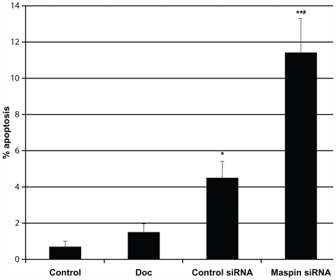
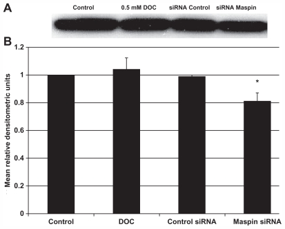
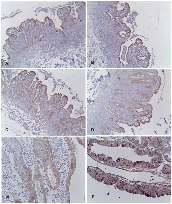
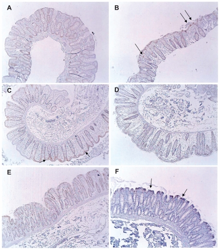
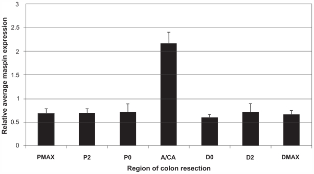
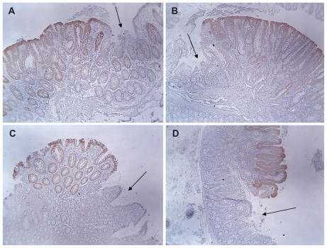
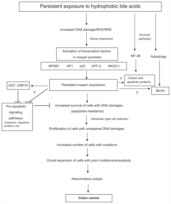
Increased maspin expression in the colon is related to colon cancer risk and patient survival. Maspin is induced by the hydrophobic bile acid, deoxycholate (DOC), which is an endogenous carcinogen and inducer of oxidative stress and DNA damage in the colon. Persistent exposure of colon epithelial cells, in vitro, to high physiologic levels of DOC results in increased constitutive levels of maspin protein expression associated with the development of apoptosis resistance. When an apoptosis-resistant colon epithelial cell line (HCT-116RC) developed in the authors' laboratory was treated with a maspin-specific siRNA probe, there was a statistically significant increase in apoptosis compared to treatment with an siRNA control probe. These results indicate, for the first time, that maspin is an anti-apoptotic protein in the colon. Immunohistochemical evaluation of maspin expression in human colonic epithelial cells during sporadic colon carcinogenesis (131 human tissues evaluated) indicated a statistically significant increase in maspin protein expression beginning at the polyp stage of carcinogenesis. There was no statistically significant difference in maspin expression between hyperplastic/adenomatous polyps and colonic adenocarcinomas. The absence of "field defects" in the non-neoplastic colonic mucosa of patients with colonic neoplasia indicates that maspin may drive the growth of tumors, in part, through its anti-apoptotic function.
Keywords: anti-apoptotic; bile acid-inducible; colon cancer; immunohistochemistry; maspin.
Figures
Similar articles
-
Deoxycholate, an endogenous cytotoxin/genotoxin, induces the autophagic stress-survival pathway: implications for colon carcinogenesis.J Toxicol. 2009;2009:785907. doi: 10.1155/2009/785907. Epub 2009 May 10. J Toxicol. 2009. PMID: 20130808 Free PMC article.
-
Development and molecular characterization of HCT-116 cell lines resistant to the tumor promoter and multiple stress-inducer, deoxycholate.Carcinogenesis. 2002 Dec;23(12):2063-80. doi: 10.1093/carcin/23.12.2063. Carcinogenesis. 2002. PMID: 12507930
-
Identification of S-nitrosylated proteins after chronic exposure of colon epithelial cells to deoxycholate.Proteomics. 2006 Mar;6(5):1654-62. doi: 10.1002/pmic.200500240. Proteomics. 2006. PMID: 16404723
-
Increased GADD gene expression in human colon epithelial cells exposed to deoxycholate.J Cell Physiol. 2005 Jan;202(1):295-303. doi: 10.1002/jcp.20135. J Cell Physiol. 2005. PMID: 15316935
-
Role of apoptosis in biology and pathology: resistance to apoptosis in colon carcinogenesis.Ultrastruct Pathol. 1995 Jul-Aug;19(4):221-48. doi: 10.3109/01913129509064227. Ultrastruct Pathol. 1995. PMID: 7571081 Review.
Cited by
-
TMEM45A, SERPINB5 and p16INK4A transcript levels are predictive for development of high-grade cervical lesions.Am J Cancer Res. 2016 Jul 1;6(7):1524-36. eCollection 2016. Am J Cancer Res. 2016. PMID: 27508094 Free PMC article.
-
Expression and localization of maspin in cervical cancer and its role in tumor progression and lymphangiogenesis.Arch Gynecol Obstet. 2014 Feb;289(2):373-82. doi: 10.1007/s00404-013-2988-4. Arch Gynecol Obstet. 2014. PMID: 23959090 Free PMC article.
-
Maspin is a marker for early recurrence in primary stage III and IV colorectal cancer.Br J Cancer. 2013 Sep 17;109(6):1636-47. doi: 10.1038/bjc.2013.489. Epub 2013 Sep 3. Br J Cancer. 2013. PMID: 24002600 Free PMC article.
-
Deficient expression of DNA repair enzymes in early progression to sporadic colon cancer.Genome Integr. 2012 Apr 11;3(1):3. doi: 10.1186/2041-9414-3-3. Genome Integr. 2012. PMID: 22494821 Free PMC article.
References
-
- Bernstein H, Bernstein C, Payne CM, Garewal H. Recent molecular evidence illuminates the decades-long debate on the role of dietary fat in colon carcinogenesis. J Cancer Integr Med. 2004;2:107–109.
-
- Bernstein H, Bernstein C, Payne CM, Dvorakova K, Garewal H. Bile acids as carcinogens in human gastrointestinal cancers. Mutat Res. 2005;589(1):47–65. - PubMed
-
- Bernstein C, Payne CM, Dvorak K, Bernstein H. Role of bile acids in gastrointestinal carcinogenesis. US Gastroenterol Hepatol Rev. 2008;4:68–72.
-
- Crowley-Weber CL, Payne CM, Gleason-Guzman M, et al. Development and molecular characterization of colon cell lines resistant to the tumor promoter and multiple stress-inducer, deoxycholate. Carcinogenesis. 2002;23(12):2063–2080. - PubMed
Grants and funding
LinkOut - more resources
Full Text Sources
