. 2012 Feb 10;287(7):4726-39.
doi: 10.1074/jbc.M111.260349. Epub 2011 Dec 9.

Regenerating islet-derived 1α (Reg-1α) protein is new neuronal secreted factor that stimulates neurite outgrowth via exostosin Tumor-like 3 (EXTL3) receptor

Affiliations

Regenerating islet-derived 1α (Reg-1α) protein is new neuronal secreted factor that stimulates neurite outgrowth via exostosin Tumor-like 3 (EXTL3) receptor

Isabelle Acquatella-Tran Van Ba et al. J Biol Chem. .

Abstract

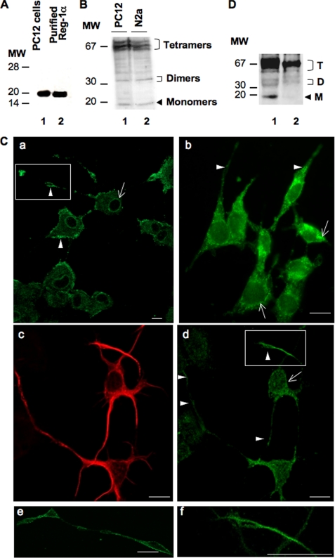
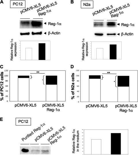
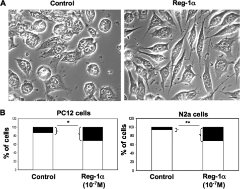
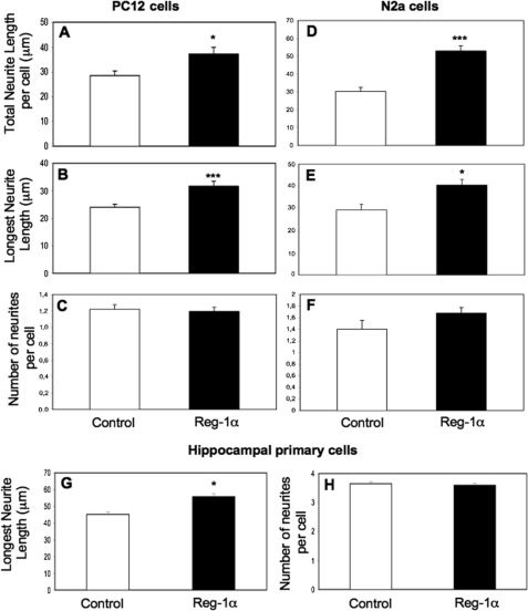
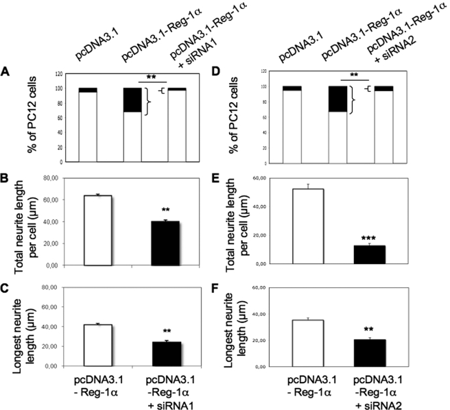
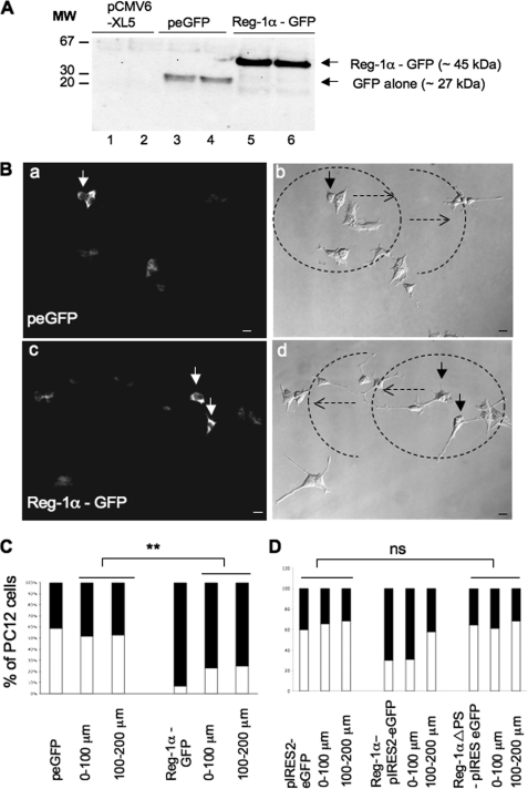
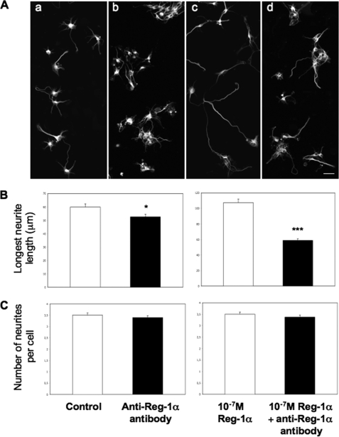
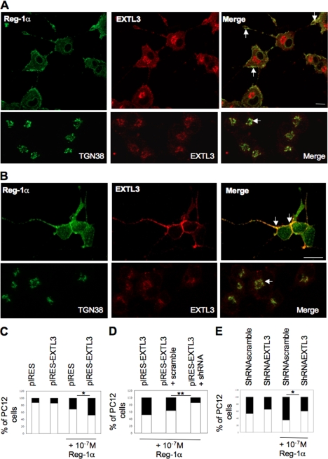
Regenerating islet-derived 1α (Reg-1α)/lithostathine, a member of a family of secreted proteins containing a C-type lectin domain, is expressed in various organs and plays a role in proliferation, differentiation, inflammation, and carcinogenesis of cells of the digestive system. We previously reported that Reg-1α is overexpressed during the very early stages of Alzheimer disease, and Reg-1α deposits were detected in the brain of patients with Alzheimer disease. However, the physiological function of Reg-1α in neural cells remains unknown. Here, we show that Reg-1α is expressed in neuronal cell lines (PC12 and Neuro-2a) and in rat primary hippocampal neurons (E17.5). Reg-1α is mainly localized around the nucleus and at the membrane of cell bodies and neurites. Transient overexpression of Reg-1α or addition of recombinant Reg-1α significantly increases the number of cells with longer neurites by stimulating neurite outgrowth. These effects are abolished upon down-regulation of Reg-1α by siRNA and following inhibition of secreted Reg-1α by antibodies. Moreover, Reg-1α colocalizes with exostosin tumor-like 3 (EXTL3), its putative receptor, at the membrane of these cells. Overexpression of EXTL3 increases the effect of recombinant Reg-1α on neurite outgrowth, and Reg-1α is not effective when EXTL3 overexpression is down-regulated by shRNA. Our findings indicate that Reg-1α regulates neurite outgrowth and suggest that this effect is mediated by its receptor EXTL3.


Figures

FIGURE 1.
FIGURE 1.
Reg-1α is expressed in PC12 and N2a cells as well as in embryonic hippocampal neurons (E17.5; 2 days in vitro). A, total cell protein extracts (20 μg) from differentiated PC12 cells grown in the presence of 50 ng/ml NGF for 48 h were separated by 15% SDS-PAGE, and Western blot analysis was performed using a rabbit polyclonal anti-Reg-1α antibody. Molecular weight markers (MW) are indicated on the left. Lane 1, Reg-1α in PC12 cells has an apparent molecular mass of ∼20 kDa; lane 2, the specificity of the anti-Reg-1α antibody was tested using purified human recombinant Reg-1α, which shows an apparent molecular mass of 18 kDa. B, PC12 (lane 1) and N2a (lane 2) cell extracts were separated by 12.5% SDS-PAGE, and Western blot analysis using the rabbit polyclonal anti-Reg-1α antibody shows three clusters of Reg-1α expression with molecular masses of ∼70, 35, and 20 kDa (tetramers, dimers, and monomers of Reg-1α). Molecular weight markers (MW) are indicated on the left. C, immunofluorescence analysis in differentiated PC12 (panels a and e), N2a (panel b) cells, and hippocampal neurons (panels c, d, and f), which express β3-tubulin (panel c), show that Reg-1α is mainly localized at the plasma membrane (panels a, b, and d, arrowheads), particularly in growth cones (panels e and f; higher magnifications of the insets in panels a and d) and in the perinuclear region (panels a, b, and d, arrows). Images were visualized by confocal microscopy (Z-projections of three confocal optical sections; intervals, 0.6 μm). Scale bars, 10 μm. D, immunoblot analysis of membrane (lane 1) and cytoplasm (lane 2) fractions from differentiated PC12 cells using the polyclonal anti-Reg-1α antibody. Tetramers (T), dimers (D), and monomers (M) of Reg-1α are indicated. Molecular weight markers (MW) are indicated on the left.
FIGURE 2.
FIGURE 2.
Overexpression of Reg-1α increases number of PC12 and N2a cells considered as differentiated and secretion of Reg-1α. Total cell protein extracts obtained from PC12 (A) and N2a (B) cells transfected with control empty vector (pCMV6-XL5) or with Reg-1α (pCMV6-XL5-Reg-1α) were resolved by SDS-PAGE and analyzed by Western blotting using the polyclonal anti-Reg-1α antibody. Actin was used as a loading control. Histograms (A and B) show the quantification of Reg-1α relative expression. Quantification of the percentage of undifferentiated (white) and differentiated (i.e. when neurite length is twice that of the cell body) (black) PC12 (C) and N2a (D) cells shows an increase in the number of cells considered as differentiated upon overexpression of Reg-1α but not of empty pCMV6-XL5 vector. Asterisks indicate significant differences from controls based on Student's t test analysis (**, p < 0.01). E, 20 mg of proteins from freeze-dried culture medium from PC12 cells transfected with control empty vector (pCMV6-XL5) or with Reg-1α (pCMV6-XL5-Reg-1α) were resolved by SDS-PAGE and analyzed by Western blotting using the polyclonal anti-Reg-1α antibody. Histograms show the quantification of Reg-1α relative expression.
FIGURE 3.
FIGURE 3.
Human recombinant Reg-1α increases number of PC12 and N2a cells with longer neurites. A, phase-contrast images of N2a cells in culture. Note the increase of neurite length 48 h after addition of 10−7 m Reg-1α in the medium (right panel) compared with untreated, control cells (left panel). Scale bars, 10 μm. B, quantification of the percentage of undifferentiated (white) and differentiated (i.e. neurite length is twice that of the cell body) (black) PC12 and N2a cells. Treatment with 10−7 m recombinant Reg-1α increases the number of cells with longer neurites (black) in comparison with untreated controls. Asterisks indicate significant differences from controls based on the Student's t test analysis (*, p < 0.05; **, p < 0.01).
FIGURE 4.
FIGURE 4.
Recombinant Reg-1α increases outgrowth of PC12 and N2a cells as well as of rat embryonic hippocampal neurons. Total neurite length per cell (A and D), length of the longest neurite (B, E, and G), and number of neurites (C, F, and H) per cell were measured and are expressed as mean ± S.E. Recombinant Reg-1α (10−7 m) significantly increases the total neurite length (A and D) and longest neurite length (B, E, and G) but does not influence the number of neurites per cell (C, F, and H). Asterisks indicate significant differences from controls based on the Student's t test analysis (*, p < 0.05; ***, p < 0.001). White, control cells; black, Reg-1α-treated cells. Results are representative of three independent experiments.
FIGURE 5.
FIGURE 5.
Down-regulation of overexpressed Reg-1α by siRNAs reduces neurite outgrowth in NGF-treated PC12 cells. Transfection of the two anti-Reg-1α siRNA duplexes (siRNA1, A–C; and siRNA2, D–F) decreased the percentage of cells considered as differentiated (i.e. neurite length is twice that of the cell body) (A and D, black rectangles), the total neurite length (B and E), and the longest neurite length (C and F) per cell. Asterisks indicate significant differences from controls (pcDNA3.1-Reg-1α) based on the Student's t test analysis (**, p < 0.01; ***, p < 0.001). Results are representative of three independent experiments.
FIGURE 6.
FIGURE 6.
Western blot and immunofluorescence analysis of PC12 cells that overexpress Reg-1α and of untransfected neighboring cells. A, PC12 cells were transfected with empty pCMV6-XL5 (lanes 1 and 2) or peGFP (lanes 3 and 4) vector or Reg-1α-GFP (lanes 5 and 6) (see “Materials and Methods”). Cell lysates were collected 48 h after transfection and immunoblotted with the anti-GFP antibody to determine the transfection efficacy. Molecular weight markers (MW) are indicated on the left. B, GFP immunostaining shows that PC12 cells that overexpress Reg-1α-GFP have longer neurites (panel c, arrows) than GFP-transfected cells (panel a, arrow). Panels b and d show the concentric circles around transfected cells (dotted arrows) every 100 μm (0–100 and 100–200 μm). Scale bars, 10 μm. C, quantification of the percentage of undifferentiated (white) and differentiated (i.e. neurite length is twice that of the cell body) (black) cells shows an increase of the number of PC12 cells considered as differentiated when they overexpress Reg-1α-GFP and also of the number of their neighboring cells considered as differentiated in comparison with control, peGFP-transfected cells. Asterisks indicate significant differences from controls following Student's t test analysis (**, p < 0.01). D, quantification of the percentage of undifferentiated (white) and differentiated (i.e. neurite length is twice that of the cell body) (black) cells shows that overexpression of the mutant Reg-1α ΔPS-pIRESeGFP, which lacks the signal peptide, has no effect on the differentiation of transfected PC12 cells and of neighboring cells. ns, no significant differences from controls (Student's t test).
FIGURE 7.
FIGURE 7.
Anti-Reg-1α polyclonal antibody blocks effects of secreted and recombinant Reg-1α on hippocampal neurite outgrowth. A, immunofluorescence analysis of hippocampal neurons stained by β3-tubulin shows that the anti-Reg-1α antibody (10−6 m) (panels b and d) hinders neurite elongation when compared with control cells (untreated) (panel a) and with cells incubated with 10−7 m recombinant Reg-1α (panel c). Scale bar, 10 μm. B and C, the anti-Reg-1α antibody (10−6 m) significantly decreases the effect of secreted (B, left panel) and recombinant (B, right panel) Reg-1α on the longest neurite length but does not influence the number of neurites per cell (C). Asterisks indicate significant differences (Student's t test; *, p < 0.05; ***, p < 0.001). White, control cells (no antibody); black, Reg-1α antibody-treated cells. Results are representative of three independent experiments.
FIGURE 8.
FIGURE 8.
EXTL3 is expressed in PC12 cells and hippocampal neurons and mediates Reg-1α effect on neurite elongation. A and B, immunofluorescence analysis in PC12 cells considered as differentiated (A, upper panels) and hippocampal neurons (B, upper panels) shows that Reg-1α (green) colocalizes at the plasma membrane with EXTL3 (red) (Merge, arrows). EXTL3 (red) (A and B, lower panels) is also expressed in the Golgi apparatus and colocalizes with TGN38 (green), a specific marker of the Golgi apparatus (Merge, arrows). Images were visualized by confocal microscopy (Z-projections of three confocal optical sections; intervals, 0.6 μm). Scale bar, 10 μm. C, quantification shows that the increase in the percentage of cells with longer neurites (black rectangles) is significantly higher when recombinant Reg-1α is added to the medium of cells that overexpress EXTL3 (pIRES-EXTL3 + Reg-1α) rather than empty vector (pIRES + Reg-1α). Overexpression of EXTL3 alone (pIRES-EXTL3) does not modify the percentage of cells with longer neurites when compared with overexpression of empty vector (pIRES). D, quantitative analysis of undifferentiated (white) and differentiated (i.e. neurite length is twice that of the cell body) (black) PC12 cells shows that Reg-1α is less effective when EXTL3 is down-regulated (pIRES-EXTL3 + shRNA) as the percentage of cells considered as differentiated is significantly lower in these cells than in PC12 cells that overexpress EXTL3 (pIRES-EXTL3) or in cells co-transfected with EXTL3 and a control shRNA (pIRES-EXTL3 + scramble). Asterisks indicate significant differences (Student's t test; *, p < 0.05; **, p < 0.01). E, quantification shows that the increase in the percentage of cells with longer neurites (black rectangles) is significantly lower when recombinant Reg-1α is added to the medium of cells when endogenous EXTL3 is down-regulated (shRNAEXTL3 + Reg-1α versus shRNAscramble + Reg-1α). Down-regulation of EXTL3 alone (shRNAEXTL3) induces a slight decrease of the percentage of cells with longer neurites when compared with transfection of a non-effective shRNA (shRNAscramble). Asterisks indicate significant differences (Student's t test; *, p < 0.05).

Similar articles

Cited by

References

    1. Watanabe T., Yonekura H., Terazono K., Yamamoto H., Okamoto H. (1990) Complete nucleotide sequence of human reg gene and its expression in normal and tumoral tissues. The reg protein, pancreatic stone protein, and pancreatic thread protein are one and the same product of the gene. J. Biol. Chem. 265, 7432–7439 - PubMed
    1. Laurine E., Manival X., Montgelard C., Bideau C., Bergé-Lefranc J. L., Erard M., Verdier J. M. (2005) PAP IB, a new member of the Reg gene family: cloning, expression, structural properties, and evolution by gene duplication. Biochim. Biophys. Acta 1727, 177–187 - PubMed
    1. De Caro A., Lohse J., Sarles H. (1979) Characterization of a protein isolated from pancreatic calculi of men suffering from chronic calcifying pancreatitis. Biochem. Biophys. Res. Commun. 87, 1176–1182 - PubMed
    1. Bimmler D., Graf R., Scheele G. A., Frick T. W. (1997) Pancreatic stone protein (lithostathine), a physiologically relevant pancreatic calcium carbonate crystal inhibitor? J. Biol. Chem. 272, 3073–3082 - PubMed
    1. Geider S., Baronnet A., Cerini C., Nitsche S., Astier J. P., Michel R., Boistelle R., Berland Y., Dagorn J. C., Verdier J. M. (1996) Pancreatic lithostathine as a calcite habit modifier. J. Biol. Chem. 271, 26302–26306 - PubMed

Publication types

MeSH terms