. 2012 Apr;40(7):3006-17.
doi: 10.1093/nar/gkr1197. Epub 2011 Dec 7.

Regulation of polysome assembly on the endoplasmic reticulum by a coiled-coil protein, p180

Affiliations

Regulation of polysome assembly on the endoplasmic reticulum by a coiled-coil protein, p180

Tomonori Ueno et al. Nucleic Acids Res. 2012 Apr.

Abstract

A coiled-coil microtubule-bundling protein, p180, was originally identified as one of the ribosome receptor candidates on the rough endoplasmic reticulum (ER) and is highly expressed in secretory tissues. Recently, we reported that p180 plays crucial roles in upregulating collagen biosynthesis, mainly by facilitating ribosome association on the ER. Here, we provide evidence that p180 is required to form translationally active polysome/translocon complexes on the ER. Assembly of highly-developed polysomes on the ER was severely perturbed upon loss of p180. p180 associates with polysome/translocon complexes through multiple contact sites: it was coimmunoprecipitated with the translocon complex independently of ribosomes, while it can also bind to ribosomal large subunit specifically. The responsible domain of p180 for membrane polysome assembly was identified in the C-terminal coiled-coil region. The degree of ribosome occupation of collagen and fibronectin mRNAs was regulated in response to increased traffic demands. This effect appears to be exerted in a manner specific for a specified set of mRNAs. Collectively, our data suggest that p180 is required to form translationally active polysome/translocon complexes on the ER membrane, and plays a pivotal role in highly efficient biosynthesis on the ER membrane through facilitating polysome formation in professional secretory cells.


Figures
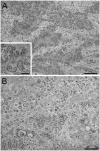
Figure 1.
Figure 1.
Transmission EM images of the rough ER surface in collagen-secreting HEL fibroblasts. Control (A) and p180 siRNA-transfected (B) HEL cells were grown in F12/DMEM containing 2% FBS in the presence of ascorbate. A large number of highly developed polysomes are clearly seen on the membrane surface in ascorbate-stimulated HEL fibroblasts, while p180 depletion results in dramatically reduced numbers of polysomes on the ER. Polysomes are frequently seen in the membrane surface view when the sections were cut parallel to the substrate. A higher magnification image of control cells (inset in A) shows spiral arrays of polysomes consisting of about 25–30 ribosomes. Bars: A and B, 500 nm: inset in A, 200 nm.
Figure 2.
Figure 2.
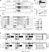
p180 facilitates polysome assembly on the ER membrane. HEL cells were cultured in the presence or absence of ascorbate (AA) and treated with a control scrambled (cont-si) or p180-specific (p180-si) siRNA. At post-transfection Day 4, sequential digitonin extractions were carried out to obtain the membrane fractions, followed by polysome analysis using a 15–50% sucrose gradient. Equal amounts of samples normalized by the total DNA contents were subjected to the polysome analysis. (A) The ribosomal RNAs in each fraction were analyzed by agarose electrophoresis. The positions of the polysome (P), monosome (M), free 40S and 60S subunits (F) and light (L) fractions are shown at the top. Top panel: cont-si-treated cells without ascorbate; middle panel: cont-si-treated cells with ascorbate; bottom panel: p180-si-treated cells with ascorbate. (B) The total RNA contents of the polysome fractions were estimated by densitometric scanning of the gels, and the relative ratios of polysomes to 80S monosome are shown (means ± SD, n = 4). (C) The relative amounts of p180 and polysomes in the membrane fractions were plotted to evaluate their correlation. The 12 samples used were obtained from five separate experiments under various conditions. The values for cont-si-treated cells without ascorbate stimulation were set as 1. (D) The distributions of p180 and other translocon-related proteins in the collected fractions were analyzed by western blotting. Non-stimulated cells with cont-si (left), ascorbate-stimulated cells with cont-si (middle), and ascorbate-stimulated p180-si treated cells (right) are shown. (E) Quantitative data for the protein markers in the polysome (P), monosome (M), free 40S and 60S subunits (F) and light (L) fractions are shown. The total amounts of each protein in the membrane fractions were estimated by densitometry using samples prior to sucrose density centrifugation, and their relative contents were assigned to the P, M, F and L fractions by densitometric scanning of the data shown in panel D. The total amounts for the membrane fractions from the cont-si-treated cells without ascorbate stimulation were set as 1. (F) Polysome fractions were collected from the cont-si treated cells after ascorbate stimulation and subjected to IP assays using an anti-p180 antibody or control rabbit IgG bound to magnetic beads conjugated with sheep anti-rabbit IgG. The samples captured by the beads were immunoblotted with antibodies against marker proteins or analyzed for rRNAs. Lane 1, input samples; lane 2, immunoprecipitates with control IgG; lane 3, immunoprecipitates with anti-p180 antibody. Sec61α was not visible because of co-existing immunoglobulin chains.
Figure 3.
Figure 3.
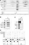
Independent association capacity of p180 with the translocon and ribosomes. (A) After removal of the cytosolic fractions with the permeabilization and wash buffers, the membrane fractions of ascorbate-treated cells were either left untreated or stripped of ribosomes by in situ EDTA treatment. Subsequently, membrane fractions were prepared with lysis buffer containing digitonin. The control (untreated) or ribosome-stripped membrane fractions were analyzed on a 5–20% sucrose gradient at 0.4 M KOAc. Marker proteins on western immunoblots and rRNAs in agarose gels analyzed for control membranes (left panel) and ribosome-stripped membranes (right panel) are shown. Flow schema for preparing the ribosome-stripped and untreated membranes are shown in Supplementary Figure S4. (B) Ribosome-stripped membrane fractions were prepared as described for (A) and subsequently centrifuged through a 0.5 M sucrose cushion to remove residual ribosomes. The resulting ribosome-free membrane fractions were subjected to IP analysis with control IgG (lane 2), an anti-p180 antibody (lane 3) and an anti-Sec61β antibody (lane 4) in the presence of 0.4 M KOAc. In the lanes containing the IP samples 10 times higher amounts were loaded compared with the input samples (lane 1). Flow schema for preparing the ribosome-free membranes are shown in Supplementary Figure S4. (C) Ribosome-stripped membrane fractions prepared with 1% NP-40 lysis buffer were further treated with 0.5% deoxycholate and 0.1% SDS at 4°C for 1 h to dissociate possible complexes in the presence of 0.4 M KOAc. After sedimentation at 100 000g for 40 min through a 0.5 M sucrose cushion, the supernatants were subjected to the IP assay with an anti-p180 antibody to prepare p180-beads. Marker proteins were analyzed for the input sample (lane 1), p180-beads (lane 2) and control-beads (lane 3) to confirm the loss of the translocon-related proteins. (D) The p180-beads or control-beads were incubated with isolated monosomes or the subunits at 4°C for 1 h at 0.4 M KOAc. After careful washing, the tested beads were recovered by a magnet and analyzed for rRNA by agarose electrophoresis and for p180 by western blotting. The monosome, 60S and 40S preparations contained no detectable levels of Sec61β, ribophorin II, TRAPα or p180 (data not shown). Lanes 1, 5 and 9: input p180-beads; lane 2: input 80S ribosomes; lanes 6 and 10: input ribosome subunits; lanes 3, 4, 7, 8, 11 and 12: recovered bead fractions after incubation.
Figure 4.
Figure 4.
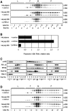
Manipulation of the p180 level affects membrane-associated polysomes. The amounts of ER-associated polysomes were analyzed using stable HeLa transfectants overexpressing p180 (HeLa/p180) and a control cell line (HeLa/puro). (A) The membrane fractions were obtained by sequential digitonin extractions from cont-si-treated HeLa/puro cells, cont-si-treated HeLa/p180 cells and p180-si-treated HeLa/p180 cells, and subjected to polysome analyses using a 15–50% sucrose gradient. (B) The RNA contents of the polysome fractions were estimated by densitometric scanning of the gels, and the relative ratios of polysome:80S are shown. (C) The distributions of p180 and other translocon-related proteins in the collected fractions were analyzed by western blotting. Data for cont-si-treated HeLa/puro cells (left), cont-si-treated HeLa/p180 cells (middle) and p180-si-treated HeLa/p180 cells (right) are presented. (D) The cytosolic fractions were subjected to polysome analyses on a 15–50% sucrose gradient, and the rRNAs were analyzed.
Figure 5.
Figure 5.
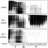
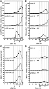
p180 depletion perturbs ascorbate-stimulated de novo biosynthesis in polysome fractions on the ER membrane. The de novo biosynthesis activity was estimated using a non-RI labeling system with AHA in HEL cells. The cells were cultured in the presence or absence of ascorbate and treated with a control or p180-specific siRNA. After incubation with AHA, the membrane fractions were fractionated on a 15–50% sucrose gradient. The AHA-incorporating proteins were labeled with biotin-alkyne by click chemistry and immunoblotted with an anti-biotin antibody. Top panel: non-stimulated cells with cont-si; middle panel: ascorbate-stimulated cells with cont-si; bottom panel: ascorbate-stimulated cells with p180-si.
Figure 6.
Figure 6.
Ribosome occupation of mRNAs for specific proteins is accelerated upon ascorbate treatment in a p180-dependent manner. HEL cells were cultured in the presence or absence of ascorbate and treated with a control siRNA or p180-specific siRNA. The membrane fractions obtained by sequential digitonin extractions were subjected to polysome analyses using a 15–50% sucrose gradient. cDNAs were synthesized from RNA samples extracted from individual tubes. The distributions of mRNAs from non-stimulated cells with cont-si, ascorbate-treated cells with cont-si and ascorbate-treated cells with p180-si are plotted. Relative signal intensity of mRNAs for procollagen type 1 alpha chain (A), fibronectin (B), MMP-2 (C) and TIMP-1 (D) are shown. M, monosome fraction; P, polysome fraction.
Figure 7.
Figure 7.
Cytoplasmic expression of the coiled-coil domain perturbs polysome assembly on the membranes. (A) Schematic representations of full-length and truncated constructs of various regions of p180. Human p180 has a predicted transmembrane domain (TM) close to the N-terminus, a highly basic tandem repeat domain (dark gray box), a microtubule binding and bundling domain (MTB-1) and a C-terminal acidic coiled-coil domain (gray boxes). The numbers on the left of each truncated mutant denote the amino acid residues of human p180 (DDBJ accession number: AB287347). (B) A series of GFP-tagged polypeptides containing different regions of human p180 were expressed in HEL cells, and the rRNAs (indicated by 28S and 18S, respectively) in the cytosolic and membrane fractions are shown. (C) The RNA contents in the cytosolic and membrane-bound fractions were estimated by densitometric scanning, and the relative amounts are shown. Data represent means ± SD (n = 3). (D) Polysome analyses were performed using the membrane fractions of cells overexpressing peptides containing amino acid residues 945–1133, 1134–1293 and 1300–1540 compared with those of control cells or cells overexpressing control peptides. Arrowheads indicate 28S (upper) and 18S (lower) rRNAs. (E) The relative ratios of polysomal RNA to 80S RNA are shown. (F) Schematic representations of wild-type and various C-terminal deletion mutants of p180. (G) Expression plasmids encoding wild-type p180 and p180 mutants were transfected into HeLa cells. The rRNAs in the cytosolic and membrane fractions were analyzed. (H) The relative RNA contents in the cytosolic and membrane-bound fractions are shown. Data represent means ± SD (n = 3). (I) Polysome analyses were performed using the membrane fractions of these cells. Arrowheads indicate 28S (upper) and 18S (lower) rRNAs. (J) The relative ratios of polysomal RNA to 80S RNA are shown.

Similar articles

Cited by

References

    1. Diehn M, Bhattacharya R, Botstein D, Brown PO. Genome-scale identification of membrane-associated human mRNAs. PLoS Genet. 2006;2:39–50. - PMC - PubMed
    1. Diehn M, Eisen MB, Botstein D, Brown PO. Large-scale identification of secreted and membrane-associated gene products using DNA microarrays. Nat. Genet. 2000;25:58–62. - PubMed
    1. Stephens SB, Nicchitta CV. Divergent regulation of protein synthesis in the cytosol and endoplasmic reticulum compartments of mammalian cells. Mol. Biol. Cell. 2008;19:623–632. - PMC - PubMed
    1. Sonenberg N, Hinnebusch AG. Regulation of translation initiation in eukaryotes: mechanisms and biological targets. Cell. 2009;136:731–745. - PMC - PubMed
    1. Gebauer F, Hentze MW. Molecular mechanisms of translational control. Nat. Rev. Mol. Cell. Biol. 2004;5:827–835. - PMC - PubMed

Publication types

MeSH terms