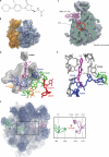
Resistance to linezolid caused by modifications at its binding site on the ribosome
- PMID: 22143525
- PMCID: PMC3264260
- DOI: 10.1128/AAC.05702-11
Resistance to linezolid caused by modifications at its binding site on the ribosome
Abstract
Linezolid is an oxazolidinone antibiotic in clinical use for the treatment of serious infections of resistant Gram-positive bacteria. It inhibits protein synthesis by binding to the peptidyl transferase center on the ribosome. Almost all known resistance mechanisms involve small alterations to the linezolid binding site, so this review will therefore focus on the various changes that can adversely affect drug binding and confer resistance. High-resolution structures of linezolid bound to the 50S ribosomal subunit show that it binds in a deep cleft that is surrounded by 23S rRNA nucleotides. Mutation of 23S rRNA has for some time been established as a linezolid resistance mechanism. Although ribosomal proteins L3 and L4 are located further away from the bound drug, mutations in specific regions of these proteins are increasingly being associated with linezolid resistance. However, very little evidence has been presented to confirm this. Furthermore, recent findings on the Cfr methyltransferase underscore the modification of 23S rRNA as a highly effective and transferable form of linezolid resistance. On a positive note, detailed knowledge of the linezolid binding site has facilitated the design of a new generation of oxazolidinones that show improved properties against the known resistance mechanisms.
Figures


Similar articles
-
Oxazolidinone resistance mutations in 23S rRNA of Escherichia coli reveal the central region of domain V as the primary site of drug action.J Bacteriol. 2000 Oct;182(19):5325-31. doi: 10.1128/JB.182.19.5325-5331.2000. J Bacteriol. 2000. PMID: 10986233 Free PMC article.
-
R chi-01, a new family of oxazolidinones that overcome ribosome-based linezolid resistance.Antimicrob Agents Chemother. 2008 Oct;52(10):3550-7. doi: 10.1128/AAC.01193-07. Epub 2008 Jul 28. Antimicrob Agents Chemother. 2008. PMID: 18663023 Free PMC article.
-
Ribosomal and non-ribosomal resistance to oxazolidinones: species-specific idiosyncrasy of ribosomal alterations.Mol Microbiol. 2002 Dec;46(5):1295-304. doi: 10.1046/j.1365-2958.2002.03242.x. Mol Microbiol. 2002. PMID: 12453216
-
Linezolid update: stable in vitro activity following more than a decade of clinical use and summary of associated resistance mechanisms.Drug Resist Updat. 2014 Apr;17(1-2):1-12. doi: 10.1016/j.drup.2014.04.002. Epub 2014 Apr 6. Drug Resist Updat. 2014. PMID: 24880801 Review.
-
Oxazolidinones: activity, mode of action, and mechanism of resistance.Int J Antimicrob Agents. 2004 Feb;23(2):113-9. doi: 10.1016/j.ijantimicag.2003.11.003. Int J Antimicrob Agents. 2004. PMID: 15013035 Review.
Cited by
-
Mycobacterial RNA isolation optimized for non-coding RNA: high fidelity isolation of 5S rRNA from Mycobacterium bovis BCG reveals novel post-transcriptional processing and a complete spectrum of modified ribonucleosides.Nucleic Acids Res. 2015 Mar 11;43(5):e32. doi: 10.1093/nar/gku1317. Epub 2014 Dec 24. Nucleic Acids Res. 2015. PMID: 25539917 Free PMC article.
-
Clinical Characteristics and Drug Resistance Mechanisms of Linezolid-Non-Susceptible Enterococcus in a Tertiary Hospital in Northwest China.Infect Drug Resist. 2024 Feb 8;17:485-494. doi: 10.2147/IDR.S442105. eCollection 2024. Infect Drug Resist. 2024. PMID: 38348228 Free PMC article.
-
Efficacy and clinical potential of phage therapy in treating methicillin-resistant Staphylococcus aureus (MRSA) infections: A review.Eur J Microbiol Immunol (Bp). 2024 Feb 2;14(1):13-25. doi: 10.1556/1886.2023.00064. Print 2024 Feb 23. Eur J Microbiol Immunol (Bp). 2024. PMID: 38305804 Free PMC article. Review.
-
Molecular Epidemiology and Mechanisms of 43 Low-Level Linezolid-Resistant Enterococcus faecalis Strains in Chongqing, China.Ann Lab Med. 2019 Jan;39(1):36-42. doi: 10.3343/alm.2019.39.1.36. Ann Lab Med. 2019. PMID: 30215228 Free PMC article.
-
The Continuing Threat of Methicillin-Resistant Staphylococcus aureus.Antibiotics (Basel). 2019 May 2;8(2):52. doi: 10.3390/antibiotics8020052. Antibiotics (Basel). 2019. PMID: 31052511 Free PMC article. Review.
References
-
- Barbachyn MR, Ford CW. 2003. Oxazolidinone structure-activity relationships leading to linezolid. Angew. Chem. Int. Ed. Engl. 42:2010–2023 - PubMed
-
- Bongiorno D, et al. 2010. DNA methylase modifications and other linezolid resistance mutations in coagulase-negative staphylococci in Italy. J. Antimicrob. Chemother. 65:2336–2340 - PubMed
Publication types
MeSH terms
Substances
LinkOut - more resources
Full Text Sources
Medical

