. 2012 Jan 1;188(1):345-57.
doi: 10.4049/jimmunol.1101703. Epub 2011 Dec 2.

Skin mast cells protect mice against vaccinia virus by triggering mast cell receptor S1PR2 and releasing antimicrobial peptides

Affiliations

Skin mast cells protect mice against vaccinia virus by triggering mast cell receptor S1PR2 and releasing antimicrobial peptides

Zhenping Wang et al. J Immunol. .

Abstract

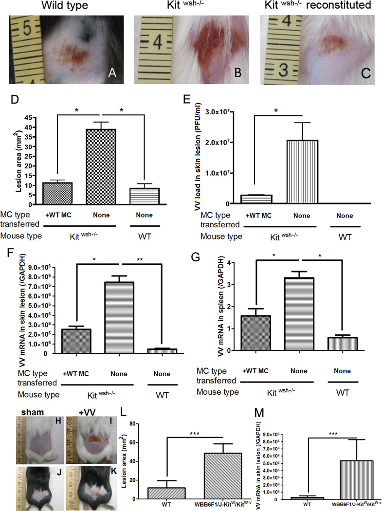
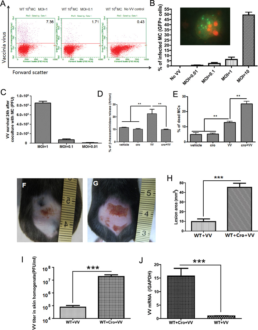
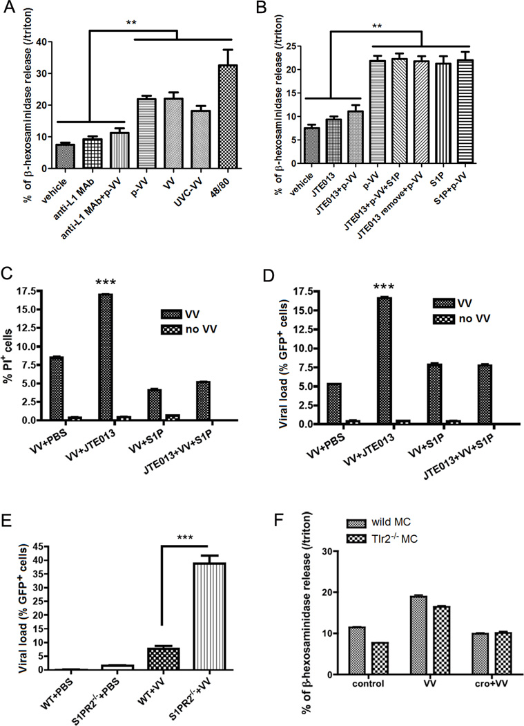
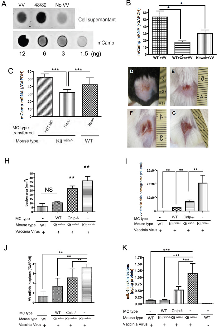
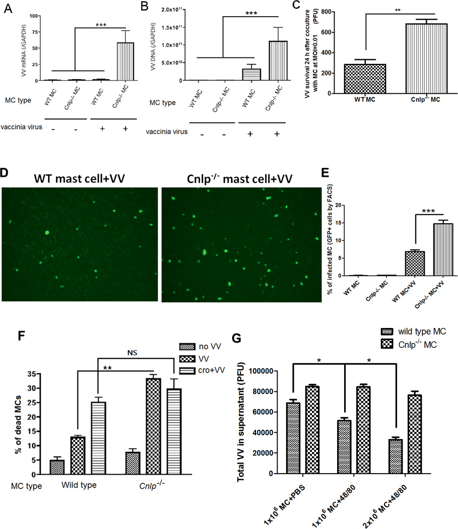
Mast cells (MCs) are well-known effectors of allergic reactions and are considered sentinels in the skin and mucosa. In addition, through their production of cathelicidin, MCs have the capacity to oppose invading pathogens. We therefore hypothesized that MCs could act as sentinels in the skin against viral infections using antimicrobial peptides. In this study, we demonstrate that MCs react to vaccinia virus (VV) and degranulate using a membrane-activated pathway that leads to antimicrobial peptide discharge and virus inactivation. This finding was supported using a mouse model of viral infection. MC-deficient (Kit(wsh-/-)) mice were more susceptible to skin VV infection than the wild type animals, whereas Kit(wsh-/-) mice reconstituted with MCs in the skin showed a normal response to VV. Using MCs derived from mice deficient in cathelicidin antimicrobial peptide, we showed that antimicrobial peptides are one important antiviral granule component in in vivo skin infections. In conclusion, we demonstrate that MC presence protects mice from VV skin infection, MC degranulation is required for protecting mice from VV, neutralizing Ab to the L1 fusion entry protein of VV inhibits degranulation apparently by preventing S1PR2 activation by viral membrane lipids, and antimicrobial peptide release from MC granules is necessary to inactivate VV infectivity.


Figures

Figure 1
Figure 1. VV infection in mast cell-deficient Kit wsh−/− mice
106 PFU of VV was applied on the lower backs of Kitwsh−/− mice and their wild-type littermate control mice. (A) Left image is representative of the reaction seen at 72 hours in wild type mice. (B) The center image is representative of the skin lesion in Kitwsh−/− mice. (C) The right image is Kitwsh−/− mice reconstituted with mast cells. (D) Quantification of lesion size (mm2) at 72 h post-infection. (E) Quantification of viral load (in PFU/ml) in the skin lesions at 72 h post-infection. (F) Expression of VV early gene at the skin lesions was quantified by real-time quantitative RT-PCR. (G) Expression of VV early gene in the spleen was quantified by real-time quantitative RT-PCR. *P<0.05. VV infection in mast cell-deficient WBB6F1/J-KitW/KitW-v double heterozygotes mice (H–M) 106 PFU of VV was applied on the lower backs of WBB6F1/J-KitW/KitW-v double heterozygotes mice and their wild-type littermate control mice. (H) Sham control of wild type mice scarified without VV application at 72 hours (I) Upper right image is representative of the skin lesion in WBB6F1/J-KitW/KitW-v double heterozygotes mice. (J) Sham control of WBB6F1/J-KitW/KitW-v double heterozygotes mice scarified without VV application at 72 hours (K) Lower right image is representative of the reaction seen at 72 hours in wild type mice. The right image is Kitwsh−/− mice reconstituted with mast cells. (L) Quantification of lesion size (mm2) at 72 h post-infection. (M) Expression of VV early gene at the skin lesions was quantified by real-time quantitative RT-PCR at 72 h post-infection. Three independent experiments with 5 mice per group were performed for each experiment
Figure 2
Figure 2. Mast cells degranulate upon contact with VV
(A) Mast cells were infected with VV at different MOI for 24 h and analyzed by FACS. The right plot shows resting mast cells before virus interaction; the two other plots show cells after contact with VV. The upper quadrant number is the percentage of infected mast cells. (B) FACS analysis of VV expression in mast cells infected with VV (GFP+ cells) at different MOI. The insert shows green fluorescence in mast cells infected with VV. The nucleus was stained in red with propidium iodide (PI). (C) VV titer 24 h after co-culture VV with mast cells at different MOI. VV titer in the supernatant was expressed as PFU by plaque assay in BS-C-1 cells. (D) Mast cell β-hexosaminidase release (as index of granule content release) 1 h after contact with VV. The granule release was blocked by adding cromolyn (cro) to the medium. **P<0.01 compared to other groups. (E) Mast cell death was measured with trypan blue 1 h after contact with VV. The number of dead cells was increased by cromolyn in the medium. *P<0.05. All the in vitro experiments have been performed in triplicate (F) Wild-type C57BL/6 mice at 72 h following VV inoculation. (G) Cromolyn pretreated wild-type C57BL/6 mice at 72 h following VV inoculation. (H) Quantification of skin lesion size (mm2) in non-pretreated and cromolyn-pretreated mice at 72 h post infection. (I) Quantification of viral load in skin lesions expressed as PFU/ml. (J) Quantification of VV early gene expression in skin lesions by real-time quantitative RT-PCR. ***P<0.001. Three independent experiments with 4 mice per group were performed for each experiment
Figure 3
Figure 3. Mast cells degranulate on contact with viral envelopes
(A) β-hexosaminidase release of mast cells upon contact with live purified VV (p-VV), UVC-inactivated VV (UVC-VV), live unpurified VV at MOI=1 for 1h. VV, p-VV and UVC-VV induced mast cell degranulation. Anti-L1 MAb control (Anti-L1 MAb) and Anti-L1 MAb pretreated virus (Anti-L1 MAb +VV) did not induced degranulation. 48/80 is the positive control for degranulation. NS: no significant difference between VV and p-VV treatment. **P<0.01 compared to other treatments. (B) Mast cell β-hexosaminidase release (as index of granule content release) on contact with purified VV (p-VV) at MOI=1 for 1h. The granule release was blocked by JTE013, the S1PR2 specific antagonists (JTE013+p-VV). When JTE013 was removed from the medium, p-VV inoculation (JTE013 remove+p-VV) can induce mast cell degranulation, which showed that mast cells were not damaged by JTE013 addition. ***P<0.001. (C) FACS analysis of mast cells stained with PI as index of cell damage 24 h after VV infection at MOI=1. The cells pretreated with JTE013 had the most damage, but addition of S1P with JTE013 prevented the cell damage. ***P<0.001 compared to all the other groups. (D) FACS analysis of mast cells inoculated with GFP+ VV at MOI=1 for 24 h (GFP+ cells as viral load marker). The cells pretreated with JTE013 (JTE013+VV) had the highest viral load, and addition of S1P with JTE013 (JTE013+VV+S1P) decreased mast cell viral load. ***P<0.001 compared to all the other columns. (E) FACS analysis of mast cell from S1PR2−/− Balb/C ByJ mice and their wild type littermate control mice. Mast cells were inoculated with GFP+ VV at MOI=1 for 24 h. The S1PR2−/− cells (S1PR2−/− +VV) had the highest viral load (GFP+ cells). ***P<0.001. (F) β-hexosaminidase release of mast cells from Tlr2−/− mice and their wild-type littermates on contact with VV at MOI=1 for 1 h with or without cromolyn (cro) pretreatment. All the in vitro experiments have been performed in triplicate
Figure 4
Figure 4. Antimicrobial peptide release is important for mast cell antiviral function
(A) Dot blot of the mCamp released in the supernatant when mast cells are exposed to VV at MOI=1 for 1 h and to compound 48/80 as positive control; the lower lane shows the standard blot of mCamp synthetic peptide. Experiments have been performed in triplicate (B) mCamp expression by real-time quantitative RT-PCR in VV induced skin lesions of Kitwsh−/− mice, their wild type littermates and cromolyn (Cro) pretreated wild type littermates 3 days post infection. *P<0.05. (C) mCamp expression by real-time quantitative RT-PCR in VV induced skin lesions of Kitwsh−/− mice, their wild type littermates, and Kitwsh−/− mice reconstituted with wild type mast cells 3 days post infection. ***P<0.001 compared to all the other groups. (D–K) Kitwsh−/− mice reconstituted with mast cells from Cnlp−/− mice and their wild type littermates. Representative lesions from wild-type littermates (D), Kitwsh−/− mice reconstituted with wild type mast cells (E) and Kitwsh−/− mice reconstituted with Cnlp−/− mast cells (F) and Kitwsh−/− mice (G) at 72 h following VV scarification. (H) Quantification of lesion size (mm2) in wild-type littermates, Kitwsh−/− mice and mast cell-reconstituted Kitwsh−/− mice at 72 h post infection. **P<0.01 compared to other groups. NS: no significant difference. (I) Quantification of VV load (PFU/ml) in skin lesions. **P<0.01. (J) Quantification of VV early gene expression in the spleen by real-time quantitative RT-PCR. **P<0.01. (K) Mouse IL-6 ELISA quantification in skin lesions homogenate expressed as pg/µg tissue protein. ***P<0.001. Three independent experiments with 5 mice per group were performed for each experiment
Figure 5
Figure 5. Cathelicidin expression is important in mast cell defense toward VV
Mast cells generated from Cnlp−/− C57BL/6 mice and their wild type littermate mice inoculated with 106 PFU VV at MOI=1 for 24 h. Quantification of VV early mRNA expression. ***P<0.001 (A) and VV DNA expression. ***P<0.001 (B) by real-time quantitative RT-PCR. (C) VV titer in supernatants from Cnlp−/− and wild-type mast cells with VV at MOI=0.01 for 24 h. **P<0.01. (D) Fluorescence visualization of VV infected Cnlp−/− and wild-type mast cells treated with VV at MOI=1 for 24 h. Infected cells are bright green. The number of infected Cnlp−/− mast cells is significantly higher than infected wild type mast cells. (E) FACS quantification of VV infected Cnlp−/− and wild-type mast cells treated with VV at MOI=1 for 24 h (F) Mast cell death was measured with trypan blue 1 h after contact with VV at MOI=1. One group is pretreated with cromolyn before adding VV (cro+VV). **P<0.01. NS: no significant difference. The total number of cells per well were 111± 12 per sqmm, in the control cells; 98± 8 in the VV treated cells and 99±7 in Cro+VV treated cells for the WT cells; The total number of cells per well were 105± 7 per sqmm, in the control cells; 90± 7 in the VV treated cells and 100±7 in Cro+VV treated cells for the Cnlp−/− mast cells (G) Wild type and Cnlp−/− mast cells purified granule activity against VV. 1×10⁶ mast cells +PBS represents baseline activity, 1×10⁶ mast cells +48/80 represents purified granules from 1×10⁶ mast cells and 2×10⁶ mast cell +48/80 represents purified granules from 2×10⁶ mast cells. With wild type mast cell granules, we observed a dose-dependent effect against VV that was not evident with granules from Cnlp−/− mast cells. *P<0.05. All the in vitro experiments have been performed in triplicate

Similar articles

Cited by

References

    1. Galli SJ, Nakae S, Tsai M. Mast cells in the development of adaptive immune responses. Nat Immunol. 2005;6:135–142. - PubMed
    1. Matsushima H, Yamada N, Matsue H, Shimada S. TLR3-, TLR7-, and TLR9-mediated production of proinflammatory cytokines and chemokines from murine connective tissue type skin-derived mast cells but not from bone marrow-derived mast cells. J Immunol. 2004;173:531–541. - PubMed
    1. Fehrenbach K, Port F, Grochowy G, Kalis C, Bessler W, Galanos C, Krystal G, Freudenberg M, Huber M. Stimulation of mast cells via FcvarepsilonR1 and TLR2: the type of ligand determines the outcome. Mol Immunol. 2007;44:2087–2094. - PubMed
    1. Malaviya R, Gao Z, Thankavel K, van der Merwe PA, Abraham SN. The mast cell tumor necrosis factor alpha response to FimH-expressing Escherichia coli is mediated by the glycosylphosphatidylinositol-anchored molecule CD48. Proc Natl Acad Sci U S A. 1999;96:8110–8115. - PMC - PubMed
    1. Krishnaswamy G, Kelley J, Johnson D, Youngberg G, Stone W, Huang SK, Bieber J, Chi DS. The human mast cell: functions in physiology and disease. Front Biosci. 2001;6:D1109–D1127. - PubMed

Publication types

MeSH terms

Substances