. 2011 Dec 2:11:42.
doi: 10.1186/1475-2867-11-42.

Down-regulation of DcR2 sensitizes androgen-dependent prostate cancer LNCaP cells to TRAIL-induced apoptosis

Affiliations

Down-regulation of DcR2 sensitizes androgen-dependent prostate cancer LNCaP cells to TRAIL-induced apoptosis

David Vindrieux et al. Cancer Cell Int. .

Abstract

Background: Dysregulation of many apoptotic related genes and androgens are critical in the development, progression, and treatment of prostate cancer. The differential sensitivity of tumour cells to TRAIL-induced apoptosis can be mediated by the modulation of surface TRAIL receptor expression related to androgen concentration. Our previous results led to the hypothesis that downregulation of TRAIL-decoy receptor DcR2 expression following androgen deprivation would leave hormone sensitive normal prostate cells vulnerable to the cell death signal generated by TRAIL via its pro-apoptotic receptors. We tested this hypothesis under pathological conditions by exploring the regulation of TRAIL-induced apoptosis related to their death and decoy receptor expression, as also to hormonal concentrations in androgen-sensitive human prostate cancer, LNCaP, cells.

Results: In contrast to androgen-insensitive PC3 cells, decoy (DcR2) and death (DR5) receptor protein expression was correlated with hormone concentrations and TRAIL-induced apoptosis in LNCaP cells. Silencing of androgen-sensitive DcR2 protein expression by siRNA led to a significant increase in TRAIL-mediated apoptosis related to androgen concentration in LNCaP cells.

Conclusions: The data support the hypothesis that hormone modulation of DcR2 expression regulates TRAIL-induced apoptosis in LNCaP cells, giving insight into cell death induction in apoptosis-resistant hormone-sensitive tumour cells from prostate cancer. TRAIL action and DcR2 expression modulation are potentially of clinical value in advanced tumour treatment.


Figures

Figure 1
Figure 1
Androgen control of LNCaP cell proliferation. (A): cells were treated with increasing concentrations of R1881 (1 pM-10 nM) or ethanol as the control vehicle. Proliferation was measured 4 days later by [3H]-thymidine incorporation with *P < 0.005 versus other concentrations and by cell number counting with **P < 0.05 versus other concentrations. (B): cell cycle was analysed by flowcytometry after treatment with R1881 at 1 pM, 0.1 nM, and 10 nM, with its ethanol vehicle as control (Ct) (0.001% was the highest concentration used), and with 300 μM Etoposide (positive control) and its DMSO diluent at 0.03%. A representative cell cycle profile is presented. (C) and (D): Cell cycle distribution analysis; the values are expressed as the mean ± SD determined from 2 independent experiments, with triplicate cultures per treatment condition. (C): percentage DNA fragmentation (S-G0), *P < 0.005 versus control and R1881 concentrations, **P < 0.005 versus DMSO. (D): percentage of cells in G0-G1 and in G2-M with *P < 0.005 and **P < 0.005 versus the other treatment conditions, respectively.
Figure 2
Figure 2
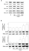
Androgen control of TRAIL and TRAIL-receptor protein expression in LNCaP cells. Cells were treated for 4 days with increasing concentrations of R1881 (1 pM-10 nM) or with ethanol vehicle as control (Ct) (0.001% was the highest concentration used). Proteins were characterized by immunoblot analysis using anti-TRAIL, anti-DR4 and anti-DcR1 antibodies (A), and with anti-DcR2 and anti-DR5 antibodies (B). Histograms represent corresponding protein levels and are normalized for β-actin expression. The values represent the mean ± SD determined from triplicate cultures of a representative experiment from 3 independent experiments. For each protein a representative autoradiograph is shown. *P < 0.005 compared with control.
Figure 3
Figure 3
DcR2 and DR5 protein expression in prostate cancer cells when androgen action had been neutralised. (A) and (B): in LNCaP cells incubated for 4 days with ethanol vehicle as control (Ct) (0.001% was the highest concentration used) or R1881 at 0.1 nM or 10 nM, without or with 25 μM antiandrogen bicalutamide. (C) and (D): in PC3 cells, devoid of androgen receptor, incubated for 4 days with ethanol vehicle as control or increasing concentrations of R1881. All the samples were run and detected together, but some films were cut and pasted to remove spots from replicate cell protein extracts. Immunoblot results and histograms represent DcR2 and DR5 levels and were normalized to β-actin expression. The values represent the mean ± SD determined from triplicate cultures of a representative experiment from 2 independent experiments. For each protein a representative autoradiograph is shown. *P < 0.05 versus the other treatment conditions.
Figure 4
Figure 4
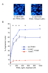
Androgen regulation of TRAIL-induced apoptosis in LNCaP cells. After 4 days of culture with ethanol vehicle (Ct: control), or R1881, LNCaP cells were treated with increasing TRAIL concentrations for 24 h. (A): fragmented nuclei were investigated after DAPI staining by fluorescence microscopy. (B): the number of fragmented nuclei was determined in LNCaP treated with increasing TRAIL concentrations for 24 h. The values represent the mean ± SD of fragmented nuclei per 1,000 cells after analysis of 5,000 randomly selected cells per culture and determined from 5 replicate cultures of a representative experiment from 3 independent experiments. *P < 0.005 versus TRAIL treated cells in androgen free and 10 nM R1881 conditions.
Figure 5
Figure 5
SiRNAs specific for DcR2 and DR5 suppress gene expression in LNCaP cells. Gene-specific siRNAs and scrambled siRNAs controls (si0) (0.5 μg) were added to the media using lipophilic transfection-enhancing reagent (lipofectamine/lipo) for 4 h, and then the cells were cultured in media containing 0.1 nM R1881. Cells were harvested after 48 or 96 h for immunoblot analyses with DcR2 (A) and DR5 (B) specific antibodies. The blots were reprobed with antibody against ß-actin to confirm equal protein loading (15 μg). All the samples were run and detected together but 96 h films were cut and pasted to remove spots from replicate cell protein extracts. Representative autoradiograph results are shown and histograms represent DcR2 (right scale: 48 h incubation; left scale: 96 h incubation) and DR5 protein levels and were normalized for ß-actin expression. (C): the number of fragmented nuclei per 1,000 cells was determined in all transfection treatment conditions (scrambled and specific siRNAs and lipofectamine) with DAPI method. The values represent the mean ± SD determined from triplicate cultures of a representative experiment from 3 independent experiments. *P < 0.005 versus the other treatment conditions.
Figure 6
Figure 6
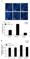
Inhibition of DcR2 and DR5 protein expression by siRNA sensitizes LNCaP cells to TRAIL-induced apoptosis in medium containing 0.1 nM R1881. LNCaP cells, untransfected (Ct: control) or transfected with scrambled siRNA (si0) or siDcR2 or siDR5 (0.5 μg) for 48 h were treated with TRAIL (100 ng/ml) for 24 h (A). White arrows show fragmented nuclei detected by the DAPI method. Histograms represent the number of fragmented nuclei per 1,000 cells from 5,000 randomly selected cells per culture. The values represent the mean ± SD determined from triplicate cultures of a representative experiment from 3 independent experiments. a, P < 0.005, *P < 0.005 and **P < 0.005 versus the other treatment conditions. TRAIL-induced apoptosis, under previous conditions, was controlled by caspase-3 activity assay (B). Histogram values represent the mean ± SD determined from triplicate cultures of a representative experiment from 2 independent experiments performed at 0.1 nM R1881, *P < 0.005 versus the other treatment conditions.
Figure 7
Figure 7
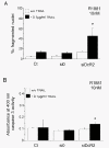
Inhibition of DcR2 protein expression by siRNA sensitizes LNCaP cells to TRAIL-induced apoptosis, in medium containing 10 nM R1881. LNCaP cells, untransfected (Ct: control) or transfected with scrambled siRNA (si0) or siDcR2 (0.5 μg) for 48 h were treated with TRAIL (100 ng/ml) for 24 h. (A) Histograms represent the number of fragmented nuclei per 1,000 cells from 5,000 randomly selected cells per culture. The values represent the mean ± SD determined from triplicate cultures of a representative experiment from 3 independent experiments. *P < 0.005 versus the other treatment conditions. (B) TRAIL-induced apoptosis, under previous conditions, was controlled by caspase-3 activity assay. Histogram values represent the mean ± SD determined from triplicate cultures of a representative experiment from 2 independent experiments performed at 10 nM R1881, *P < 0.05 with the Fisher post-test versus the other treatment conditions, except the control with TRAIL.

Similar articles

Cited by

References

    1. Jemal A, Siegel R, Ward E, Hao Y, Xu J, Murray T, Thun MJ. Cancer statistics, 2008. CA: A Cancer J Clin. 2008;58:71–96. doi: 10.3322/CA.2007.0010. - DOI - PubMed
    1. Guseva NV, Taghiyev AF, Rokhlin OW, Cohen MB. Death receptor-induced cell death in prostate cancer. J Cell Biochem. 2004;91:70–99. doi: 10.1002/jcb.10707. - DOI - PubMed
    1. Zornig M, Hueber AO, Baum W, Evan G. Apoptosis regulators and their role in tumorigenesis. Biochem Biophys Acta. 2001;1551:F1–F37. - PubMed
    1. Ashkenazi A. Targeting death and decoy receptors of the tumour-necrosis factor superfamily. Nat Rev Cancer. 2002;2:420–430. doi: 10.1038/nrc821. - DOI - PubMed
    1. Wiley SR, Schooley K, Smolak PJ, Din WS, Huang CP, Nicholl JK, Sutherland GR, Smith TD, Rauch C, Smith CA, Goodwin RG. Identification and characterization of a new member of the TNF family that induces apoptosis. Immunity. 1995;3:673–682. doi: 10.1016/1074-7613(95)90057-8. - DOI - PubMed

LinkOut - more resources