Associated expressions of FGFR-2 and FGFR-3: from mouse mammary gland physiology to human breast cancer
- PMID: 22124578
- PMCID: PMC7511987
- DOI: 10.1007/s10549-011-1883-6
Associated expressions of FGFR-2 and FGFR-3: from mouse mammary gland physiology to human breast cancer
Abstract
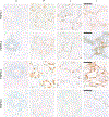
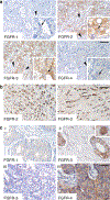
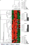
Fibroblast growth factor receptors (FGFRs) are tyrosine kinase receptors which have been implicated in breast cancer. The aim of this study was to evaluate FGFR-1, -2, -3, and -4 protein expressions in normal murine mammary gland development, and in murine and human breast carcinomas. Using immunohistochemistry and Western blot, we report a hormonal regulation of FGFR during postnatal mammary gland development. Progestin treatment of adult virgin mammary glands resulted in changes in localization of FGFR-3 from the cytoplasm to the nucleus, while treatment with 17-β-estradiol induced changes in the expressions and/or localizations of FGFR-2 and -3. In murine mammary carcinomas showing different degrees of hormone dependence, we found progestin-induced increased expressions, mainly of FGFR-2 and -3. These receptors were constitutively activated in hormone-independent variants. We studied three luminal human breast cancer cell lines growing as xenografts, which particularly expressed FGFR-2 and -3, suggesting a correlation between hormonal status and FGFR expression. Most importantly, in breast cancer samples from 58 patients, we found a strong association (P < 0.01; Spearman correlation) between FGFR-2 and -3 expressions and a weaker correlation of each receptor with estrogen receptor expression. FGFR-4 correlated with c-erbB2 over expression. We conclude that FGFR-2 and -3 may be mechanistically linked and can be potential targets for treatment of estrogen receptor-positive breast cancer patients.
Conflict of interest statement
Figures


Similar articles
-
Interaction between FGFR-2, STAT5, and progesterone receptors in breast cancer.Cancer Res. 2011 May 15;71(10):3720-31. doi: 10.1158/0008-5472.CAN-10-3074. Epub 2011 Apr 4. Cancer Res. 2011. PMID: 21464042
-
Futibatinib Is a Novel Irreversible FGFR 1-4 Inhibitor That Shows Selective Antitumor Activity against FGFR-Deregulated Tumors.Cancer Res. 2020 Nov 15;80(22):4986-4997. doi: 10.1158/0008-5472.CAN-19-2568. Epub 2020 Sep 24. Cancer Res. 2020. PMID: 32973082
-
Carcinoma-associated fibroblasts activate progesterone receptors and induce hormone independent mammary tumor growth: A role for the FGF-2/FGFR-2 axis.Int J Cancer. 2008 Dec 1;123(11):2518-31. doi: 10.1002/ijc.23802. Int J Cancer. 2008. PMID: 18767044
-
Fibroblast growth factors in development and cancer: insights from the mammary and prostate glands.Curr Drug Targets. 2009 Jul;10(7):632-44. doi: 10.2174/138945009788680419. Curr Drug Targets. 2009. PMID: 19601767 Review.
-
Morphogenic and tumorigenic potentials of the mammary growth hormone/growth hormone receptor system.Mol Cell Endocrinol. 2002 Nov 29;197(1-2):153-65. doi: 10.1016/s0303-7207(02)00259-9. Mol Cell Endocrinol. 2002. PMID: 12431808 Review.
Cited by
-
New Method for Joint Network Analysis Reveals Common and Different Coexpression Patterns among Genes and Proteins in Breast Cancer.J Proteome Res. 2016 Mar 4;15(3):743-54. doi: 10.1021/acs.jproteome.5b00925. Epub 2016 Feb 2. J Proteome Res. 2016. PMID: 26733076 Free PMC article.
-
Identifying subtype-specific associations between gene expression and DNA methylation profiles in breast cancer.BMC Med Genomics. 2017 May 24;10(Suppl 1):28. doi: 10.1186/s12920-017-0268-z. BMC Med Genomics. 2017. PMID: 28589855 Free PMC article.
-
Establishment of primary mixed cell cultures from spontaneous canine mammary tumors: Characterization of classic and new cancer-associated molecules.PLoS One. 2017 Sep 25;12(9):e0184228. doi: 10.1371/journal.pone.0184228. eCollection 2017. PLoS One. 2017. PMID: 28945747 Free PMC article.
-
Receptor tyrosine kinases in breast cancer treatment: unraveling the potential.Am J Cancer Res. 2024 Sep 15;14(9):4172-4196. doi: 10.62347/KIVS3169. eCollection 2024. Am J Cancer Res. 2024. PMID: 39417188 Free PMC article. Review.
-
Nuclear Fibroblast Growth Factor Receptor Signaling in Skeletal Development and Disease.Curr Osteoporos Rep. 2019 Jun;17(3):138-146. doi: 10.1007/s11914-019-00512-2. Curr Osteoporos Rep. 2019. PMID: 30982184 Free PMC article. Review.
References
-
- Morrison RS, Yamaguchi F, Bruner JM et al. (1994) Fibroblast growth factor receptor gene expression and immunoreactivity are elevated in human glioblastoma multiforme. Cancer Res 54:2794–2799 - PubMed
-
- Yoshimura N, Sano H, Hashiramoto A et al. (1998) The expression and localization of fibroblast growth factor-1 (FGF-1) and FGF receptor-1 (FGFR-1) in human breast cancer. Clin Immunol Immunopathol 89:28–34 - PubMed
-
- Giri D, Ropiquet F, Ittmann M (1999) Alterations in expression of basic fibroblast growth factor (FGF) 2 and its receptor FGFR-1 in human prostate cancer. Clin Cancer Res 5:1063–1071 - PubMed
-
- Ahmed NU, Ueda M, Ito AM et al. (1997) Expression of fibroblast growth factor receptors in naevus-cell naevus and malignant melanoma. Melanoma Res 7:299–305 - PubMed
-
- Myoken Y, Myoken Y, Okamoto T et al. (1996) Expression of fibroblast growth factor-1 (FGF-1), FGF-2 and FGF receptor-1 in a human salivary-gland adenocarcinoma cell line: evidence of growth. Int J Cancer 65:650–657 - PubMed
Publication types
MeSH terms
Substances
Grants and funding
LinkOut - more resources
Full Text Sources
Medical
Research Materials
Miscellaneous

