

TGF-β promotes proliferation of thyroid epithelial cells in IFN-γ(-/-) mice by down-regulation of p21 and p27 via AKT pathway
- PMID: 22119715
- PMCID: PMC3349845
- DOI: 10.1016/j.ajpath.2011.10.009
TGF-β promotes proliferation of thyroid epithelial cells in IFN-γ(-/-) mice by down-regulation of p21 and p27 via AKT pathway
Abstract
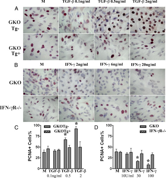
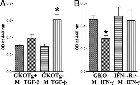
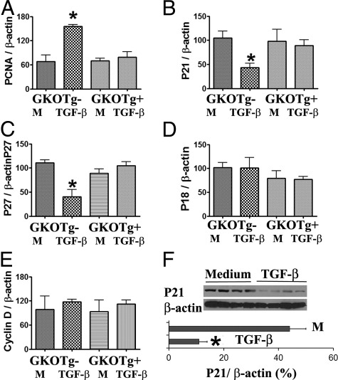
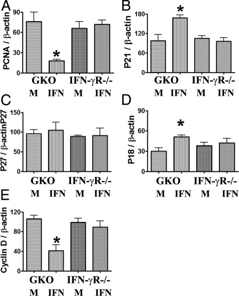
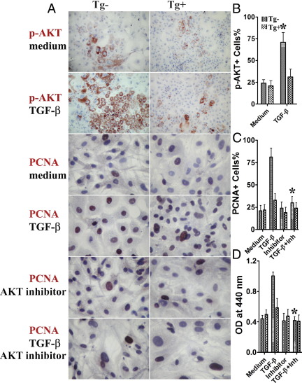
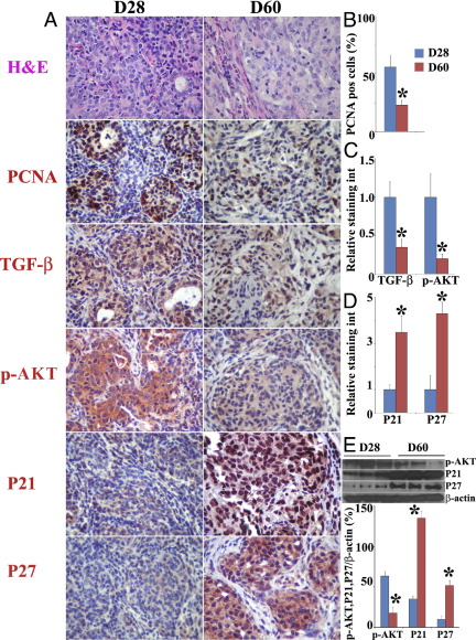
IFN-γ(-/-) NOD.H-2h4 mice develop an autoimmune disease characterized by hyperplasia and proliferation of thyroid epithelial cells (TEC H/P). Proliferating TECs produce TGF-β, and IFN-γ inhibits TEC H/P. In the present study, cultured TECs were used to directly determine the mechanisms by which these cytokines act on TECs to result in proliferation or inhibition of proliferation. With TECs from IFN-γ(-/-) NOD.H-2h4 mice or mice expressing the dominant negative TGF-β type II receptor on TECs, TGF-β was shown to promote TEC proliferation and IFN-γ was shown to inhibit TEC proliferation in vitro. TGF-β may promote TEC proliferation by down-regulating antiproliferative molecules p21 and p27, whereas IFN-γ may inhibit proliferation by up-regulating antiproliferative molecules p18 and p21 and down-regulating the pro-proliferative molecule cyclin D. Inhibition of AKT abolished the effect of TGF-β on p21 and p27, resulting in similar proliferation of TGF-β-treated and control TECs. Increased expression of proliferating cell nuclear antigen (PCNA), TGF-β, and p-AKT and decreased expression of p21 and p27 by proliferating TECs correlated with the proliferative state of TEC H/P. Taken together, the results suggest that TGF-β promotes TEC proliferation by down-regulating p21 and p27 via the AKT pathway in IFN-γ(-/-) NOD.H-2h4 mice, which may have significant implications for development of effective therapeutic strategies targeting the TGF-β and AKT pathways for treatment of hyperplasia and/or neoplasia.
Copyright © 2012 American Society for Investigative Pathology. Published by Elsevier Inc. All rights reserved.
Figures

Similar articles
-
TGF-beta promotes thyroid epithelial cell hyperplasia and fibrosis in IFN-gamma-deficient NOD.H-2h4 mice.J Immunol. 2008 Aug 1;181(3):2238-45. doi: 10.4049/jimmunol.181.3.2238. J Immunol. 2008. PMID: 18641364
-
Suppression of human lens epithelial cell proliferation by proteasome inhibition, a potential defense against posterior capsular opacification.Invest Ophthalmol Vis Sci. 2006 Oct;47(10):4482-9. doi: 10.1167/iovs.06-0139. Invest Ophthalmol Vis Sci. 2006. PMID: 17003443
-
Agonistic anti-CD40 induces thyrocyte proliferation and promotes thyroid autoimmunity by increasing CD40 expression on thyroid epithelial cells.J Immunol. 2013 Apr 15;190(8):3928-38. doi: 10.4049/jimmunol.1202929. Epub 2013 Mar 15. J Immunol. 2013. PMID: 23509363 Free PMC article.
-
IFN-γ and TGF-β, Crucial Players in Immune Responses: A Tribute to Howard Young.J Interferon Cytokine Res. 2022 Dec;42(12):643-654. doi: 10.1089/jir.2022.0132. J Interferon Cytokine Res. 2022. PMID: 36516375 Free PMC article. Review.
-
Cytokines and Regulating Epithelial Cell Division.Curr Drug Targets. 2024;25(3):190-200. doi: 10.2174/0113894501279979240101051345. Curr Drug Targets. 2024. PMID: 38213162 Review.
Cited by
-
Blueberry as a Potential Radiosensitizer for Treating Cervical Cancer.Pathol Oncol Res. 2019 Jan;25(1):81-88. doi: 10.1007/s12253-017-0319-y. Epub 2017 Sep 30. Pathol Oncol Res. 2019. PMID: 28963664
-
IL-39 acts as a friend to pancreatic cancer.Med Oncol. 2018 Dec 1;36(1):12. doi: 10.1007/s12032-018-1236-y. Med Oncol. 2018. PMID: 30506430
-
Culture promotes transfer of thyroid epithelial cell hyperplasia and proliferation by reducing regulatory T cell numbers.Cell Immunol. 2013 Sep-Oct;285(1-2):84-91. doi: 10.1016/j.cellimm.2013.09.003. Epub 2013 Sep 18. Cell Immunol. 2013. PMID: 24135055 Free PMC article.
-
Functional assessment of the BMPR2 gene in lymphoblastoid cell lines from Graves' disease patients.J Cell Mol Med. 2018 Mar;22(3):1538-1547. doi: 10.1111/jcmm.13425. Epub 2017 Dec 20. J Cell Mol Med. 2018. PMID: 29266775 Free PMC article.
-
Species-specific sensitivity to TGFβ signaling and changes to the Mmp13 promoter underlie avian jaw development and evolution.Elife. 2022 Jun 6;11:e66005. doi: 10.7554/eLife.66005. Elife. 2022. PMID: 35666955 Free PMC article.
References
-
- Derynck R., Zhang Y.E. Smad-dependent and Smad-independent pathways in TGF-β family signaling. Nature. 2003;425:577–584. - PubMed
Publication types
MeSH terms
Substances
Grants and funding
LinkOut - more resources
Full Text Sources
Molecular Biology Databases
Research Materials
Miscellaneous
