. 2011 Nov 23;480(7375):63-8.
doi: 10.1038/nature10658.

Mutations causing syndromic autism define an axis of synaptic pathophysiology

Affiliations

Mutations causing syndromic autism define an axis of synaptic pathophysiology

Benjamin D Auerbach et al. Nature. .

Abstract

Tuberous sclerosis complex and fragile X syndrome are genetic diseases characterized by intellectual disability and autism. Because both syndromes are caused by mutations in genes that regulate protein synthesis in neurons, it has been hypothesized that excessive protein synthesis is one core pathophysiological mechanism of intellectual disability and autism. Using electrophysiological and biochemical assays of neuronal protein synthesis in the hippocampus of Tsc2(+/-) and Fmr1(-/y) mice, here we show that synaptic dysfunction caused by these mutations actually falls at opposite ends of a physiological spectrum. Synaptic, biochemical and cognitive defects in these mutants are corrected by treatments that modulate metabotropic glutamate receptor 5 in opposite directions, and deficits in the mutants disappear when the mice are bred to carry both mutations. Thus, normal synaptic plasticity and cognition occur within an optimal range of metabotropic glutamate-receptor-mediated protein synthesis, and deviations in either direction can lead to shared behavioural impairments.


Figures

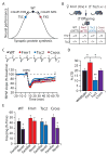
Figure 1
Figure 1. Tsc2+/− mice have a specific deficit in mGluR-LTD
(A) DHPG induces significantly less LTD in slices from Tsc2+/− mice vs. littermate WT mice (WT: 74.3 ± 1.4%, n = 5 animals, 10 slices; Tsc2+/−: 86.3 ± 3.1%, n = 6 animals, 12 slices; *p = 0.004). In this and all subsequent electrophysiology figures, representative field potential traces (average of 10 sweeps) were taken at times indicated by numerals and scale bars equal 0.5 mV, 5 ms, unless stated otherwise. Error bars represent SEM. (B) Synaptically-induced mGluR-LTD, elicited by delivering pairs of pulses (50 ms interstimulus interval) at 1 Hz for 20 minutes (PP-LFS, 1200 pulses) in the presence of the NMDA receptor antagonist D-(−)-2-Amino-5-phosphonopentanoic acid (D-AP5, 50 μM), is also deficient in slices from Tsc2+/− mice (WT: 65.1 ± 2.1%, n = 3 animals, 9 slices; Tsc2+/−: 85.0 ± 2.5%, n = 4 animals, 11 slices; *p = 0.003). (C) The magnitude of NMDA receptor-dependent LTD evoked by low frequency stimulation (LFS, 900 pulses at 1 Hz) does not differ between genotypes (WT: 79.8 ± 1.6%, n = 4 animals, 6 slices; Tsc2+/−: 79.4 ± 1.9%, n = 6 animals, 6 slices; p = 0.610). (D) Hippocampal slices were stimulated with 50 μM DHPG for 5 min, and ERK1/2 activation (phosphorylation) assessed via immunoblot (normalized WT: 100.0 ± 6.1%, WT DHPG: 119.6 ± 5.5%, Tsc2+/−: 97.5 ± 5.6%, Tsc2+/− DHPG: 116.2 ± 3.9%; ANOVA: genotype p = 0.623, treatment *p = 0.0008, genotype × treatment p = 0.923; n = 9 animals). Results reveal that DHPG significantly increases ERK1/2 activation in both WT (*p = 0.040) and Tsc2+/− (*p = 0.003). Error bars represent SEM.
Figure 2
Figure 2. Excessive mTOR activity suppresses the protein-synthesis-dependent component of mGluR-LTD
(A) LTD is significantly attenuated by pretreatment with the protein synthesis inhibitor cycloheximide (CHX, 60 μM, gray bar) in slices from WT animals (control: 74.3 ± 1.4%, n = 5 animals, 10 slices; CHX: 85.2 ± 2.8%, n = 4 animals, 7 slices; *p = 0.014). (B) CHX treatment has no effect on LTD in slices from Tsc2+/− mice (control: 86.3 ± 3.1%, n = 6 animals, 12 slices; CHX: 85.3 ± 3.2%, n = 4 animals, 7 slices, p = 0.796). ANOVA: genotype *p = 0.041, treatment p = 0.089, genotype × treatment *p = 0.045. (C) Presynaptic LTD is not affected by genotype or CHX (see also Fig. S2). DHPG significantly increased PPF in slices from both WT and Tsc2+/− mice (PPF with a 50 ms inter-stimulus interval: WT baseline: 1.37 ± 0.02, WT DHPG: 1.59 ± 0.06, n = 5 animals, 9 slices, *p = 0.003; Tsc2+/− baseline: 1.39 ± 0.02, Tsc2+/− DHPG: 1.64 ± 0.03, n = 5 animals, 9 slices, *p = 0.001) and this effect was not blocked by CHX (WT DHPG + CHX: 1.58 ± 0.06, n = 7 animals, 11 slices, p = 0.89; Tsc2+/− DHPG + CHX: 1.64 ± 0.04, n = 6 animals, 7 slices, p = 0.94). (D) Metabolic labeling of hippocampal slices reveals a significant reduction of basal protein synthesis in Tsc2+/− mice (WT: 100.0 ± 3.1%, Tsc2+/−: 88.2 ± 3.3%, n = 13 animals; *p = 0.043). Differences in protein synthesis are exemplified by representative autoradiograph and total protein stain of the same membrane. (E) Immunoblotting experiments show that Arc expression is significantly reduced in Tsc2+/− hippocampal slices (WT: 100.0 ± 4.7%, Tsc2+/−: 76.6 ± 6.4%, n = 12 animals; *p = 0.005). (F) Arc translation was measured by metabolic labeling of hippocampal slices, followed by immunoprecipitation of Arc. Comparison of the ratios of 35S-incorporated-to-total Arc reveals a significant reduction in Arc translation in the Tsc2+/− hippocampus (WT: 100.0 ± 11.5%, Tsc2+/−: 74.7 ± 6.8%, n = 19 animals; *p = 0.049). (G) Pretreatment of slices with the mTORC1 inhibitor rapamycin (RAP, 20 nM, gray bar) significantly enhances DHPG-induced LTD in slices from Tsc2+/− mice (DMSO: 85.7 ± 2.1%, n = 8 animals, 17 slices; RAP: 72.9 ± 1.8%, n = 7 animals, 18 slices; *p = 0.002). (H) The rescue by rapamycin of DHPG-induced LTD in Tsc2+/− mice is prevented by the protein synthesis inhibitor cycloheximide (DMSO: 87.1 ± 4.7%, n = 6 animals, 10 slices; RAP: 88.1 ± 2.4%, n = 7 animals, 9 slices; p = 0.796). ANOVA: rapamycin treatment *p = 0.043, cycloheximide treatment *p = 0.004, rapamycin × cycloheximide *p = 0.018. (I) Metabolic labeling experiments show that rapamycin (20 nM) normalizes protein synthesis in the Tsc2+/− hippocampus to WT levels (WT DMSO: 100.0 ± 2.5%, WT RAP: 106.5 ± 3.6%, Tsc2+/− DMSO: 88.8 ± 2.6%, Tsc2+/− RAP: 100.4 ± 3.9%; ANOVA: genotype *p = 0.008, treatment *p = 0.006, genotype × treatment p = 0.430; t-test: WT vs. Tsc2+/− DMSO *p = 0.003; WT vs. Tsc2+/− RAP p = 0.344; Tsc2+/− DMSO vs. RAP *p = 0.037; n = 22 animals). Error bars represent SEM.
Figure 3
Figure 3. Positive modulation of mGluR5 reverses synaptic and behavioral deficits in Tsc2+/− mice
(A) Model to account for effects of Tsc2+/− and Fmr1−/y mutations on mGluR5- and protein synthesis-dependent LTD. This model predicts that the impairment in Tsc2+/− mice can be overcome either by inhibiting mTOR with rapamycin or by augmenting mGluR5 signaling with an mGluR5 PAM. (B) Consistent with the model, pretreatment of slices from Tsc2+/− mice with CDPPB (10μM, gray bar) significantly enhances DHPG-induced LTD (DMSO: 86.4 ± 2.5%, n = 8 animals, 13 slices; CDPPB: 71.7 ± 3.9%, n = 7 animals, 12 slices; *p < 0.001). (C) CDPPB treatment fails to enhance DHPG-induced LTD in Tsc2+/− mice when co-applied with the protein synthesis inhibitor cycloheximide (DMSO: 89.0 ± 4.4% n = 8 animals, 10 slices; CDPPB: 83.9 ± 2.1%, n = 7 animals, 9 slices; p = 0.64). ANOVA: CDPPB treatment *p = 0.008, CHX treatment p = 0.087, CDPPB × CHX *p = 0.034. (D) CDPPB (10 μM) restores protein synthesis in the Tsc2+/− hippocampus to WT levels (WT DMSO: 100.0 ± 3.2%, WT CDPPB: 97.2 ± 1.9%, Tsc2+/− DMSO: 86.1 ± 2.7%, Tsc2+/− CDPPB: 94.9 ± 3.0%; ANOVA: genotype *p = 0.006, treatment p = 0.275, genotype × treatment *p = 0.041; t-test: WT vs. Tsc2+/− DMSO *p = 0.012; WT vs. Tsc2+/− CDPPB p = 0.538; Tsc2+/− DMSO vs. CDPPB *p = 0.049; n = 17 animals). (E) CDPPB exposure significantly increases Arc translation in the Tsc2+/− hippocampus (WT DMSO 100.0 ± 28.2%, WT CDPPB 121.0 ± 21.2%, Tsc2+/− DMSO 59.2 ± 7.0%, Tsc2+/− CDPPB 129.4 ± 20.3%; ANOVA genotype p = 0.554, treatment *p = 0.009, genotype × treatment p = 0.114; t-test: Tsc2+/− DMSO vs. CDPPB *p = 0.026; n = 6 animals). Error bars represent SEM. (F) Experimental design of context discrimination task. (G) WT mice display intact memory by freezing more in the familiar context than the novel context (Black bars; Familiar: 50 ± 7.7%, n = 12; Novel: 34.1 ± 3.2%, n = 14; *p = 0.003). A single injection of CDPPB (10 mg/kg, i.p.) 30 minutes prior to training has no effect on WT context discrimination (Familiar: 42.3 ± 3.7%, n = 12; Novel: 26.4 ± 3.6%, n = 12; *p = 0.005). Control Tsc2+/− mice display an impairment in context discrimination (Blue bars; Familiar: 40.9 ± 5.3%, n = 11; Novel: 39.3 ± 5.2%, n = 14; p = 0.501), but this deficit is corrected by a single injection of CDPPB (Familiar: 44.5 ± 4.3%, n = 11; Novel: 31.6 ± 3%, n = 12; *p = 0.034). Error bars represent SEM.
Figure 4
Figure 4. Genetic cross of Tsc2+/− and Fmr1−/y mice rescues synaptic and behavioral impairments present in both single mutants
(A) The data suggest that optimal synaptic function requires a narrow and tightly regulated level of synaptic protein synthesis and that deviations in either direction can impair function,. TSC and FXS fall on different ends of this spectrum and respond to opposite alterations of mGluR5 signaling. These results raise the possibility that introducing both mutations to a mouse may normalize aspects of neural function. (B) Genetic rescue strategy. Heterozygous Tsc2 male mice (Tsc2+/−) were bred with heterozygous Fmr1 females (Fmr1 x+/x) to obtain male offspring of four genotypes: wild type (Tsc2+/+, Fmr1+/y), Fmr1 KO (Tsc2+/+, Fmr1−/y), Tsc2 Het (Tsc2+/−, Fmr1+/y), and Cross (Tsc2+/−, Fmr1−/y). (C) DHPG-induced LTD is significantly decreased in slices from Tsc2+/− mice (*p = 0.002) and significantly increased in slices from Fmr1−/y mice (*p = 0.017), as compared to WT slices. DHPG-LTD in slices from Tsc2+/− × Fmr1−/y mice is comparable in magnitude to WT slices (p = 0.558). (WT: 78.9 ± 2.1%, n = 7 animals, 17 slices; Fmr1: 71.2 ± 2.7%, n = 7 animals, 21 slices; Tsc2: 89.5 ± 2.6%, n = 7 animals, 15 slices; Cross: 77.4 ± 1.8%, n = 9 animals, 19). (D) Summary of LTD data. Bar graphs represent percent decrease from baseline in fEPSP (average of last 5 minutes of recording ± SEM); *p < 0.05, **p < 0.01. (E) Both mutations cause a deficit in context discrimination that is rescued in the double mutant. WT mice (Familiar: 42.9 ± 4.6%, n = 11; Novel: 27.8 ± 3.4%, n = 12; *p = 0.024), Fmr1−/y mice (Familiar: 49.0 ± 5.6%, n = 11; Novel: 43.5 ± 6.7%, n = 12; p = 0.483), Tsc2+/−(Familiar: 42.1 ± 6.8%, n = 12; Novel: 35.6 ± 6.0%, n = 12; p = 0.395) and Tsc2+/− x Fmr1−/y mice (Familiar: 50.5 ± 5.2%, n = 11; Novel: 29.8 ± 5.2%, n = 11; *p = 0.011). Error bars represent SEM.

Comment in

Similar articles

Cited by

References

    1. Newschaffer CJ, et al. The epidemiology of autism spectrum disorders. Annu Rev Public Health. 2007;28:235–258. - PubMed
    1. Krueger DD, Bear MF. Toward fulfilling the promise of molecular medicine in fragile X syndrome. Annu Rev Med. 2011;62:411–429. - PMC - PubMed
    1. Kelleher RJ, 3rd, Bear MF. The autistic neuron: troubled translation? Cell. 2008;135:401–406. - PubMed
    1. Ehninger D, et al. Reversal of learning deficits in a Tsc2+/− mouse model of tuberous sclerosis. Nat Med. 2008;14:843–848. - PMC - PubMed
    1. Meikle L, et al. Response of a neuronal model of tuberous sclerosis to mammalian target of rapamycin (mTOR) inhibitors: effects on mTORC1 and Akt signaling lead to improved survival and function. J Neurosci. 2008;28:5422–5432. - PMC - PubMed

Publication types

MeSH terms

Substances