. 2011 Dec;121(12):4889-902.
doi: 10.1172/JCI59259. Epub 2011 Nov 21.

Herpes simplex encephalitis in children with autosomal recessive and dominant TRIF deficiency

Affiliations

Herpes simplex encephalitis in children with autosomal recessive and dominant TRIF deficiency

Vanessa Sancho-Shimizu et al. J Clin Invest. 2011 Dec.

Abstract

Herpes simplex encephalitis (HSE) is the most common sporadic viral encephalitis of childhood. Autosomal recessive (AR) UNC-93B and TLR3 deficiencies and autosomal dominant (AD) TLR3 and TRAF3 deficiencies underlie HSE in some children. We report here unrelated HSE children with AR or AD TRIF deficiency. The AR form of the disease was found to be due to a homozygous nonsense mutation that resulted in a complete absence of the TRIF protein. Both the TLR3- and the TRIF-dependent TLR4 signaling pathways were abolished. The AD form of disease was found to be due to a heterozygous missense mutation, resulting in a dysfunctional protein. In this form of the disease, the TLR3 signaling pathway was impaired, whereas the TRIF-dependent TLR4 pathway was unaffected. Both patients, however, showed reduced capacity to respond to stimulation of the DExD/H-box helicases pathway. To date, the TRIF-deficient patients with HSE described herein have suffered from no other infections. Moreover, as observed in patients with other genetic etiologies of HSE, clinical penetrance was found to be incomplete, as some HSV-1-infected TRIF-deficient relatives have not developed HSE. Our results provide what we believe to be the first description of human TRIF deficiency and a new genetic etiology for HSE. They suggest that the TRIF-dependent TLR4 and DExD/H-box helicase pathways are largely redundant in host defense. They further demonstrate the importance of TRIF for the TLR3-dependent production of antiviral IFNs in the CNS during primary infection with HSV-1 in childhood.


Figures

Figure 1
Figure 1. AR TRIF deficiency in P1.
(A) Family pedigree of kindred A with allele segregation of the mutation. The HSE patient is shaded in black. Roman numerals (left margin) indicate generations. An arrow indicates the proband. (B) Automated sequencing profiles for the TRIF C421>T mutation in gDNA isolated from leukocytes from a healthy unrelated control; the patient, P1; and the father, I.2. The arrow indicates the position of the mutation. (C) A schematic representation of the TRIF protein (1–712 amino acids) indicating the amino acid position, R141X, affected by the C421>T mutation. Proline-rich domains (pro rich) are shaded in gray; functional domains are shaded in black (TRAF6 binding, TIR, RIP homotypic interacting motif [RHIM]). (D) TRIF protein expression by immunoblot analysis of SV40 fibroblast cell lysates from a healthy control (C+) and P1. Samples were migrated on the same blot. TRIF expression levels were quantified by densitometry results normalized with respect to ACTIN levels and expressed as relative intensity of TRIF. This is a representative blot from 3 independent experiments (mean ± SEM). (E) RT-PCR of full-length TRIF cDNA is shown with ACTB cDNA as an internal control. TRIF cDNA levels were assessed by real-time PCR in control fibroblasts (C+) and P1. Data are represented as relative fold change (ΔΔCt units), where GUS was used for normalization. An average of 3 independent experiments is represented (mean ± SEM).
Figure 2
Figure 2. AD TRIF deficiency in P2.
(A) Family pedigree of kindred B with allele segregation of the mutation. The HSE patient is shaded in black. Roman numerals (left margin) indicate generations. An arrow indicates the proband, and “E?” indicates an undetermined genotype. (B) Automated sequencing profiles for the TRIF C557>T mutation in gDNA isolated from leukocytes from a healthy unrelated control and in the patient, P2. The arrow indicates the position of the mutation. (C) A representation of the TRIF protein (1–712 amino acids) indicating the amino acid position, S186L, affected by the C557>T mutation. Proline-rich domains are shaded in gray; functional domains are shaded in black (TRAF6 binding, TIR, and RHIM). (D) Multiple alignment of TRIF amino acid sequence surrounding the mutation S186L. (E) TRIF protein expression by immunoblot analysis of SV40 fibroblast cell lysates from a healthy control (C+) and P2. TRIF expression levels were quantified by densitometry results normalized with respect to ACTIN levels and expressed as relative intensity of TRIF. This is a representative blot from 3 independent experiments (mean ± SEM). (F) TRIF cDNA levels were assessed by real-time PCR in control fibroblasts and P2. Data are represented as relative fold change (ΔΔCt units), where GUS was used for normalization. An average of 3 independent experiments is represented (mean ± SEM).
Figure 3
Figure 3. Response to TLR3 and TLR4 ligands.
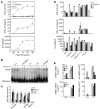
(A) SV40 fibroblasts were stimulated with increasing doses of poly(I:C) for 24 hours, and production of cytokines was assessed. C+ is a healthy control; UNC93B1–/– served as a negative control. Values (mean ± SEM) were calculated from 3 independent experiments. (B) Native gel Western blot showing IRF3 dimers from total fibroblast cell lysates after stimulation with poly(I:C) for 1 or 2 hours. C+ and MYD88–/– cells were used as positive controls to poly(I:C) stimulation, and UNC93B1–/– was used as a negative control. (C) Nuclear protein extracts from SV40 fibroblasts stimulated for 30 minutes with IL-1β or TNF-α and 120 minutes with poly(I:C) were tested for the presence of the p65 subunit of NF-κB by ELISA. NEMO–/– cells were used as a negative control for all stimuli; MyD88–/– was used as a negative control for IL-1β; and UNC93B1–/– was used for poly(I:C) stimulation. Values (mean ± SEM) were calculated from 3 independent experiments. NS, nonstimulated. (D) SV40 fibroblasts were stimulated with a TLR3-specific ligand, poly(A:U), transfected poly(I:C), RIG-I–specific ligand (7sk-as), or lipofectamine alone (L) for 24 hours and tested for cytokine production. Values (mean ± SEM) were calculated from 3 independent experiments. (E) PBMCs from healthy controls, various family members from kindred A (AII.1 and AII.3) and B (BII.3), and P1 and P2 were stimulated with LPS for 2 hours. Induction of IFNB1 and IL6 mRNA was assessed by real-time PCR. Values are expressed as relative fold change using the ΔΔCt method, where GUS was used for normalization; values (mean ± SEM) from 3 independent experiments were calculated.
Figure 4
Figure 4. Response to viral infections.
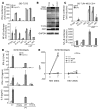
(A) IFN-β, IFN-λ1/3, and IL-6 production in SV40 fibroblasts 24 hours after infection with VSV. Values (mean ± SEM) were calculated from 3 independent experiments. (B) Cell viability of SV40 fibroblasts after 24 hours of infection with VSV in the absence (left) or presence (right) of recombinant IFN-α. The percentage of surviving cells was assessed using resazurin. C+ is a healthy control; UNC93B1–/– and STAT1–/– cells were used as negative controls. Values (mean ± SEM) were calculated from 3 independent experiments. (C) VSV replication in SV40 fibroblasts was estimated at various time points after infection using an MOI of 10. Cells were pretreated with IFN-α or media alone. VSV titer estimation was carried out on Vero cells. Values (mean ± SEM) were calculated from 3 independent experiments. (D) IFN-β, IFN-λ1/3, and IL-6 production after infection with 1 MOI of HSV-1 in SV40 fibroblasts after 24 hours. Values (mean ± SEM) were calculated from 3 independent experiments. (E) Cell viability of SV40 fibroblasts was assessed using resazurin after 24 hours of HSV-1 infection. C+ is a healthy control; UNC93B1–/– and STAT1–/– cells were used as negative controls. Cells were pretreated with media (left) or recombinant IFN-α (right). Values (mean ± SEM) were calculated from 3 independent experiments. (F) Replication of HSV-1 GFP was assessed in SV40 fibroblasts after 24 hours of infection. Cells were pretreated with media (left) or recombinant IFN-α (right). Values (mean ± SEM) were calculated from 3 independent experiments.
Figure 5
Figure 5. Molecular characterization of the R141X AR TRIF mutation.
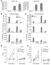
(A) 293 HEK-TLR3 cells were transfected with 10 ng empty vector, WT, R141X, or ΔNC TRIF along with IFN-β–Luc/NF-κB–Luc and RL-TK vectors to assess induction upon overexpression of TRIF. ΔNC TRIF serves as a dominant-negative construct of TRIF, containg only the TIR domain. Transfected cells were left unstimulated or stimulated with 50 μg/ml poly(I:C) for 4 hours. Firefly luciferase values were normalized using renilla values. Total transfected DNA was held constant by adding empty vector. Values (mean ± SEM) were calculated from 3 independent experiments. (B) Immunoblot analysis of protein lysates from 293HEK-TLR3–transfected cells, using an antibody recognizing the N-terminal of TRIF. CFP was cotransfected as a control for transfection efficiency. Samples were migrated on the same blot. (C) 293HEK-TLR4-MD2-CD14 cells were transfected with 10 ng TRIF vectors, including WT, R141X, and ΔNC TRIF, and the induction of IFN-β–Luc/NF-κB–Luc was assessed by luciferase. Values (mean ± SEM) were calculated from 3 independent experiments. (D) Control (C+) SV40 fibroblasts and P1’s fibroblasts retrovirally transduced with empty vector, WT TRIF, or R141X TRIF were stimulated with 25 μg/ml poly(I:C) for 24 hours, and production of cytokines was assessed by ELISA. Values (mean ± SEM) were calculated from 3 independent experiments. (E) HSV-1 GFP replication was assessed after 24 hours of infection in control SV40 fibroblasts (C+) and SV40 fibroblasts from P1 retrovirally transduced with empty vector, WT, or R141X TRIF. Cells were pretreated with media (left) or recombinant IFN-α (right).Values (mean ± SEM) were calculated from 3 independent experiments.
Figure 6
Figure 6. Functional characterization of the S186L AD TRIF mutation.
(A) 293HEK-TLR3 cells were transfected with empty vector, WT, or S186L TRIF plasmids along with IFN-β–Luc/NF-κB–Luc and RL-TK vectors to assess IFN-β and NF-κB promoter induction upon overexpression of TRIF. Values (mean ± SEM) were calculated from at least 3 independent experiments. (B) SV40 fibroblasts from control (C+) and P1’s fibroblasts retrovirally transduced with either empty vector, WT TRIF, or S186L TRIF were stimulated with 25 μg/ml poly(I:C) for 24 hours, and the production of cytokines was assessed by ELISA. Values (mean ± SEM) were calculated from at least 3 independent experiments. (C) SV40 fibroblasts from control (C+) and P2’s fibroblasts retrovirally transduced with empty vector, WT TRIF, or S186L TRIF were stimulated with 25 μg/ml poly(I:C) for 24 hours, and the production of cytokines was assessed by ELISA. Values (mean ± SEM) were calculated from at least 3 independent experiments. (D) Replication of HSV-1 GFP was assessed after 24 hours of infection in control SV40 fibroblasts (C+); SV40 fibroblasts from P1 retrovirally transduced with empty vector, WT, or S186L TRIF; and (E) SV40 fibroblasts from P2 retrovirally transduced with empty vector, WT, or S186L. Cells were pretreated with media (left) or recombinant IFN-α (right). Values (mean ± SEM) were calculated from at least 3 independent experiments.

Similar articles

Cited by

References

    1. Whitley RJ. Herpes simplex virus in children. Curr Treat Options Neurol. 2002;4(3):231–237. doi: 10.1007/s11940-002-0040-2. - DOI - PubMed
    1. Whitley RJ, et al. Vidarabine versus acyclovir therapy in herpes simplex encephalitis. N Engl J Med. 1986;314(3):144–149. doi: 10.1056/NEJM198601163140303. - DOI - PubMed
    1. Whitley RJ, Lakeman F. Herpes simplex virus infections of the central nervous system: therapeutic and diagnostic considerations. Clin Infect Dis. 1995;20(2):414–420. doi: 10.1093/clinids/20.2.414. - DOI - PubMed
    1. McGrath N, Anderson NE, Croxson MC, Powell KF. Herpes simplex encephalitis treated with acyclovir: diagnosis and long term outcome. J Neurol Neurosurg Psychiatry. 1997;63(3):321–326. doi: 10.1136/jnnp.63.3.321. - DOI - PMC - PubMed
    1. Gordon B, Selnes OA, Hart J, Jr, Hanley DF, Whitley RJ. Long-term cognitive sequelae of acyclovir–treated herpes simplex encephalitis. Arch Neurol. 1990;47(6):646–647. - PubMed

Publication types

MeSH terms

Associated data