EBV tegument protein BNRF1 disrupts DAXX-ATRX to activate viral early gene transcription
- PMID: 22102817
- PMCID: PMC3213115
- DOI: 10.1371/journal.ppat.1002376
EBV tegument protein BNRF1 disrupts DAXX-ATRX to activate viral early gene transcription
Abstract
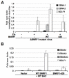
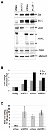
Productive infection by herpesviruses involve the disabling of host-cell intrinsic defenses by viral encoded tegument proteins. Epstein-Barr Virus (EBV) typically establishes a non-productive, latent infection and it remains unclear how it confronts the host-cell intrinsic defenses that restrict viral gene expression. Here, we show that the EBV major tegument protein BNRF1 targets host-cell intrinsic defense proteins and promotes viral early gene activation. Specifically, we demonstrate that BNRF1 interacts with the host nuclear protein Daxx at PML nuclear bodies (PML-NBs) and disrupts the formation of the Daxx-ATRX chromatin remodeling complex. We mapped the Daxx interaction domain on BNRF1, and show that this domain is important for supporting EBV primary infection. Through reverse transcription PCR and infection assays, we show that BNRF1 supports viral gene expression upon early infection, and that this function is dependent on the Daxx-interaction domain. Lastly, we show that knockdown of Daxx and ATRX induces reactivation of EBV from latently infected lymphoblastoid cell lines (LCLs), suggesting that Daxx and ATRX play a role in the regulation of viral chromatin. Taken together, our data demonstrate an important role of BNRF1 in supporting EBV early infection by interacting with Daxx and ATRX; and suggest that tegument disruption of PML-NB-associated antiviral resistances is a universal requirement for herpesvirus infection in the nucleus.
Conflict of interest statement
The authors have declared that no competing interests exist.
Figures


Similar articles
-
Viral reprogramming of the Daxx histone H3.3 chaperone during early Epstein-Barr virus infection.J Virol. 2014 Dec;88(24):14350-63. doi: 10.1128/JVI.01895-14. Epub 2014 Oct 1. J Virol. 2014. PMID: 25275136 Free PMC article.
-
Stimulation of the Replication of ICP0-Null Mutant Herpes Simplex Virus 1 and pp71-Deficient Human Cytomegalovirus by Epstein-Barr Virus Tegument Protein BNRF1.J Virol. 2016 Oct 14;90(21):9664-9673. doi: 10.1128/JVI.01224-16. Print 2016 Nov 1. J Virol. 2016. PMID: 27535048 Free PMC article.
-
Structural basis underlying viral hijacking of a histone chaperone complex.Nat Commun. 2016 Sep 1;7:12707. doi: 10.1038/ncomms12707. Nat Commun. 2016. PMID: 27581705 Free PMC article.
-
Human B cells on their route to latent infection--early but transient expression of lytic genes of Epstein-Barr virus.Eur J Cell Biol. 2012 Jan;91(1):65-9. doi: 10.1016/j.ejcb.2011.01.014. Epub 2011 Mar 29. Eur J Cell Biol. 2012. PMID: 21450364 Review.
-
New players in heterochromatin silencing: histone variant H3.3 and the ATRX/DAXX chaperone.Nucleic Acids Res. 2016 Feb 29;44(4):1496-501. doi: 10.1093/nar/gkw012. Epub 2016 Jan 14. Nucleic Acids Res. 2016. PMID: 26773061 Free PMC article. Review.
Cited by
-
Role of Epitranscriptomic and Epigenetic Modifications during the Lytic and Latent Phases of Herpesvirus Infections.Microorganisms. 2022 Aug 30;10(9):1754. doi: 10.3390/microorganisms10091754. Microorganisms. 2022. PMID: 36144356 Free PMC article. Review.
-
Baculovirus IE2 Interacts with Viral DNA through Daxx To Generate an Organized Nuclear Body Structure for Gene Activation in Vero Cells.J Virol. 2019 Apr 3;93(8):e00149-19. doi: 10.1128/JVI.00149-19. Print 2019 Apr 15. J Virol. 2019. PMID: 30728268 Free PMC article.
-
Viral FGARAT ORF75A promotes early events in lytic infection and gammaherpesvirus pathogenesis in mice.PLoS Pathog. 2018 Feb 1;14(2):e1006843. doi: 10.1371/journal.ppat.1006843. eCollection 2018 Feb. PLoS Pathog. 2018. PMID: 29390024 Free PMC article.
-
Enhancing safety of cytomegalovirus-based vaccine vectors by engaging host intrinsic immunity.Sci Transl Med. 2019 Jul 17;11(501):eaaw2603. doi: 10.1126/scitranslmed.aaw2603. Sci Transl Med. 2019. PMID: 31316006 Free PMC article.
-
Contribution of Epstein-Barr Virus Lytic Proteins to Cancer Hallmarks and Implications from Other Oncoviruses.Cancers (Basel). 2023 Apr 2;15(7):2120. doi: 10.3390/cancers15072120. Cancers (Basel). 2023. PMID: 37046781 Free PMC article. Review.
References
-
- Cohen JI. Epstein-Barr virus infection. N Engl J Med. 2000;343:481–492. - PubMed
-
- Rickinson AB, Kieff E. Epstein-Barr Virus. In: Fields BN, Knipe DM, Howley PM, editors. Philadelphia: Wolters Kluwer Health/Lippincott Williams & Wilkins.; 2007. 2 v. (xix, 3091, 3086 p.) p.
-
- Young LS, Rickinson AB. Epstein-Barr virus: 40 years on. Nat Rev Cancer. 2004;4:757–768. - PubMed
-
- Kieff E. Epstein-Barr Virus and its replication. In: Fields BN, Knipe DM, Howley PM, editors. Philadelphia: Wolters Kluwer Health/Lippincott Williams & Wilkins.; 2007. 2 v. (xix, 3091, 3086 p.) p.
-
- Gottschalk S, Rooney CM, Heslop HE. Post-Transplant Lymphoproliferative Disorders. Annu Rev Med. 2005;56:29–44. - PubMed
Publication types
MeSH terms
Substances
Grants and funding
LinkOut - more resources
Full Text Sources
Molecular Biology Databases
Research Materials
Miscellaneous

