Open Wound Healing In Vivo: Monitoring Binding and Presence of Adhesion/Growth-Regulatory Galectins in Rat Skin during the Course of Complete Re-Epithelialization
- PMID: 22096259
- PMCID: PMC3210424
- DOI: 10.1267/ahc.11014
Open Wound Healing In Vivo: Monitoring Binding and Presence of Adhesion/Growth-Regulatory Galectins in Rat Skin during the Course of Complete Re-Epithelialization
Abstract
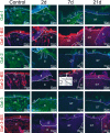
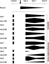
Galectins are a family of carbohydrate-binding proteins that modulate inflammation and immunity. This functional versatility prompted us to perform a histochemical study of their occurrence during wound healing using rat skin as an in vivo model. Wound healing is a dynamic process that exhibits three basic phases: inflammation, proliferation, and maturation. In this study antibodies against keratins-10 and -14, wide-spectrum cytokeratin, vimentin, and fibronectin, and non-cross-reactive antibodies to galectins-1, -2, and -3 were applied to frozen sections of skin specimens two days (inflammatory phase), seven days (proliferation phase), and twenty-one days (maturation phase) after wounding. The presence of binding sites for galectins-1, -2, -3, and -7 as a measure for assessing changes in reactivity was determined using labeled proteins as probes. Our study detected a series of alterations in galectin parameters during the different phases of wound healing. Presence of galectin-1, for example, increased during the early phase of healing, whereas galectin-3 rapidly decreased in newly formed granulation tissue. In addition, nuclear reactivity of epidermal cells for galectin-2 occurred seven days post-trauma. The dynamic regulation of galectins during re-epithelialization intimates a role of these proteins in skin wound healing, most notably for galectin-1 increasing during the early phases and galectin-3 then slightly increasing during later phases of healing. Such changes may identify a potential target for the development of novel drugs to aid in wound repair and patients' care.
Keywords: differentiation; lectin; migration; proliferation; repair.
Figures


Similar articles
-
Early stages of trachea healing process: (immuno/lectin) histochemical monitoring of selected markers and adhesion/growth-regulatory endogenous lectins.Folia Biol (Praha). 2012;58(4):135-43. Folia Biol (Praha). 2012. PMID: 22980504
-
Role of galectins in re-epithelialization of wounds.Ann Transl Med. 2014 Sep;2(9):89. doi: 10.3978/j.issn.2305-5839.2014.09.09. Ann Transl Med. 2014. PMID: 25405164 Free PMC article. Review.
-
Differential regulation of galectin expression/reactivity during wound healing in porcine skin and in cultures of epidermal cells with functional impact on migration.Physiol Res. 2009;58(6):873-884. doi: 10.33549/physiolres.931624. Epub 2008 Dec 17. Physiol Res. 2009. PMID: 19093745
-
Galectins-3 and -7, but not galectin-1, play a role in re-epithelialization of wounds.J Biol Chem. 2002 Nov 1;277(44):42299-305. doi: 10.1074/jbc.M200981200. Epub 2002 Aug 22. J Biol Chem. 2002. PMID: 12194966
-
Regulation of wound healing and fibrosis by galectins.J Mol Med (Berl). 2022 Jun;100(6):861-874. doi: 10.1007/s00109-022-02207-1. Epub 2022 May 19. J Mol Med (Berl). 2022. PMID: 35589840 Review.
Cited by
-
Galectin-2 induces a proinflammatory, anti-arteriogenic phenotype in monocytes and macrophages.PLoS One. 2015 Apr 17;10(4):e0124347. doi: 10.1371/journal.pone.0124347. eCollection 2015. PLoS One. 2015. PMID: 25884209 Free PMC article.
-
Pharmacological activation of estrogen receptors-α and -β differentially modulates keratinocyte differentiation with functional impact on wound healing.Int J Mol Med. 2016 Jan;37(1):21-8. doi: 10.3892/ijmm.2015.2351. Epub 2015 Sep 21. Int J Mol Med. 2016. PMID: 26397183 Free PMC article.
-
How Signaling Molecules Regulate Tumor Microenvironment: Parallels to Wound Repair.Molecules. 2017 Oct 26;22(11):1818. doi: 10.3390/molecules22111818. Molecules. 2017. PMID: 29072623 Free PMC article. Review.
-
Characterization of Immunogenicity Associated with the Biocompatibility of Type I Collagen from Tilapia Fish Skin.Polymers (Basel). 2022 Jun 6;14(11):2300. doi: 10.3390/polym14112300. Polymers (Basel). 2022. PMID: 35683972 Free PMC article.
-
Galectin-3/Gelatin Electrospun Scaffolds Modulate Collagen Synthesis in Skin Healing but Do Not Improve Wound Closure Kinetics.Bioengineering (Basel). 2024 Sep 25;11(10):960. doi: 10.3390/bioengineering11100960. Bioengineering (Basel). 2024. PMID: 39451336 Free PMC article.
References
-
- André S., Sanchez-Ruderisch H., Nakagawa H., Buchholz M., Kopitz J., Forberich P., Kemmner W., Böck C., Deguchi K., Detjen K. M., Wiedenmann B., von Knebel Doeberitz M., Gress T. M., Nishimura S-I., Rosewicz S., Gabius H.-J. Tumor suppressor p16INK4a: modulator of glycomic profile and galectin-1 expression to increase susceptibility to carbohydrate-dependent induction of anoikis in pancreatic carcinoma cells. FEBS J. 2007;274:3233–3256. - PubMed
-
- Barbul A., Regan M. C. In “Surgical Basic Science”, ed. by J. A. Fischer. Mosby-Yearbook; St. Louis: 1993. Biology of wound healing; pp. 68–88.
-
- Barrientos S., Stojadinovic O., Golinko M. S., Brem H., Tomic-Canic M. Growth factors and cytokines in wound healing. Wound Repair Regen. 2008;16:585–601. - PubMed
-
- Cada Z., Chovanec M., Smetana K., Betka J., Lacina L., Plzák J., Kodet R., Stork J., Lensch M., Kaltner H., André S., Gabius H.-J. Galectin-7: will the lectin’s activity establish clinical correlations in head and neck squamous cell and basal cell carcinomas? Histol. Histopathol. 2009;24:41–48. - PubMed
-
- Cada Z., Smetana K., Jr., Lacina L., Plzáková Z., Stork J., Kaltner H., Russwurm R., Lensch M., André S., Gabius H.-J. Immunohistochemical fingerprinting of the network of seven adhesion/growth-regulatory lectins in human skin and detection of distinct tumour-associated alterations. Folia Biol. (Praha). 2009;55:145–152. - PubMed
LinkOut - more resources
Full Text Sources
Other Literature Sources
Research Materials

