. 2011 Nov 18;334(6058):977-82.
doi: 10.1126/science.1210915.

Structural basis of silencing: Sir3 BAH domain in complex with a nucleosome at 3.0 Å resolution

Affiliations

Structural basis of silencing: Sir3 BAH domain in complex with a nucleosome at 3.0 Å resolution

Karim-Jean Armache et al. Science. .

Abstract

Gene silencing is essential for regulating cell fate in eukaryotes. Altered chromatin architectures contribute to maintaining the silenced state in a variety of species. The silent information regulator (Sir) proteins regulate mating type in Saccharomyces cerevisiae. One of these proteins, Sir3, interacts directly with the nucleosome to help generate silenced domains. We determined the crystal structure of a complex of the yeast Sir3 BAH (bromo-associated homology) domain and the nucleosome core particle at 3.0 angstrom resolution. We see multiple molecular interactions between the protein surfaces of the nucleosome and the BAH domain that explain numerous genetic mutations. These interactions are accompanied by structural rearrangements in both the nucleosome and the BAH domain. The structure explains how covalent modifications on H4K16 and H3K79 regulate formation of a silencing complex that contains the nucleosome as a central component.


Figures

Figure 1
Figure 1. Crystal structure of Sir3 BAH domain with nucleosome core particle
a) General overview of the structure. Two different views of the complex; front view and view rotated by 90° around y axis. The structure is color coded (BAH domain is depicted in orange, H2A in yellow, H2B in light pink, H3 in blue, H4 in green and DNA in light grey). b) Histone H4 K16 and histone H3 K79. Both residues that are critical in the regulation of SIR complex mediated silencing are shown in this figure. Histone H4 K16 is depicted as stick in green, histone H3 K79 in blue, the BAH domain surface is rendered in orange. c) Folding transitions in the complex. Both the nucleosome and BAH domain are depicted in grey and regions that get folded upon interaction are shown in red. d) Correlation between structural and genetic contacts. ‘Open book’ view of the complex. NCP surface is shown on the bottom and BAH domain on top of the figure. Surfaces colored in red represent physical contacts as seen in the structure. Surfaces colored in green represent residues both in the NCP and the BAH domains where mutation has been shown to impact silencing. Yellow surfaces represent the overlay of physical (structure-derived) and genetic contacts.
Figure 2
Figure 2. Overview of interactions in the complex
a) Overview of interactions. Same view as in Fig1a (front) BAH domain is depicted in orange, H2A in yellow, H2B in light pink, H3 in blue, H4 in green and DNA in light grey. Secondary structure elements are also depicted here. Secondary structure was assigned using KSDSSP. b) Primary and secondary structure of D205N BAH. Residues with black shading were mapped previously in genetic screens. Spheres above the sequence show histone interactions (within 4.1 Å) that are ordered and visible in the electron density. Colors of spheres represent which histone makes interaction with this residue of BAH domain. Bars above secondary structure inform which histone is interacting with this particular region of BAH domain. There are no spheres over residues in loop1 (residues 17-37) as this region is poorly ordered.
Figure 3
Figure 3. Overview of H4 tail interactions
a) General view of BAH structural elements that interact with histone H4. BAH surface depicted here in orange interacts with H4 tail in green. The interaction surface is in between 2 domains of BAH, the helical H-domain and the β-sheet and that loops 2 and 4 play a crucial role in this interaction. b) Detailed view of the H4 tail interface. Same view as Fig. 3a. All H4 tail residues (13-23) are shown as sticks whereas in BAH only residues that make contacts are depicted as sticks. Magenta dashes connect residues forming potential hydrogen bonds (≤3.5Å). There are 6 possible hydrogen bonds in this interface; K16 forms one, H18 two, the R23 side chain two and L22 main chain carbonyl forms one. Other H4 residues that could participate in these polar interactions are K20 (with E182), and R23 (E178 HB and S212). G13, A15, V21 and R19 all make vander Waals interactions with numerous BAH residues (K97, F94, V62, T63, E95, L91, P179, T180, S212, and E178). Side-chain density for the majority of the H4 tail residues is visible (see SOM), the exceptions being side-chains of R17 and R19, which are apparently more flexible and display weak side-chain density. c) Charge complementarity of the interface. Basic histone H4 tail interaction with a negatively charged BAH domain surface. APBS-calculated electrostatics (−5kT to 5kT). Red surface represents negative and blue positive charge respectively. d) Close up view of H4K16 and H4H18 binding in the charged pocket. e) K16 binding pocket in BAH. Detailed view of K16 and H18 side chain interactions. The K16 ε-amino group interacts with polar or negatively charged side chains of the BAH domain (D60, Y69, E95 and S67). K16 appears to form a hydrogen bond with S67 (3.1Å) and potentially a weak electrostatic interaction with the Y90 main chain carbonyl. Methyl groups of V62 and T63 could stabilize the alkyl chain of K16. Side-chain carbonyls of E137 and E95 and the main- chain carbonyl of P179 can form hydrogen bonds and an electrostatic interaction with the imidazole moiety of H18, respectively. H18 is additionally coordinated through van der Waals contacts.
Figure 4
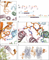
Figure 4. Interactions of BAH domain with a NCP body
a) General view of interactions in this region. Folded loop 3 and the β-strands that interact with regions of histones H3, H4 and H2B are shown. b) Sequence alignment of regions of Xenopus and yeast histones (color coded the same as structure) that interact with BAH domain. Shaded residues were described in previous genetic screens. Orange spheres above the sequence depict which residues interact with BAH domain. The region of histone H4 that is disordered in the structure is depicted in grey. c) Detailed interactions of BAH with H3. A magnified view of a top part of panel a. Magenta dashes connect residues forming potential hydrogen bonds. Five LRS (Q76, D77, F78, K79 and T80) residues in helix α1 and loop L1 of H3 that contact the BAH domain in the structure. BAH W86 is within 4 Å of the H3Q76 carbonyl, T80 side chain and K79 Cα. There is a potential hydrogen bond between the H3D77 side chain carbonyl and the BAH N205 side chain amide. K79 could potentially form 3 hydrogen bonds with the BAH domain, one to the side chain of E84 and two to the E140 side chain. K79 conformation is further stabilized by van der Waals interactions with BAH W86 and H4E74. T80 side chain interacts with main chain of L138 and S139. BAH R75 forms polar interactions, one of which is a potential hydrogen bond with main chain of H3 residues D77 and F78. Additionally a hydrogen bond might also be formed between the BAH E140 side chain carbonyl and main chain amide of H3 T80. d) Detailed interactions of BAH with H4 and H2B. F78. Additionally a hydrogen bond might also be formed between the BAH A magnified view of a bottom part of panel a. Magenta dashes connect residues forming potential hydrogen bonds. Two residues at the tip of loop 3 (L79 and N80) interact with histones H4 and histone H2B. They make van der Waals contacts with histone H4 residues E74, H75 and K77. Additionally the BAH N80 side-chain could form hydrogen bonds with main chain carbonyl of H4 E74 and side chain of H2B R89. L79 interacts with 3 H2B residues in helix a3 (R89, T93 and Q92). BAH N77 and T78 main chain carbonyls make charged interactions and a potential hydrogen bond with side-chains of H2B residues R96 and Q92, respectively. The side-chain of BAH N77 can additionally interact with four residues of H2B located in helix αC. e) Interaction of the BAH domain with the acidic patch, same view as in Fig.1 (front). A positively charged BAH patch (residues 28-34) is in close proximity to acidic residues E61, E64, D90 and E92 of H2A as well as residue E110 of H2B. f) Crystal packing interaction.

Similar articles

Cited by

References

    1. Rusche LN, Kirchmaier AL, Rine J. Annu Rev Biochem. 2003;72:481. - PubMed
    1. Loo S, Rine J. Annu Rev Cell Dev Biol. 1995;11:519. - PubMed
    1. McBryant SJ, Krause C, Woodcock CL, Hansen JC. Mol Cell Biol. 2008 Jun;28:3563. - PMC - PubMed
    1. Martino F, et al. Mol Cell. 2009 Feb 13;33:323. - PubMed
    1. Johnson A, et al. Mol Cell. 2009 Sep 24;35:769. - PMC - PubMed

Publication types

MeSH terms

Substances

Associated data

LinkOut - more resources