. 2012 Jan;86(2):1129-44.
doi: 10.1128/JVI.00306-11. Epub 2011 Nov 16.

Duck Hepatitis A virus possesses a distinct type IV internal ribosome entry site element of picornavirus

Affiliations

Duck Hepatitis A virus possesses a distinct type IV internal ribosome entry site element of picornavirus

Meng Pan et al. J Virol. 2012 Jan.

Abstract

Sequence analysis of duck hepatitis virus type 1 (DHV-1) led to its classification as the only member of a new genus, Avihepatovirus, of the family Picornaviridae, and so was renamed duck hepatitis A virus (DHAV). The 5' untranslated region (5' UTR) plays an important role in translation initiation and RNA synthesis of the picornavirus. Here, we provide evidence that the 651-nucleotide (nt)-long 5' UTR of DHAV genome contains an internal ribosome entry site (IRES) element that functions efficiently in vitro and within BHK cells. Comparative sequence analysis showed that the 3' part of the DHAV 5' UTR is similar to the porcine teschovirus 1 (PTV-1) IRES in sequence and predicted secondary structure. Further mutational analyses of the predicted domain IIId, domain IIIe, and pseudoknot structure at the 3' end of the DHAV IRES support our predicted secondary structure. However, unlike the case for the PTV-1 IRES element, analysis of various deletion mutants demonstrated that the optimally functional DHAV IRES element with a size of approximately 420 nt is larger than that of PTV-1 and contains other peripheral domains (Id and Ie) that do not exist within the type IV IRES elements. The domain Ie, however, could be removed without significant loss of activity. Surprisingly, like the hepatitis A virus (HAV) IRES element, the activity of DHAV IRES could be eliminated by expression of enterovirus 2A protease. These findings indicate that the DHAV IRES shares common features with type IV picornavirus IRES elements, whereas it exhibits significant differences from type IV IRESs. Therefore, we propose that DHAV possesses a distinct type IV IRES element of picornavirus.


Figures

Fig 1
Fig 1
Sequence and predicted secondary structure of the 5′ UTR and IRES element of DHAV. (A) Alignment of the DHAV 5′ UTR and PTV-1 IRES sequences. Sequences were aligned with ClustalW and manually edited in light of the predicted secondary structure models. Dots indicate conserved bases (relative to the C-GY sequence); dashes indicate deletions. The initiation codon (AUG) is underlined. Boxes indicate approximate locations of the proposed helical structures. From the 5′ terminus, complementary sequences involved in potential helices are labeled sequentially by major domain (Ia, Ia′; Ib, Ib′, etc.). Potential helices that are not supported by the presence of covariant base substitutions have labels enclosed by parentheses, but their sequences are completely conserved among different strains. The regions of the PTV-1 IRES corresponding to domain II and domain III are indicated. The sequences involved in base pairing to form the pseudoknot (domain IIIf) structure within the PTV-1 IRES and DHAV IRES elements are marked with xxxx. (B) Predicted secondary structure of the DHAV C-GY 5′ UTR RNA. The structure was predicted by comparative sequence analysis and by using Pfold and Mfold, as described in the text. Domain II and domain III are labeled according to corresponding domains in the PTV-1 IRES. The two stems (S1 and S2) and loop regions (L1 and L2) that form the pseudoknot are shown. The stem of L1 is labeled S3; individual helical segments in domain III are labeled III-1 and III-2. The initiation codon (AUG) is underlined. Lightly shaded rectangles indicate base pairs that are maintained despite sequence variation between types, strains, and isolates of designated viruses; single-site substitutions that do not disrupt base pairing are indicated by asterisks, and base pairs maintained by paired covariant substitutions are indicated by lozenges. The conserved apical loops of domain IIIe and the apical GGG element of the domain IIId loop conserved in the HCV IRES are indicated by gray shading. (C) Comparison of predicted secondary structures for the domains IIId, IIIe, and IIIf among HCV, PTV-1, AEV, and DHAV IRES elements. The two stems (S1 and S2) and loop regions (L1 and L2) that form the pseudoknot are indicated. The stem of L1 within DHAV IRES is labeled S3. Initiation codons (AUG) are underlined. Note that for HCV, this codon is within a stem-loop structure (domain IV) but no such structure is predicted within the PTV-1, AEV, or DHAV sequences. Note the complete identity of domain IIIe in DHAV, HCV, and AEV, and the conserved GGG motif within domain IIId. Within the DHAV sequence, the nucleotides indicated in bold type are those that were modified in the experiments shown in Fig. 6.
Fig 1
Fig 1
Sequence and predicted secondary structure of the 5′ UTR and IRES element of DHAV. (A) Alignment of the DHAV 5′ UTR and PTV-1 IRES sequences. Sequences were aligned with ClustalW and manually edited in light of the predicted secondary structure models. Dots indicate conserved bases (relative to the C-GY sequence); dashes indicate deletions. The initiation codon (AUG) is underlined. Boxes indicate approximate locations of the proposed helical structures. From the 5′ terminus, complementary sequences involved in potential helices are labeled sequentially by major domain (Ia, Ia′; Ib, Ib′, etc.). Potential helices that are not supported by the presence of covariant base substitutions have labels enclosed by parentheses, but their sequences are completely conserved among different strains. The regions of the PTV-1 IRES corresponding to domain II and domain III are indicated. The sequences involved in base pairing to form the pseudoknot (domain IIIf) structure within the PTV-1 IRES and DHAV IRES elements are marked with xxxx. (B) Predicted secondary structure of the DHAV C-GY 5′ UTR RNA. The structure was predicted by comparative sequence analysis and by using Pfold and Mfold, as described in the text. Domain II and domain III are labeled according to corresponding domains in the PTV-1 IRES. The two stems (S1 and S2) and loop regions (L1 and L2) that form the pseudoknot are shown. The stem of L1 is labeled S3; individual helical segments in domain III are labeled III-1 and III-2. The initiation codon (AUG) is underlined. Lightly shaded rectangles indicate base pairs that are maintained despite sequence variation between types, strains, and isolates of designated viruses; single-site substitutions that do not disrupt base pairing are indicated by asterisks, and base pairs maintained by paired covariant substitutions are indicated by lozenges. The conserved apical loops of domain IIIe and the apical GGG element of the domain IIId loop conserved in the HCV IRES are indicated by gray shading. (C) Comparison of predicted secondary structures for the domains IIId, IIIe, and IIIf among HCV, PTV-1, AEV, and DHAV IRES elements. The two stems (S1 and S2) and loop regions (L1 and L2) that form the pseudoknot are indicated. The stem of L1 within DHAV IRES is labeled S3. Initiation codons (AUG) are underlined. Note that for HCV, this codon is within a stem-loop structure (domain IV) but no such structure is predicted within the PTV-1, AEV, or DHAV sequences. Note the complete identity of domain IIIe in DHAV, HCV, and AEV, and the conserved GGG motif within domain IIId. Within the DHAV sequence, the nucleotides indicated in bold type are those that were modified in the experiments shown in Fig. 6.
Fig 2
Fig 2
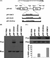
Identification of an IRES element within the DHAV 5′ UTR in vitro and in vivo. (A) The structures of dicistronic luciferase plasmids used in this study are shown. The fragments of the 5′ UTR of the DHAV C-GY genome were inserted between the Rluc and Fluc genes at XhoI and NcoI sites of plasmid pRL-NO in sense or antisense orientation as described in Materials and Methods. Nucleotide numbers corresponding to the fragments are shown. The EMCV IRES was cloned similarly to yield the construct pRF-EMCV and was used as a positive control. (B) In vitro translation reactions containing RRL and biotinylated lysine-tRNA complex were programmed with RNA transcripts derived from the dicistronic plasmids containing the indicated virus sequences. Reaction products were analyzed by Transcend nonradioactive translation detection systems (Promega). The positions of the Rluc and Fluc proteins are indicated. (C) Transient-expression assay in BHK cells. The dicistronic plasmids containing the indicated virus sequences were transfected into vTF7-3-infected BHK cells. After 20 h, cell lysates were prepared and analyzed for Rluc and Fluc expression by Western blotting. The Rluc and Fluc activities were measured. IRES activity was calculated as the mean of three independent experiments, and the results were standardized to the values for the Fluc/Rluc ratio from the pRL-EMCV, which was set at 100%. The mean values (plus standard errors of the means [error bars]) are shown.
Fig 3
Fig 3
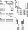
Delimitation of the DHAV 5′ UTR sequences required for IRES activity in BHK cells. The cDNA fragments indicated were generated by using the PCR as described in Materials and Methods and inserted into the XhoI and NcoI-digested vector pRL-NO. Dicistronic luciferase plasmids with 5′-terminal (A), internal (B), and 3′-terminal (C) deletions made in the DHAV 5′ UTR and inclusion of 30 nt of coding sequence downstream of the DHAV 5′ UTR were used to transfect vTF7-3-infected BHK cells. After 20 h, cell extracts were prepared and analyzed for the expression of Rluc and Fluc. (C and D) The Rluc and Fluc activities were measured and IRES activity was calculated as the mean of three independent experiments. The results were standardized to the values for the Fluc/Rluc ratio directed by the plasmid pRL-DHAV, which was set at 100%. The mean values (plus standard errors of the means [error bars]) are shown.
Fig 4
Fig 4
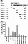
Schematic representation of transcriptional units present in monocistronic plasmid constructs used as templates for synthesis of RNAs transiently expressed in BHK cells. (A) Monocistronic constructs pFluc and pDHAV and 5′-terminal deletion mutants made from pDHAV. (B) Uncapped or capped mRNA transcribed from the indicated monocistronic plasmids were transfected into BHK cells. After 20 h, cell extracts were prepared and analyzed for Fluc expression. The Fluc expression was measured using the Promega assay kit and a luminometer. Results are the mean of three independent transfections. The results were standardized to the values for Fluc activity in pDHAV, which was set at 100%. The mean values (plus standard errors of the means [error bars]) are shown. pFluc-cap is capped and the rest are uncapped.
Fig 5
Fig 5
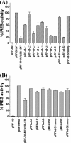
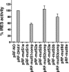
Mutational analysis of domain Id and domain Ie. Dicistronic plasmids containing the indicated mutations within the predicted stem-loops regions of domain Id (A) and Ie (B) were transfected into vTF7-3-infected BHK cells and analyzed for the expression of Rluc and Fluc by using the Dual Glo luciferase kit (Promega). The IRES activities were standardized to the values for Fluc/Rluc ratio directed by the plasmid pRL-DHAV, which was set at 100%. The results are the mean values for Fluc/Rluc ratios from three experiments.
Fig 6
Fig 6
Analysis of DHAV IRES activity by mutation of the loop of domain IIId and IIIe or pseudoknot structure. Dicistronic plasmids containing the indicated mutations within the predicted pseudoknot region and the loop IIId and IIIe were transfected into vTF7-3-infected BHK cells and analyzed for the expression of Rluc and Fluc as described above. The IRES activities were standardized to the values for Fluc/Rluc ratio directed by the plasmid pRL-DHAV, which was set at 100%. The results are the mean values for Fluc/Rluc ratios from three experiments.
Fig 7
Fig 7
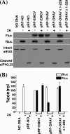
Inhibition of the DHAV IRES activity in the presence of cleaved eIF4G. (A) Dicistronic plasmids (4 μg) containing the indicated IRES sequences were transfected into BHK cells in the absence (−) or presence (+) of plasmid pT7-2A encoding SVDV 2A protease (1 μg). After 20 h, cell extracts were prepared and analyzed for Rluc and Fluc expression as described in the legend for Fig. 2. Samples were also analyzed by immunoblotting to analyze the status of eIF4G. The position of the C-terminal cleavage product of eIF4G is indicated (Ct). (B) Dual luciferase assays were performed on cell extracts from three separate transfections. The relative luciferase activity of Rluc (open bars) and Fluc (solid bars) from each DNA construct was normalized to that from the plasmid pRL-EMCV in the absence of pT7-2A, with the value set at 100%. Error bars indicate standard errors of the means.

Similar articles

Cited by

References

    1. Ali IK, McKendrick L, Morley SJ, Jackson RJ. 2001. Activity of the hepatitis A virus IRES requires association between the cap-binding translation initiation factor (eIF4E) and eIF4G. J. Virol. 75:7854–7863 - PMC - PubMed
    1. Bakhshesh M, et al. 2008. The picornavirus avian encephalomyelitis virus possesses a hepatitis C virus-like internal ribosome entry site element. J. Virol. 82:1993–2003 - PMC - PubMed
    1. Belsham GJ. 2009. Divergent picornavirus IRES elements. Virus Res. 139:183–192 - PubMed
    1. Belsham GJ, Jackson RJ. 2000. Translation initiation on picornavirus RNA, p 869–900 In Sonenberg N, Hershey JWB, Mathews MB. (ed), Translational control of gene expression. Cold Spring Harbor monograph 39. Cold Spring Harbor Laboratory Press, Cold Spring Harbor, NY
    1. Bochkov YA, Palmenberg AC. 2006. Translational efficiency of EMCV IRES in bicistronic vectors is dependent upon IRES sequence and gene location. Biotechniques 41:283–284, 286, 288 passim - PubMed

Publication types