. 2012 Jan 2;287(1):607-618.
doi: 10.1074/jbc.M111.310987. Epub 2011 Nov 14.

Nitric oxide storage and transport in cells are mediated by glutathione S-transferase P1-1 and multidrug resistance protein 1 via dinitrosyl iron complexes

Affiliations

Nitric oxide storage and transport in cells are mediated by glutathione S-transferase P1-1 and multidrug resistance protein 1 via dinitrosyl iron complexes

Hiu Chuen Lok et al. J Biol Chem. .

Abstract

Nitrogen monoxide (NO) plays a role in the cytotoxic mechanisms of activated macrophages against tumor cells by inducing iron release. We showed that NO-mediated iron efflux from cells required glutathione (GSH) (Watts, R. N., and Richardson, D. R. (2001) J. Biol. Chem. 276, 4724-4732) and that the GSH-conjugate transporter, multidrug resistance-associated protein 1 (MRP1), mediates this release potentially as a dinitrosyl-dithiol iron complex (DNIC; Watts, R. N., Hawkins, C., Ponka, P., and Richardson, D. R. (2006) Proc. Natl. Acad. Sci. U.S.A. 103, 7670-7675). Recently, glutathione S-transferase P1-1 (GST P1-1) was shown to bind DNICs as dinitrosyl-diglutathionyl iron complexes. Considering this and that GSTs and MRP1 form an integrated detoxification unit with chemotherapeutics, we assessed whether these proteins coordinately regulate storage and transport of DNICs as long lived NO intermediates. Cells transfected with GSTP1 (but not GSTA1 or GSTM1) significantly decreased NO-mediated 59Fe release from cells. This NO-mediated 59Fe efflux and the effect of GST P1-1 on preventing this were observed with NO-generating agents and also in cells transfected with inducible nitric oxide synthase. Notably, 59Fe accumulated in cells within GST P1-1-containing fractions, indicating an alteration in intracellular 59Fe distribution. Furthermore, electron paramagnetic resonance studies showed that MCF7-VP cells transfected with GSTP1 contain significantly greater levels of a unique DNIC signal. These investigations indicate that GST P1-1 acts to sequester NO as DNICs, reducing their transport out of the cell by MRP1. Cell proliferation studies demonstrated the importance of the combined effect of GST P1-1 and MRP1 in protecting cells from the cytotoxic effects of NO. Thus, the DNIC storage function of GST P1-1 and ability of MRP1 to efflux DNICs are vital in protection against NO cytotoxicity.


Figures
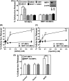
FIGURE 1.
FIGURE 1.
MRP1 mediates NO-stimulated 59Fe release. A, WT- and MRP1 KO-MEFs were labeled with 59Fe-Tf (0.75 μm) for 24 h at 37 °C, washed on ice, and then incubated with control medium with or without MK571 (20 μm) for 30 min at 37 °C. Cells were then reincubated with control medium containing either GSNO (0.5 mm) or GSH (0.5 mm) with or without MK571 (20 μm) for 6 h at 37 °C. The inset shows MRP1 protein expression in WT- and MRP1 KO-MEFs (typical of 3 experiments). B and C, WT- and MRP1 KO-MEFs were labeled with 59Fe-Tf (0.75 μm) for 24 h at 37 °C, washed on ice, and then incubated with control medium containing either GSNO (0.01, 0.05, 0.1, and 0.5 mm) (B) or SperNONOate (0.01, 0.05, 0.1, and 0.5 mm) (C) for 6 h at 37 °C. D, WT- and MRP1 KO-MEFs cells were transfected with either iNOS or the relevant empty vector, labeled with 59Fe-Tf as described for B and C, and then reincubated with control medium for 24 h at 37 °C. The activity of iNOS was assessed by nitrite accumulation in the incubation medium (see “Results”). Results are mean ± S.D. (3 experiments). *, p < 0.05; **, p < 0.01; ***, p < 0.001.
FIGURE 2.
FIGURE 2.
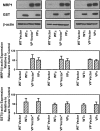
Western blot analysis demonstrating MRP1 expression in MCF7-VP and MCF7-WT cells as well as the pronounced protein expression of GST P1-1, GST A1-1, or GST M1-1 in cells transfected with GSTP1, GSTA1, or GSTM1, respectively, relative to cells transfected with the appropriate empty vectors alone. Photographs of blots are typical of 3 experiments, and densitometry is mean ± S.D. (3 experiments).
FIGURE 3.
FIGURE 3.
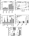
Only GST P1-1, but not GST A1-1 or M1-1, significantly inhibits NO-mediated 59Fe release from MRP1 hyper-expressing VP cells. A, cells were labeled with 59Fe-Tf (0.75 μm) for 3 h at 37 °C, washed, and then reincubated for 3 h at 37 °C with or without GSNO (0.5 mm) at 37 °C. B, cells were labeled and washed as in A and then reincubated with or without GSNO (0.5 mm) for up to 24 h at 37 °C. Significance values compare VPπ and VP Vector cells treated with GSNO at the relevant time points. C, cells were labeled and washed as in A and then reincubated with control medium or medium containing GSNO, SperNO, GSH, or spermine (Sper) (all at 0.5 mm) or the iron chelators desferrioxamine (DFO), Exjade®, or di-2-pyridylketone 4,4-dimethyl-3-thiosemicarbazone (Dp44mT) (all at 25 μm) for 3 h at 37 °C. D, VP Vector and VPπ cells transfected with either iNOS or vector were labeled with 59Fe-Tf (0.75 μm) for 24 h at 37 °C, washed on ice, and then incubated with control medium for 24 h at 37 °C. Activity of iNOS was assessed by nitrite accumulation in the incubation medium (see “Results”). E, HaCaT cells treated with either si-GSTP1 (siRNA) or scrambled siRNA (Scrambled) were labeled, washed, and treated as in A. Western analysis was then done to assess GST P1-1 expression (see inset that is typical of 3 experiments). Results are mean ± S.D. (3 experiments). *, p < 0.05; **, p < 0.01; ***, p < 0.001.
FIGURE 4.
FIGURE 4.
Increased intracellular 59Fe accumulation is identified in a peak that contains GST P1-1 in VPπ cells relative to VP Vector cells after fractionation of the cytosol using non-denaturing FPLC. VP cells (transfected with vector alone or GSTP1) were incubated with 59Fe-Tf (0.75 μm) for 3 h at 37 °C with GSNO (0.5 mm). The cells were then washed and lysed, and the cytosolic fraction was analyzed by FPLC. The fractions were then assessed by Western blot, and GST P1-1 expression was observed in fractions 20, 21, and 22 only. Results are typical of 3 experiments.
FIGURE 5.
FIGURE 5.
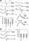
Transfection of WT or VP cells with GSTP1, but not GSTA1 or GSTM1, resulted in a unique EPR signal after incubation with GSNO (0.5 mm) for 3 h at 37 °C. A, panels i–iv, low temperature (77 K) EPR spectra of a synthetic dinitrosyl diglutathionyl iron complex (DNDGIC; 0.5 mm) (panel i); WT Vector cells (1010 cells) incubated with either control medium (panel ii) or GSNO (0.5 mm) (panel iii); and WTπ cells after incubation with GSNO (0.5 mm) (panel iv). Panel v, quantification of EPR signals from panels iii and iv and from panels vii and viii. EPR signal intensity is represented as normalized peak height in arbitrary units. Results are mean ± S.D. (3–5 experiments), ***, p < 0.001. Panels vi and vii, low temperature (77 K) EPR spectra of VP Vector cells incubated with either control medium (panel vi) or GSNO (0.5 mm) (panel vii). Panel viii, VPπ cells after incubation with GSNO (0.5 mm). Panels ix and x, room temperature (293 K) EPR spectra of VP Vector (panel ix) or VPπ cells (panel x) after incubation with GSNO (0.5 mm) or control medium for 3 h at 37 °C. Arrows highlight the additional features resolved in the VPπ cells, consistent with the formation of different DNICs when compared with VP Vector cells. B, panels i–iii, low temperature (77 K) EPR spectra of VP Vector* (panel i), VPα (panel ii), or VPμ (panel iii) cells incubated with GSNO (0.5 mm) for 3 h at 37 °C. Panel iv, quantification of EPR signals from panels i–iii. EPR signal intensity is represented as normalized peak height in arbitrary units. Results are typical scans of 3–5 experiments, and the quantification represents mean ± S.D. (3–5 experiments).
FIGURE 6.
FIGURE 6.
The MRP1 inhibitor, MK571, markedly increases EPR signal intensity in VP Vector cells. Panels i and ii, low temperature (77 K) EPR spectra of VP Vector cells (1010 cells) preincubated with or without MK571 (20 μm) for 30 min at 37 °C followed by incubation for 3 h at 37 °C with either GSNO (0.5 mm) (panel i) or MK571 (20 μm) and GSNO (0.5 mm) (panel ii), respectively. Panel iii, quantification of EPR signals from VP Vector or VPπ cells in the presence or absence of MK571. EPR signal intensity is represented as normalized peak height in arbitrary units. Results are typical scans of 3–5 experiments, and the quantification represents mean ± S.D. (3–5 experiments), *, p < 0.05. Panels iv and v, low temperature (77 K) EPR spectra of VPπ cells (1010 cells) incubated with GSNO (0.5 mm) for 3 h in the presence and absence of MK571 (20 μm). Results are typical from 5 experiments.
FIGURE 7.
FIGURE 7.
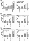
MRP1 and GST P1-1 cooperate in preventing the cytotoxic activity of NO. A, panel i, co-expression of both MRP1 and GST P1-1 (but not GST A1-1 or GST M1-1) is required for maximum resistance against NO as GSNO. WT Vector cells, VP Vector/Vector* cells, and WT/VPα, VPμ, or VPπ cells were incubated with GSNO (0.02–10 mm) for 72 h at 37 °C, and proliferation was assessed. The expression of both GST P1-1 and MRP1 in VPπ cells leads to maximum resistance to the cytotoxicity of GSNO. Panel ii, co-expression of MRP1 and GST P1-1 in VPπ cells leads to greatest sensitivity to GSNO when cells were preincubated with BSO (0.1 mm) for 20 h at 37 °C and then incubated with GSNO (0.02–10 mm) for 72 h at 37 °C; proliferation was assessed after these experiments. B, the cytotoxic effect of GSNO and MRP1 inhibitors on cell proliferation in MRP1 hyper-expressing cells in the absence or presence of GST P1-1. The WTπ and VPπ cells and their relevant vector control cells were incubated with either GSNO (0.02–10 mm) or MK571 (20 μm) and GSNO (0.02–10 mm) (panel i) or GSNO (0.02–10 mm) or sulfinpyrazone (2 mm) and GSNO (0.02–10 mm) (panel ii) for 72 h at 37 °C, and proliferation was assessed. C, the effect of GST inhibitors in cells hyper-expressing MRP1 in the absence or presence of GST P1-1. The WTπ and VPπ cells and their relevant vector control cells were incubated with either GSNO (0.02–10 mm) or ethacrynic acid (200 μm) and GSNO (0.02–10 mm) (panel i) or GSNO (0.02–10 mm) or dicumarol (1.25 mm) and GSNO (0.02–10 mm) (panel ii) for 72 h at 37 °C, and proliferation was assessed. Results are mean ± S.D. (3–5 experiments). ***, p < 0.001.
FIGURE 8.
FIGURE 8.
Schematic illustrating the respective roles of GST P1-1 and MRP1 in NO storage and transport. NO can diffuse through the membrane or may be actively transported into cells by protein disulfide isomerase (PDI) (37). Due to the ability of NO to act as a ligand, it can bind iron transported into cells and released from transferrin. GSH completes the NO-Fe complex to form a DNIC. DNICs can be bound by GST P1-1 or effluxed out of cells via MRP1.

Similar articles

Cited by

References

    1. Watts R. N., Ponka P., Richardson D. R. (2003) Biochem. J. 369, 429–440 - PMC - PubMed
    1. Hibbs J. B., Jr., Taintor R. R., Vavrin Z. (1984) Biochem. Biophys. Res. Commun. 123, 716–723 - PubMed
    1. Bosworth C. A., Toledo J. C., Jr., Zmijewski J. W., Li Q., Lancaster J. R., Jr. (2009) Proc. Natl. Acad. Sci. U.S.A. 106, 4671–4676 - PMC - PubMed
    1. Drapier J. C., Hibbs J. B., Jr. (1986) J. Clin. Invest. 78, 790–797 - PMC - PubMed
    1. Harrop T. C., Tonzetich Z. J., Reisner E., Lippard S. J. (2008) J. Am. Chem. Soc. 130, 15602–15610 - PubMed

Publication types

MeSH terms