. 2012 Feb;30(2):197-209.
doi: 10.1002/stem.783.

Nitric oxide sustains long-term skeletal muscle regeneration by regulating fate of satellite cells via signaling pathways requiring Vangl2 and cyclic GMP

Affiliations
Free PMC article

Nitric oxide sustains long-term skeletal muscle regeneration by regulating fate of satellite cells via signaling pathways requiring Vangl2 and cyclic GMP

Roberta Buono et al. Stem Cells. 2012 Feb.
Free PMC article

Abstract

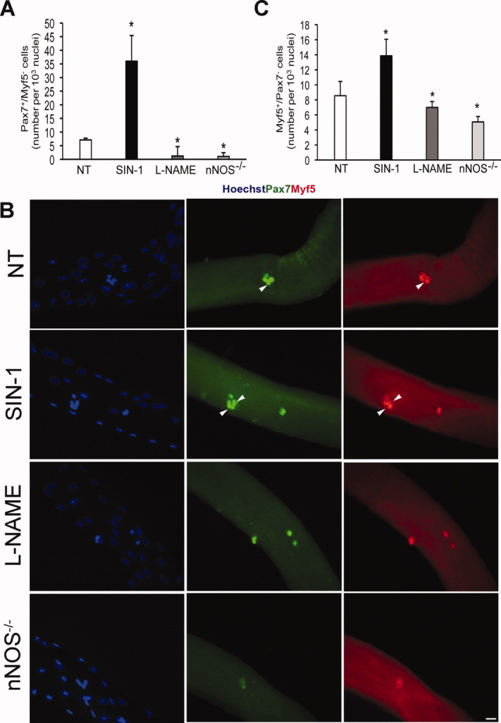
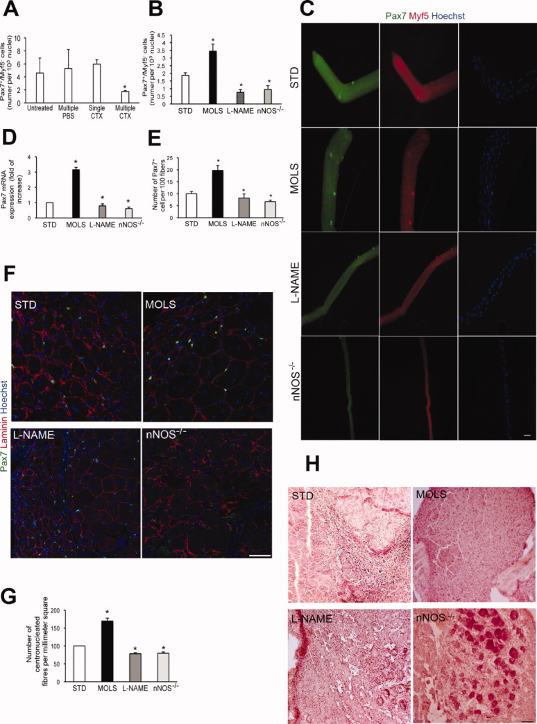
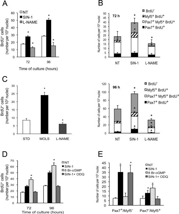
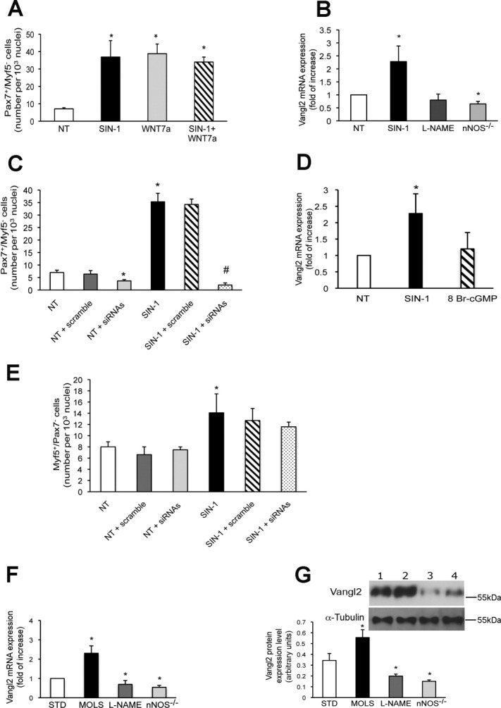
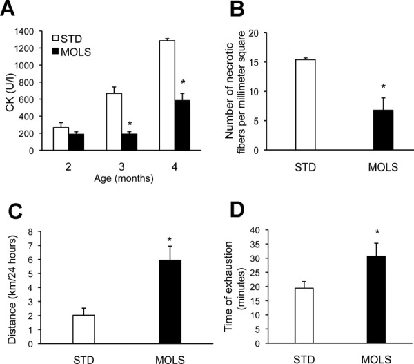
Satellite cells are myogenic precursors that proliferate, activate, and differentiate on muscle injury to sustain the regenerative capacity of adult skeletal muscle; in this process, they self-renew through the return to quiescence of the cycling progeny. This mechanism, while efficient in physiological conditions does not prevent exhaustion of satellite cells in pathologies such as muscular dystrophy where numerous rounds of damage occur. Here, we describe a key role of nitric oxide, an important signaling molecule in adult skeletal muscle, on satellite cells maintenance, studied ex vivo on isolated myofibers and in vivo using the α-sarcoglycan null mouse model of dystrophy and a cardiotoxin-induced model of repetitive damage. Nitric oxide stimulated satellite cells proliferation in a pathway dependent on cGMP generation. Furthermore, it increased the number of Pax7(+)/Myf5(-) cells in a cGMP-independent pathway requiring enhanced expression of Vangl2, a member of the planar cell polarity pathway involved in the Wnt noncanonical pathway. The enhanced self-renewal ability of satellite cells induced by nitric oxide is sufficient to delay the reduction of the satellite cell pool during repetitive acute and chronic damages, favoring muscle regeneration; in the α-sarcoglycan null dystrophic mouse, it also slowed disease progression persistently. These results identify nitric oxide as a key messenger in satellite cells maintenance, expand the significance of the Vangl2-dependent Wnt noncanonical pathway in myogenesis, and indicate novel strategies to optimize nitric oxide-based therapies for muscular dystrophy.


Figures

Figure 1
Figure 1
Nitric oxide (NO) increases the number of satellite cells in ex vivo isolated myofibers. (A): Number of Pax7+/Myf5 cells in single fibers cultured in floating conditions. Fibers were obtained from wild-type mice and treated for 96 hours with the NO donor SIN-1 (3 μM), the NOS inhibitor L-NAME (3 mM), or vehicle (NT), or obtained from nNOS−/− mice. Data are expressed as mean ± SEM and normalized for fiber nuclei number. Results are from at least 50 fibers for each experiment, n = 4; *, p ≤ .05 versus NT. Representative images of the results reported in (A) are shown in (B). Pax7+ cells are green, Myf5+ are red. Blue staining shows nuclei. Scale bar = 25 μm. (C): Number of Pax7/Myf5+ in single fibers cultured in floating conditions. Data are expressed as mean ± SEM after normalization for fiber nuclei number. Results are from at least 50 fibers for each experiment, n = 4; *, p ≤ .05 versus NT. Abbreviations: L-NAME, Nωnitro-L-arginine methylester; NT, untreated; nNOS, nitric oxide synthase; SIN-1, 3-morpholinosydnonimine.
Figure 2
Figure 2
Nitric oxide (NO) increases the satellite cell pool after repeated CTX injections in vivo. (A): Number of Pax7+/Myf5 cells in single fibers isolated 10 days after single or three (multiple) CTX injections. Data are expressed as mean ± SEM and normalized for fiber nuclei number. At least 50 fibers were analyzed for each experiment, n = 3; *, p ≤ .05 versus single damage. The number of Pax7+/Myf5 cells counted in fibers isolated prior to CTX injection (untreated) or in mice receiving three (multiple) injections of PBS as control are also shown. (B): Number of Pax7+/Myf5 cells counted in single fibers isolated from Extensor Digitorum Longus muscle (EDL) muscle after multiple injections of CTX. Fibers were either obtained from wild-type mice that received STD or a diet containing the NO donor MOLS (3 mg/kg) or with L-NAME (1 mg/ml) in drinking water, or isolated from nNOS−/− mice. Data are expressed as mean ± SEM (≥100 fibers for each experiment, n = 3) and normalized for fiber nuclei number; *, p ≤ .05 versus STD. (C): Representative images of data shown in (B). Pax7+ cells are green, Myf5+ cells are red. Blue staining shows nuclei staining by Hoechst. Scale bar = 25 μm. (D): Real-time PCR analysis of Pax7 expression in myofibers isolated after CTX injection. Results are from three independent experiments and are reported as fold increases over STD, set arbitrarily at 1; *, p ≤ .05 versus STD. (E): Number of Pax7+ satellite cells counted in sections of isolated Tibialis Anterior (TA) muscles. Data are expressed as mean ± SEM and normalized for fiber number, n = 5; *, p ≤ .05 versus STD. (F): Representative images of Pax7-stained sections. Pax7 is green while laminin is red. Nuclei are blue. Scale bar = 75 μm. (G): Number of centronucleated fibers counted in hematoxylin and eosin (H&E) sections from TA muscles. Results are expressed as mean ± SEM and reported as fold increases over STD, set arbitrarily at 100, n = 5; *, p ≤ .05. (H): Representative images of H&E sections. Scale bar = 100 μm. Abbreviations: CTX, cardiotoxin; L-NAME, Nωnitro-L-arginine methylester; MOLS, molsidomine; nNOS, nitric oxide synthase; PBS, phosphate-buffered saline; PCR, polymerase chain reaction; STD, standard diet.
Figure 3
Figure 3
Nitric oxide (NO) stimulates satellite cells proliferation via cyclic GMP (cGMP). (A): Number of 5 bromo-2′-deoxyuridine (BrdU+) cells measured in single fibers cultured in floating conditions for 72 or 96 hours and treated with the NO donor SIN-1 or the NOS inhibitor L-NAME or vehicle (NT). Data are expressed as mean ± SEM and normalized on fiber nuclei number. Results from at least 50 fibers for each experiment, n = 4; *, p ≤ .05 versus NT. (B): Number of cells expressing Pax7 and/or Myf5 and/or labeled by BrdU, as indicated in the caption, in single fibers cultured in floating conditions for 72 (upper) or 96 (lower) hours. Data are expressed as mean ± SEM and normalized on fiber nuclei number. Results from at least 50 fibers for each experiment, n = 4; *, p ≤ .05 versus NT. (C): Number of BrdU+ cells in single fibers isolated from EDL muscle of wild-type mice that received STD or a diet containing MOLS or with L-NAME in drinking water. Data are expressed as mean ± SEM (≥100 fibers for each experiment, n = 3) and normalized on fiber nuclei number; *, p ≤ .05 versus STD. (D): Number of BrdU+ cells measured in single fibers cultured in floating conditions for 72 or 96 hours and treated with SIN-1 or the cell-permeable cGMP analog 8-Br cGMP (0.5 mM), SIN1 plus the inhibitor of the guanylate cyclase 1H-[1, 2, 4]oxadiazolo[4,3-a]quinoxalin-1-one (3 μM) or vehicle (NT). Data are expressed as mean ± SEM and normalized on fiber nuclei number. Results from at least 30 fibers for each experiment, n = 4; *, p ≤ .05 versus NT. (E): Number of Pax7+/Myf5 and Pax7/Myf5+ SC measured in single fibers cultured in floating conditions for 96 hours. Data are expressed as mean ± SEM and normalized on fiber nuclei number. Results are from at least 30 fibers for each experiment, n = 4; *, p ≤ .05 versus NT. Abbreviations: L-NAME, Nωnitro-L-arginine methylester; MOLS, molsidomine; NT, untreated; nNOS, nitric oxide synthase; SIN1, 3-morpholinosydnonimine;STD, standard diet; siRNA, small interfering RNA.
Figure 4
Figure 4
Vangl2 mediates the effect of nitric oxide (NO) on regulation of Pax7+/Myf5 satellite cell numbers. (A): Number of Pax7+/Myf5 in single fibers cultured in floating conditions and treated with SIN-1 or Wnt7a (25 ng/ml) alone or in combination. Vehicle-treated fibers are shown for comparison (NT). Data are expressed as mean ± SEM and normalized on fiber nuclei number. At least 50 fibers for each experiment, n = 4, were analyzed, *, p ≤ .05 versus NT. (B): Real-time polymerase chain reaction (PCR) analysis of Vangl2 expression in single fibers from wild-type or from nNOS−/− mice, cultured in floating conditions with SIN-1, L-NAME, or vehicle (NT). Results are expressed as fold changes over the values in NT set arbitrarily at 1, n = 3, *, p ≤ .05 versus NT. (C): Number of Pax7+/Myf5 SC in single fibers treated with SIN-1 or vehicle (NT) and transfected with scramble sequence or with the Vangl2 siRNAs (siRNA). Data are expressed as mean ± SEM and normalized on fiber nuclei number. Number of fibers analyzed was ≥50 for each treatment, n = 3, *, p ≤ .05 versus NT; #, p ≤ .05 versus scramble-transfected SIN-1 treated fibers. (D): Real-time PCR analysis of Vangl2 expression in single fibers cultured in floating conditions and treated with SIN-1, 8-Br cGMP, or vehicle (NT). Results are expressed as fold changes over the values in NT set arbitrarily at 1, n = 3, *, p ≤ .05 versus NT. (E): Number of Pax7/Myf5+ SC in single fibers. Data are expressed as mean ± SEM and normalized on fiber nuclei number. The number of fibers analyzed was ≥50 for each treatment, n = 3, *, p ≤ .05 versus NT. (F): Real-time PCR analysis of Vangl2 expression in myofibers isolated after CTX injection. Fibers were from wild-type mice that received STD or a diet containing the NO donor MOLS or with L-NAME in drinking water, or isolated from nNOS−/− mice. Results are expressed as fold changes over the values in STD set arbitrarily at 1, n = 3, *, p ≤ .05 versus STD. (G): Western blotting data showing expression of Vangl2 protein and α-tubulin, used as loading control, in lysates of myoblasts isolated from limb muscles, n = 3. *, p ≤ .05 versus STD. Representative images are shown in the inset (lane 1: STD, lane 2: MOLS, lane 3: L-NAME, lane 4: nNOS−/). Abbreviations: NT, untreated; L-NAME, Nωnitro-L-arginine methylester; MOLS, molsidomine; nNOS, nitric oxide synthase; siRNA, small interfering RNA; SIN-1, 3-morpholinosydnonimine; STD, standard diet.
Figure 5
Figure 5
MOLS delays the reduction of the satellite cell pool in dystrophic mice. (A): Number of Pax7+/Myf5 cells counted in single fibers isolated from α-SG null mice at 2, 5, or 10 months of age, compared with wild-type mice. Data are expressed as mean ± SEM and normalized on fiber nuclei number. Fiber number was ≥100, n = 3, *, p ≤ .05 versus 5 months. (B, C): Representative images and number of Pax7+/Myf5 cells counted in single fibers isolated from α-SG null mice that received STD or treatment with MOLS. Data are expressed as mean ± SEM and normalized on fiber nuclei number. Fiber number was ≥100, n = 3, *, p ≤ .05 versus STD. Images in (B) show the staining for Pax7 (green), Myf5+ (red), and nuclei (Hoechst, blue). Scale bar = 75 μm. (D): Real-time polymerase chain reaction (PCR) analysis of Pax7 expression in myofibers isolated from α-SG null mice treated with STD or MOLS. Results are expressed as fold changes over the values in STD set arbitrarily at 1, n = 3, *, p ≤ .05 versus STD. (E): Number of centronucleated fibers counted in hematoxylin and eosin sections (representative images in F) and number of Pax7+ SC (G) in isolated diaphragm muscles of α-SG null mice. Data are expressed as mean ± SEM and normalized on the section area, n = 15. *, p ≤ .05 versus STD. Scale bar = 100 μM. (H): Real-time PCR analysis of Vangl2 expression in fibers isolated from α-SG null mice treated with STD or MOLS. Results are expressed as fold changes over the values in STD set arbitrarily at 1, n = 3, *, p ≤ .05 versus STD. Abbreviations: MOLS, molsidomine; α-SG, α-sarcoglycan; STD, standard diet; WT, wild type.
Figure 6
Figure 6
Nitric oxide induces functional recovery in dystrophic mice. CK plasma levels (A), number of necrotic fibers counted in hematoxylin and eosin sections of isolated Tibialis Anterior (B), free wheel running (C), and treadmill test results (D) measured in α-sarcoglycan null mice that received STD or treatment with MOLS. Data are expressed as mean ± SEM, n = 15, *, p ≤ .05 versus STD. Abbreviations: CK, creatine phosphokinase; MOLS, molsidomine; STD, standard diet.
Figure 7
Figure 7
Nitric oxide reduces stem cell loss in dystrophic muscles during late embryogenesis. (A): Western blot analysis of Pax7 expression and of α-tubulin used as an internal control in homogenates of α-sarcoglycan null embryos from pregnant mothers receiving STD or treatment with MOLS. The left panel shows a representative Western blot, the right graph the Pax7 expression values, normalized against α-tubulin expression, n = 15, *, p ≤ .05 versus STD. (B): Representative images of Pax7 immunostaining of embryo intercostal sections obtained from the same groups of embryos. Pax7 staining is in red while nuclei are in blue. Scale bar = 75 μm. (C): Number of intercostal myoblasts expressing Pax7 counted in embryo intercostal sections obtained from the same groups of embryos. Data are expressed as mean ± SEM, n = 15, *, p ≤ .05 versus STD. Abbreviations: MOLS, molsidomine; STD, standard diet.

Similar articles

Cited by

References

    1. Paylor B, Natarajan A, Zhang RH, et al. Nonmyogenic cells in skeletal muscle regeneration. Curr Top Dev Biol. 2011;96:139–165. - PubMed
    1. Collins CA, Olsen I, Zammit PS, et al. Stem cell function, self-renewal, and behavioral heterogeneity of cells from the adult muscle satellite cell niche. Cell. 2005;122:289–301. - PubMed
    1. Buckingham M, Bajard L, Daubas P, et al. Myogenic progenitor cells in the mouse embryo are marked by the expression of Pax3/7 genes that regulate their survival and myogenic potential. Anat Embryol. 2006;211(suppl 1):51–56. - PubMed
    1. Kuang S, Gillespie MA, Rudnicki MA. Niche regulation of muscle satellite cell self-renewal and differentiation. Cell Stem Cell. 2008;2:22–31. - PubMed
    1. Fukada S, Uezumi A, Ikemoto M, et al. Molecular signature of quiescent satellite cells in adult skeletal muscle. Stem Cells. 2007;25:2448–2459. - PubMed

Publication types

MeSH terms