

Cytoarchitecture of mouse and human subventricular zone in developing cerebral neocortex
- PMID: 22080150
- PMCID: PMC3258402
- DOI: 10.1007/s00221-011-2933-3
Cytoarchitecture of mouse and human subventricular zone in developing cerebral neocortex
Abstract
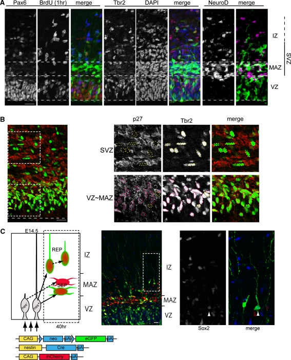
During cerebral neocortical development, excitatory neurons are generated from radial glial cells in the ventricular zone (VZ) or from secondary progenitor cells in the subventricular zone (SVZ); these neurons then migrate toward the pial surface. We have observed that post-mitotic neurons generated directly in the VZ accumulated just above the VZ with a multipolar morphology, while secondary progenitor cells having a long ascending process left the VZ faster than the post-mitotic neurons. Recent observations of human developing neocortex have revealed the existence of radial glia-like progenitors (oRG cells) in the SVZ. This type of progenitor was first thought to be human specific; however, similar cells have also been found in mouse neocortex, and the morphology of these cells resembled that of some of the secondary progenitor cells that we had previously observed, suggesting the existence of a common architecture for the developing neocortex among mammals. In this review, we discuss the nature of the SVZ and its similarities and differences between humans and mice.
Figures

Similar articles
-
Oblique radial glial divisions in the developing mouse neocortex induce self-renewing progenitors outside the germinal zone that resemble primate outer subventricular zone progenitors.J Neurosci. 2011 Mar 9;31(10):3683-95. doi: 10.1523/JNEUROSCI.4773-10.2011. J Neurosci. 2011. PMID: 21389223 Free PMC article.
-
Postnatal development of radial glia and the ventricular zone (VZ): a continuum of the neural stem cell compartment.Cereb Cortex. 2003 Jun;13(6):580-7. doi: 10.1093/cercor/13.6.580. Cereb Cortex. 2003. PMID: 12764031 Review.
-
The 5'-flanking region of the RP58 coding sequence shows prominent promoter activity in multipolar cells in the subventricular zone during corticogenesis.Neuroscience. 2012 Jan 10;201:67-84. doi: 10.1016/j.neuroscience.2011.11.006. Epub 2011 Nov 11. Neuroscience. 2012. PMID: 22119643
-
Pyramidal neurons of upper cortical layers generated by NEX-positive progenitor cells in the subventricular zone.Proc Natl Acad Sci U S A. 2005 Nov 22;102(47):17172-7. doi: 10.1073/pnas.0508560102. Epub 2005 Nov 11. Proc Natl Acad Sci U S A. 2005. PMID: 16284248 Free PMC article.
-
[The behaviors of proliferative cells in the subventricular zone during cortical development].Nihon Shinkei Seishin Yakurigaku Zasshi. 2013 Jun;33(3):131-6. Nihon Shinkei Seishin Yakurigaku Zasshi. 2013. PMID: 25069247 Review. Japanese.
Cited by
-
Of Men and Mice: Modeling the Fragile X Syndrome.Front Mol Neurosci. 2018 Mar 15;11:41. doi: 10.3389/fnmol.2018.00041. eCollection 2018. Front Mol Neurosci. 2018. PMID: 29599705 Free PMC article.
-
Radial Migration Dynamics Is Modulated in a Laminar and Area-Specific Manner During Primate Corticogenesis.Front Cell Dev Biol. 2020 Oct 16;8:588814. doi: 10.3389/fcell.2020.588814. eCollection 2020. Front Cell Dev Biol. 2020. PMID: 33178700 Free PMC article.
-
Hippocampal pyramidal neurons switch from a multipolar migration mode to a novel "climbing" migration mode during development.J Neurosci. 2014 Jan 22;34(4):1115-26. doi: 10.1523/JNEUROSCI.2254-13.2014. J Neurosci. 2014. PMID: 24453304 Free PMC article.
-
Transmitter self-regulation by extracellular glutamate in fresh human cortical slices.J Neural Transm (Vienna). 2014 Nov;121(11):1321-7. doi: 10.1007/s00702-014-1215-1. Epub 2014 Jul 10. J Neural Transm (Vienna). 2014. PMID: 25008583
-
Cognitive Impairment and Brain and Peripheral Alterations in a Murine Model of Intraventricular Hemorrhage in the Preterm Newborn.Mol Neurobiol. 2018 Jun;55(6):4896-4910. doi: 10.1007/s12035-017-0693-1. Epub 2017 Jul 28. Mol Neurobiol. 2018. PMID: 28755273
References
Publication types
MeSH terms
LinkOut - more resources
Full Text Sources
