

Review
doi: 10.2217/nnm.11.141.
Improving delivery and efficacy of nanomedicines in solid tumors: role of tumor priming
Affiliations
- PMID: 22077464
- PMCID: PMC3655409
- DOI: 10.2217/nnm.11.141
Item in Clipboard
Review
Improving delivery and efficacy of nanomedicines in solid tumors: role of tumor priming
Nanomedicine (Lond).
2011 Nov.
Abstract
Effectiveness of nanomedicines in cancer therapy is limited in part by inadequate delivery and transport in tumor interstitium. This article reviews the experimental approaches to improve nanomedicine delivery and transport in solid tumors. These approaches include tumor vasculature normalization, interstitial fluid pressure modulation, enzymatic extracellular matrix degradation, and apoptosis-inducing tumor priming technology. We advocate the latter approach due to its ease and practicality (accomplished with standard-of-care chemotherapy, such as paclitaxel) and tumor selectivity. Examples of applying tumor priming to deliver nanomedicines and to design drug/RNAi-loaded carriers are discussed.
Figures
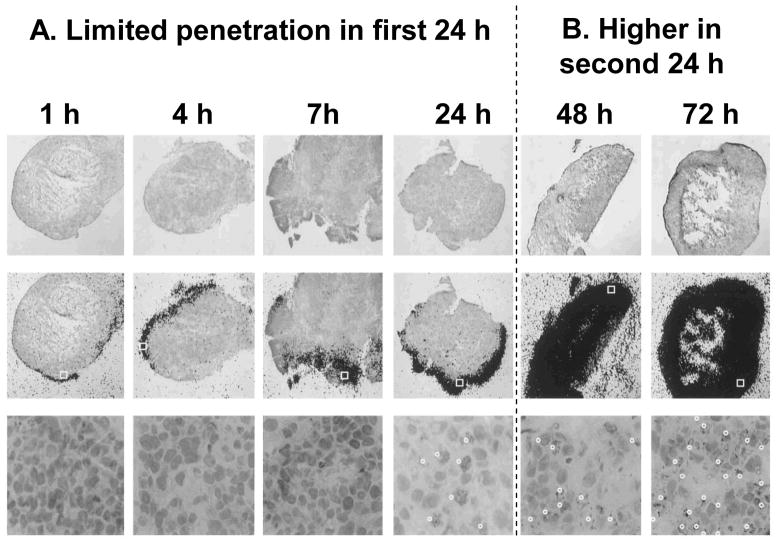
Histocultures of FaDu tumor were treated with 120 nM [3H]paclitaxel. Note changes in the kinetics during the first and second 24 h. Top: Histologic images (stained with hematoxylin and eosin), Magnification, 25×. Middle: images of autoradiographic film overlaid on histologic images, Magnification, 25×. Bottom: enlargement of the indicated boxed region of the slide in the middle panel, to demonstrate the presence of apoptotic cells (indicated by white dots), Magnification, 400×. The fractions of apoptotic cells were ~30% and ~50% at 24 and 72 h, respectively. Reproduced with permission (68).

FaDu tumor histocultures were treated with 10 and 50 nM [3H]paclitaxel continuously. In each figure, the top panels are autoradiographic images overlaid on histological image (253 magnification), and the bottom panels are histological images of the indicated boxed region in the autoradiographic images, at 400× magnification. The indicated times refer to the times after initiation of the [3H]paclitaxel treatment. Reproduced with permission(66).

Tumor-bearing mice were given an intravenous injection of the tumor priming agent (40 mg/kg paclitaxel in Cremophor EL®/ethanol) or the vehicle (control), followed by an injection of red fluorescent latex beads (100 nm diameter) at 48 h and an injection of the green fluorescent perfusion marker 3,3′-diheptyloxacarbocyanine iodide at 72 h. Two minutes after the injection of the perfusion marker, tumor and normal tissues were excised and evaluated using computer assisted image analysis. At least five images per section and at least three sections per tumor were analyzed. Four tumors per data point. Bar, 100 μm. The original color pictures, as well as the quantitative image analysis results are shown in our earlier publication (73). The current black-and-white pictures depict the fluorescence signals in white color. (A) Effect of tumor priming on tumor perfusion. Note the increase in the perfused area (white color) after priming in tumor tissues but not in other host normal tissues. (B) Effect of tumor priming on nanoparticle dispersion in tumor matrix. A tumor section was viewed under fluorescence microscope. The fluorescence signals corresponding to nanoparticles are shown in the left figures and the fluorescence signals corresponding to the perfusion marker (of the same tumor tissue section) are shown in the right figures. In the control group, the location of nanoparticles superimposes the location of perfusion marker. In contrast, the priming group shows (a) greater amount of nanoparticles throughout the tumor section and (b) dispersion of nanoparticles away from vessels. Reproduced with permission (18).
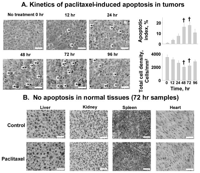
A mouse was given an intravenous injection of paclitaxel (40 mg/kg in Cremophor EL®/ethanol) or the vehicle (control). Tumors and normal tissues were excised and evaluated morphologically. (A) Kinetics of paclitaxel-induced apoptosis in tumors. Changes in apoptotic index and tumor cell density with time were measured. At least five randomly selected regions and at least 3000 cells per tumor and five tumors per time point were evaluated. Arrows, examples of apoptotic cells. (B) No apoptosis in normal tissues (72 h samples). H&E staining (hematoxylin staining for nuclei appeared black in the black-and-white micrograph). Bar, 50 μm. Mean±S.D. Differences between control and tumor priming groups were analyzed using two-sided Student’s t test. †, p < 0.001 compared with time 0 samples. Reproduced with permission (18).
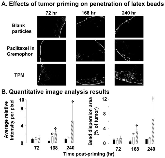
Mice bearing intraperitoneal SKOV3 tumors were given an intraperitoneal injection of a tumor priming treatment with either Cremophor EL®/ethanol or the priming TPM (40 mg/kg), followed by an intraperitoneal injection of fluorescent latex beads (2 μm diameter) given 48, 144, and 216 h later. The dose of latex beads was 40 mg/kg (2% solid, 10-fold dilution in normal saline, 0.5 ml per 25 g mice). Control group received blank, drug-free microparticles (i.e., no tumor priming pretreatment). (A) Representative tumor sections showing amount and dispersion of latex beads in tumors. Beads showed red fluorescence (shown as white dots in the black-and-white pictures). White lines indicate the outer perimeter of tumor nodules. 100× magnification. (B) Quantitative image analysis results. The amounts of latex beads in tumors are expressed as (total fluorescence intensity) normalized by (tumor area); a higher value indicates a greater amount. The bead dispersion results are expressed as percentages of tumor occupied by beads; a higher value indicates a greater dispersion. Solid bars, blank particles; open bars, paclitaxel in castor oil/ethanol; hatched bars, TPM. Error bars show 95% confidence intervals. *, elevated bead amounts or dispersion in paclitaxel in castor oil/ethanol group compared with blank particle group at corresponding time points (p <0.05, Student’s t test). †, elevated bead amounts or dispersion in TPM group compared with paclitaxel in castor oil/ethanol or blank particle groups (p <0.05, ANOVA with post hoc Tukey’s test). Reproduced with permission (92).
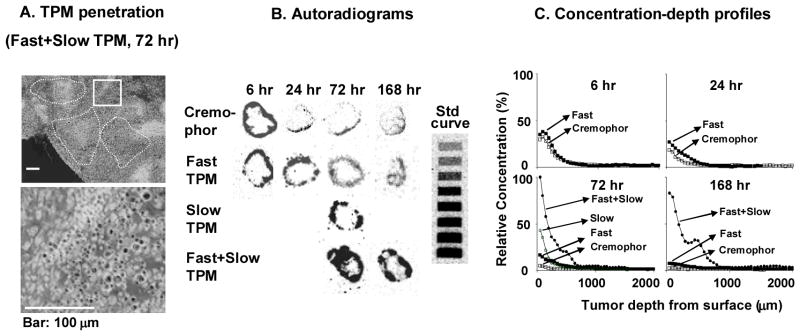
Mice bearing intraperitoneal SKOV3 tumors were given intraperitoneal injections of either paclitaxel/Cremophor EL or priming TPM or sustaining TPM (all at 20 mg/kg) or two-component TPM (40 mg/kg) (1:1 priming/sustaining). (A) TPM penetration into tumor interior. An omental tumor was removed from a mouse at 72 h after treatment with two-component TPM, sectioned, and stained with hematoxylin and eosin. The image was converted using Adobe Photoshop; TPM appeared as black dots. Top panel shows areas with clusters of TPM (circumscribed with dotted lines). Bottom panel shows the enlarged picture of the boxed area. (B) autoradiograms of tumor sections (see Materials and Methods in reference (92)). (C) Concentration-depth profiles. Autoradiograms shown in B were processed to obtain measurements of total radioactivity using computer-assisted densitometric analysis. Radioactivity was expressed as paclitaxel equivalents, with the highest level set at 100%. Reproduced with permission (92).
Similar articles
-
Tumor priming enhances delivery and efficacy of nanomedicines.J Pharmacol Exp Ther. 2007 Jul;322(1):80-8. doi: 10.1124/jpet.107.121632. Epub 2007 Apr 9. J Pharmacol Exp Ther. 2007. PMID: 17420296
-
Combining Nanomedicine and Immunotherapy.Acc Chem Res. 2019 Jun 18;52(6):1543-1554. doi: 10.1021/acs.accounts.9b00148. Epub 2019 May 23. Acc Chem Res. 2019. PMID: 31120725 Free PMC article.
-
Hyperthermia approaches for enhanced delivery of nanomedicines to solid tumors.Biotechnol Bioeng. 2015 Oct;112(10):1967-83. doi: 10.1002/bit.25653. Epub 2015 Jul 14. Biotechnol Bioeng. 2015. PMID: 25995079 Review.
-
Strategies to improve tumor penetration of nanomedicines through nanoparticle design.Wiley Interdiscip Rev Nanomed Nanobiotechnol. 2019 Jan;11(1):e1519. doi: 10.1002/wnan.1519. Epub 2018 Apr 16. Wiley Interdiscip Rev Nanomed Nanobiotechnol. 2019. PMID: 29659166 Review.
-
Recent Advances in Targeted Tumor Chemotherapy Based on Smart Nanomedicines.Small. 2018 Nov;14(45):e1802417. doi: 10.1002/smll.201802417. Epub 2018 Sep 3. Small. 2018. PMID: 30247806 Review.
Cited by
-
Complex effects of tumor microenvironment on the tumor disposition of carrier-mediated agents.Nanomedicine (Lond). 2017 Aug;12(16):2021-2042. doi: 10.2217/nnm-2017-0101. Epub 2017 Jul 26. Nanomedicine (Lond). 2017. PMID: 28745129 Free PMC article. Review.
-
Paclitaxel-loaded polymeric microparticles: quantitative relationships between in vitro drug release rate and in vivo pharmacodynamics.J Control Release. 2013 Dec 28;172(3):737-44. doi: 10.1016/j.jconrel.2013.09.011. Epub 2013 Sep 20. J Control Release. 2013. PMID: 24056144 Free PMC article.
-
Multiscale tumor spatiokinetic model for intraperitoneal therapy.AAPS J. 2014 May;16(3):424-39. doi: 10.1208/s12248-014-9574-y. Epub 2014 Feb 26. AAPS J. 2014. PMID: 24570339 Free PMC article.
-
Systemic Bioequivalence Is Unlikely to Equal Target Site Bioequivalence for Nanotechnology Oncologic Products.AAPS J. 2019 Feb 1;21(2):24. doi: 10.1208/s12248-019-0296-z. AAPS J. 2019. PMID: 30710324 Free PMC article. Review.
-
Target Site Delivery and Residence of Nanomedicines: Application of Quantitative Systems Pharmacology.Pharmacol Rev. 2019 Apr;71(2):157-169. doi: 10.1124/pr.118.016816. Pharmacol Rev. 2019. PMID: 30846487 Free PMC article. Review.
References
-
- Kim BY, Rutka JT, Chan WC. Nanomedicine. N Engl J Med. 2010;363(25):2434–2443. - PubMed
-
- Moghimi SM, Hunter AC, Murray JC. Nanomedicine: current status and future prospects. FASEB J. 2005;19(3):311–330. - PubMed
-
- Ferrari M. Cancer nanotechnology: opportunities and challenges. Nat Rev Cancer. 2005;5(3):161–171. - PubMed
-
- Ibrahim NK, Desai N, Legha S, et al. Phase I and pharmacokinetic study of ABI-007, a Cremophor-free, protein-stabilized, nanoparticle formulation of paclitaxel. Clin Cancer Res. 2002;8(5):1038–1044. - PubMed
-
- Zhang JA, Xuan T, Parmar M, et al. Development and characterization of a novel liposome-based formulation of SN-38. Int J Pharm. 2004;270(1–2):93–107. - PubMed
Publication types
MeSH terms
Substances
Grants and funding
- R43CA121744/CA/NCI NIH HHS/United States
- R01CA123159/CA/NCI NIH HHS/United States
- R43CA134047/CA/NCI NIH HHS/United States
- R44CA103133/CA/NCI NIH HHS/United States
- R44 CA103133/CA/NCI NIH HHS/United States
- R43 CA121744/CA/NCI NIH HHS/United States
- R01EB015253/EB/NIBIB NIH HHS/United States
- R21 CA100553/CA/NCI NIH HHS/United States
- R43 CA134047/CA/NCI NIH HHS/United States
- R01 CA158300/CA/NCI NIH HHS/United States
- R01CA158300/CA/NCI NIH HHS/United States
- R01 EB015253/EB/NIBIB NIH HHS/United States
- R01 CA123159/CA/NCI NIH HHS/United States
LinkOut - more resources
Full Text Sources