. 2011 Nov 14;195(4):643-56.
doi: 10.1083/jcb.201105098. Epub 2011 Nov 7.

Tim50's presequence receptor domain is essential for signal driven transport across the TIM23 complex

Affiliations

Tim50's presequence receptor domain is essential for signal driven transport across the TIM23 complex

Christian Schulz et al. J Cell Biol. .

Abstract

N-terminal targeting signals (presequences) direct proteins across the TOM complex in the outer mitochondrial membrane and the TIM23 complex in the inner mitochondrial membrane. Presequences provide directionality to the transport process and regulate the transport machineries during translocation. However, surprisingly little is known about how presequence receptors interact with the signals and what role these interactions play during preprotein transport. Here, we identify signal-binding sites of presequence receptors through photo-affinity labeling. Using engineered presequence probes, photo cross-linking sites on mitochondrial proteins were mapped mass spectrometrically, thereby defining a presequence-binding domain of Tim50, a core subunit of the TIM23 complex that is essential for mitochondrial protein import. Our results establish Tim50 as the primary presequence receptor at the inner membrane and show that targeting signals and Tim50 regulate the Tim23 channel in an antagonistic manner.


Figures

Figure 1.
Figure 1.
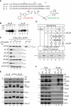
Engineering presequence probes for receptor screening. (A) Primary structure of presequence probes and rat ALDH presequence (pALDH). (B) Presequence probes were imported into isolated mitochondria in the presence or absence of a Δψ for the indicated times at 25°C. After import, mitochondria were treated with proteinase K and samples analyzed by Western blotting using streptavidin-HRP (horseradish peroxidase). (C) Radiolabeled precursor was imported for 15 min at 25°C into isolated mitochondria in the presence of indicated amounts of peptides. After proteinase K treatment, import reactions were analyzed by SDS-PAGE and digital autoradiography. i, intermediate. (D) Mitochondria were incubated with and without presequence peptide in the presence or absence of Δψ. After re-isolation of mitochondria, protein import of a radiolabeled precursor protein was assessed in a second incubation. Therefore, radiolabeled precursor was added to treated or untreated mitochondria and import performed for 15 min. A control import was performed in the presence of peptide. Samples were analyzed by SDS-PAGE and digital autoradiography as in C. i, intermediate. (E) Isolated mitochondria were incubated with 2 µM of the respective photo-peptide, equilibrated for 10 min, and subjected to UV irradiation for 30 min. PA, photo-adduct. (F) After in organello photo cross-linking for 30 min, photo-adducts (PA) were purified from mitochondria by streptavidin agarose chromatography. Samples were analyzed by Western blotting with indicated antibodies. Total, 5%; Eluate, 100%. PA, photo-adduct.
Figure 2.
Figure 2.
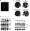
Tim50 contains a C-terminal presequence-binding domain. (A) Schematic representation of Tim50 and truncation constructs used in this study. PS, presequence; TM, transmembrane domain; NIF, NIF domain; PBD, presequence-binding domain. (B) Purified Tim50IMS was incubated with presequence probes under UV irradiation for 30 min, samples analyzed by SDS-PAGE, and proteins subjected to in-gel digest before LC MALDI MS/MS analysis. PA, photo-adduct. (C) Indicated Tim50 constructs were purified from E. coli and analyzed by SDS-PAGE and stained with colloidal Coomassie. Asterisk denotes proteolytic Tim50PBD fragment. (D) Purified Tim50 variants were subjected to presequence photo cross-linking for 30 min and analyzed by Western blotting using anti-Tim50 antibodies. PA, photo-adduct. (E) Chemical cross-linking of pCox4 and SynB2 to isolated Tim50 variants using 100 µM DFDNB for 30 min on ice. Samples were analyzed as in D. (F) Photo cross-linking of presequences to Tim50PBD analyzed as in B. PA, photo-adduct. (G) Fragment ion mass spectrum of peptide Tim50422-435 cross-linked to pL19B18-24. Photo-adduct of pL19B with Tim50PBD (F) was digested with trypsin and subjected to LC-MALDI-MS/MS analysis. The indicated series of y- and b-ions revealed Met427 as the cross-link site. Signals of m/z 837.53 and 903.49 are frequently observed after fragmentation of cross-linked methionine side chains. (H) Alignment of Tim50 using ClustalW 2.0.11. Shown is the PBD domain; black, identical residues in four species, similar residues in at least four or three species are colored in dark or light gray, respectively. Similarity rules according to Erdmann et al. (1991). S.c., Saccharomyces cerevisiae; C.g., Candida glabrata; S.p., Schizosaccharomyces pombe; Y.l., Yarrowia lipolytica; N.c., Neurospora crassa. (I) Chemical cross-linking of 1 µM Tim50IMS to 5 µM pCox4 or SynB2 in the presence of increasing salt concentrations using 100 µM DFDNB for 30 min on ice. Samples were analyzed as in D.
Figure 3.
Figure 3.
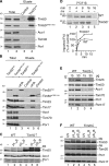
The presequence-binding domain of Tim50 is essential. (A) Diploid yeast cells carrying a single chromosomal deletion of TIM50366-476 were sporulated and subjected to tetrad dissection. (B) Yeast cells containing a chromosomal deletion of TIM50, complemented by a TIM50-containing plasmid carrying URA3, were transformed with plasmid constructs encoding the indicated Tim50 versions and subjected to plasmid loss on 5-FOA–containing medium. (C) Western blot analysis of mitochondria isolated from the cells described in A with the indicated antibodies. (D) In organello photo cross-linking of presequence probes using mitochondria from indicated strains for 30 min. Samples were analyzed as in C.
Figure 4.
Figure 4.
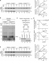
Tim50 contains two separate functional domains for presequence and Tim23 binding. (A) Ni-NTA agarose with purified Tim23IMS or without (control) was incubated with mitochondrial detergent extracts, bound proteins eluted, and analyzed by Western blotting with the indicated antibodies. Total, 5%; Eluate, 100%. (B) Immunoprecipitation from solubilized mitochondria containing reduced levels of Tim50 and Tim50HA1 or Tim50ΔPBD HA1 using anti-HA or -6xHis (control) antibodies. Samples were analyzed as in A. Total, 5%; Eluate, 100%. (C) Isolated mitochondria from indicated strains were incubated with presequence probes and subjected to photo cross-linking for 30 min. Samples were analyzed as in A. PA, photo-adduct. Asterisk denotes cross-reactive protein. (D) Radiolabeled F1β was imported into wild-type and Tim50↑ mitochondria for the indicated times at 25°C. Subsequent to proteinase K digestion samples were analyzed by SDS-PAGE and digital autoradiography. Amounts of processed and protease-protected protein were quantified. The amount after import for 16 min in wild-type mitochondria was set to 100%. m, mature protein. (E) Steady-state protein analysis of isolated wild-type and Tim23 down-regulated mitochondria analyzed by Western blotting with the indicated antibodies. (F) Isolated mitochondria were incubated with 2 µM of the indicated presequence probes and subjected UV irradiation for 30 min. Samples were analyzed as in A. PA, photo-adduct.
Figure 5.
Figure 5.
Presequence recognition by Tim50 is required for inner membrane transport. (A) Radiolabeled precursor was imported at 25°C into isolated mitochondria in the presence or absence of Δψ. Mitochondria were treated with proteinase K and analyzed by SDS-PAGE and digital autoradiography. Amounts of processed protein were quantified (right). The amount after import for 16 min in wild-type mitochondria was set to 100% (n = 3, SEM). p, precursor; m, mature. (B) Radiolabeled AAC was imported into indicated mitochondria at 25°C. After import and proteinase K treatment, mitochondria were solubilized and analyzed by BN-PAGE and digital autoradiography. (C) [35S]Su9-DHFR precursor was imported into mitochondria in the absence of Δψ, and subsequently diluted in buffer containing methotrexate and NADPH. After re-isolation, mitochondria were treated with proteinase K or left untreated as indicated. Samples were analyzed as in A. P, pellet; S, supernatant; p, precursor; f, protease-resistant DHFR fragment. (D) Radiolabeled precursor proteins were imported into mitochondria or mitoplasts for 15 min at 25°C in the presence of affinity-purified anti-Tim50PBD or control antibodies. Samples were analyzed as in A. 100%, import in the absence of antibody. Import into mitoplasts was calculated as percentage of the corresponding import into mitochondria. (E) Radiolabeled precursor was imported into mitoplasts in the presence or absence of Δψ. After import, mitoplasts were treated with proteinase K and analyzed and quantified as in A. p, precursor; m, mature.
Figure 6.
Figure 6.
Presequence and Tim50 binding to Tim23 are mutually exclusive. (A) 1 µM purified Tim23IMS was incubated with excess pCox4 in the absence or presence of the indicated amounts of Tim50IMS and subjected to chemical cross-linking by 100 µM DFDNB for 30 min. Samples were analyzed by Western blotting using the indicated antibodies (left). Quantification of Tim23–Tim50 adduct. 100%, adduct formed with maximal amount of Tim50 (SEM; n = 5; top right). Quantification of the Tim23–pCox4 adduct in response to increasing amounts of Tim50IMS or Tim21IMS (control). 100%, adduct formed without additional protein (SEM; n = 5; bottom right). Asterisks denote degradation products of Tim50IMS; Asterisk, Coomassie-stained degradation product of Tim50IMS detected as bleed-through using a fluorescence scanner at 685 nm. (B) 1 µM purified Tim23IMS was incubated with 10 µM pCox4 in the presence of 5 µM of indicated Tim50 constructs or Tim21IMS (control) and subjected to chemical cross-linking. The Tim23IMS–pCox4 adduct was quantified. 100%, adduct formed in the absence of additional protein (SEM; n = 5 for control and Tim50IMS, n = 3 for Tim50PBD, and n = 7 for Tim50ΔPBD). (C) Equimolar amounts (1 µM) of Tim50IMS and Tim23IMS were incubated with the indicated amounts of biotin-labeled pALDH. After chemical cross-linking using 100 µM DFDNB for 30 min the samples were analyzed by SDS-PAGE and Western blotting with the indicated antibodies.

Similar articles

Cited by

References

    1. Abe Y., Shodai T., Muto T., Mihara K., Torii H., Nishikawa S., Endo T., Kohda D. 2000. Structural basis of presequence recognition by the mitochondrial protein import receptor Tom20. Cell. 100:551–560 10.1016/S0092-8674(00)80691-1 - DOI - PubMed
    1. Alder N.N., Jensen R.E., Johnson A.E. 2008a. Fluorescence mapping of mitochondrial TIM23 complex reveals a water-facing, substrate-interacting helix surface. Cell. 134:439–450 10.1016/j.cell.2008.06.007 - DOI - PubMed
    1. Alder N.N., Sutherland J., Buhring A.I., Jensen R.E., Johnson A.E. 2008b. Quaternary structure of the mitochondrial TIM23 complex reveals dynamic association between Tim23p and other subunits. Mol. Biol. Cell. 19:159–170 10.1091/mbc.E07-07-0669 - DOI - PMC - PubMed
    1. Bauer M.F., Sirrenberg C., Neupert W., Brunner M. 1996. Role of Tim23 as voltage sensor and presequence receptor in protein import into mitochondria. Cell. 87:33–41 10.1016/S0092-8674(00)81320-3 - DOI - PubMed
    1. Bömer U., Meijer M., Maarse A.C., Hönlinger A., Dekker P.J., Pfanner N., Rassow J. 1997. Multiple interactions of components mediating preprotein translocation across the inner mitochondrial membrane. EMBO J. 16:2205–2216 10.1093/emboj/16.9.2205 - DOI - PMC - PubMed

Publication types

MeSH terms

Substances