. 2012 Jan;32(2):376-84.
doi: 10.1128/MCB.06227-11. Epub 2011 Nov 7.

TIN2 stability is regulated by the E3 ligase Siah2

Affiliations

TIN2 stability is regulated by the E3 ligase Siah2

Monica Bhanot et al. Mol Cell Biol. 2012 Jan.

Abstract

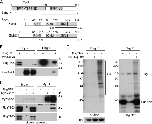
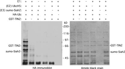
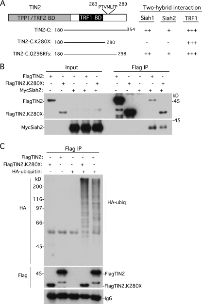
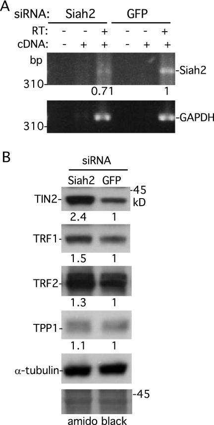
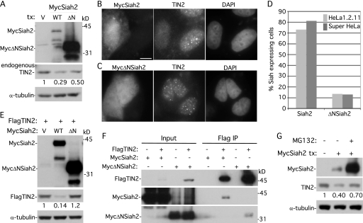
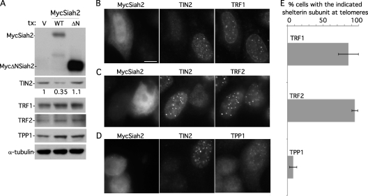
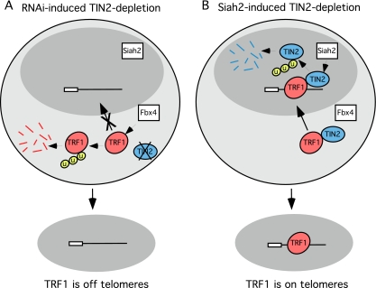
Telomeres are coated by shelterin, a six-subunit complex that is required for protection and replication of chromosome ends. The central subunit TIN2, with binding sites to three subunits (TRF1, TRF2, and TPP1), is essential for stability and function of the complex. Here we show that TIN2 stability is regulated by the E3 ligase Siah2. We demonstrate that TIN2 binds to Siah2 and is ubiquitylated in vivo. We show using purified proteins that Siah2 acts as an E3 ligase to directly ubiquitylate TIN2 in vitro. Depletion of Siah2 led to stabilization of TIN2 protein, indicating that Siah2 regulates TIN2 protein levels in vivo. Overexpression of Siah2 in human cells led to loss of TIN2 at telomeres that was dependent on the presence of the catalytic RING domain of Siah2. In contrast to RNAi-mediated depletion of TIN2 that led to loss of TRF1 and TRF2 at telomeres, Siah2-mediated depletion of TIN2 allowed TRF1 and TRF2 to remain on telomeres, indicating a different fate for shelterin subunits when TIN2 is depleted posttranslationally. TPP1 was lost from telomeres, although its protein level was not reduced. We speculate that Siah2-mediated removal of TIN2 may allow dynamic remodeling of the shelterin complex and its associated factors during the cell cycle.


Figures

Fig 1
Fig 1
TIN2 binds Siah and is ubiquitylated in vivo. (A) Schematic representation of TIN2, with the TPP1, TRF2, and TRF1 binding domains (BD) indicated and Siah1 and Siah2 and the RING domain, cysteine-rich (C-rich) domain, substrate-binding domain (SBD), and nuclear localization signal (NLS) indicated. The lines indicate the TIN2 baits and Siah prey. (B and C) TIN2 binds Siah2 in human cells. HeLaI.2.11 cells were transfected with FlagTIN2 and/or MycSiah2. Cell lysates were immunoprecipitated (IP) with anti-Flag (B) or anti-Myc (C) beads and analyzed by immunoblotting with anti-Myc or anti-Flag antibody. (D) TIN2 is ubiquitylated in vivo. HeLaI.2.11 cells were transfected for 16 h with FlagTIN2 and/or HA-ubiquitin. Cell lysates were immunoprecipitated with anti-Flag beads and analyzed by immunoblotting with anti-HA antibody (left panel) or anti-Flag antibody (right panel).
Fig 2
Fig 2
Siah2 directly ubiquitylates TIN2 in vitro. Reaction mixtures containing the indicated combinations of E1, E2 (UbcH5), E3 (Sumo-Siah2), GST-TIN2, and HA-ubiquitin were incubated, fractionated on SDS-PAGE, and transferred to nitrocellulose. Proteins were visualized by staining with Amido black (right panel), and ubiquitylated proteins were detected by probing with anti-HA antibody (left panel).
Fig 3
Fig 3
Two-hybrid analysis identified a C-terminal Siah-binding site in TIN2, but it is not essential for Siah2 binding and TIN2 ubiquitylation in vivo. (A) Schematic representation of TIN2 and the indicated TIN2-C alleles. The potential Siah-binding motif PTVMLFP is indicated. Two-hybrid interactions were scored according to the number of minutes required for the color change: +++, 15 min; ++, 30 min; +, 45 min; −, no color change. (B) Full-length TIN2 lacking the C-terminal Siah2-binding site binds to Siah2 by coimmunoprecipitation. HeLaI.2.11 cells were transfected with FlagTIN2, FlagTIN2.K280X, and/or MycSiah2. Cell lysates were immunoprecipitated with anti-Flag beads and analyzed by immunoblotting with anti-Flag or anti-Myc antibody. (C) TIN2 lacking the C-terminal Siah-binding site is ubiquitylated in vivo. HeLaI.2.11 cells were transfected with FlagTIN2, FlagTIN2.K280X, and/or HA-ubiquitin. Cell lysates were immunoprecipitated with anti-Flag beads and analyzed by immunoblotting with anti-HA or anti-Flag antibody.
Fig 4
Fig 4
Depletion of Siah2 leads to stabilization of TIN2. (A) RT-PCR analysis of RNA isolated from HeLaI.2.11 cells transfected with GFP or Siah2 siRNA. PCR was performed with primers specific for Siah2 or GAPDH in reactions without cDNA (−) or with cDNA (+) prepared without (−) or with (+) RT. Siah2 DNA levels relative to GAPDH and normalized to the GFP control are indicated below the upper gel. (B) Immunoblot analysis of extracts from HeLaI.2.11 cells transfected with Siah2 or GFP siRNA. The blot was stained with Amido black (bottom panel), and proteins were detected by probing with anti-TIN2 701, anti-TRF1 415, anti-TRF2, anti-TPP1 911, or anti-α-tubulin. Protein levels relative to α-tubulin and normalized to the vector control are indicated between the blots.
Fig 5
Fig 5
Overexpression of Siah2 leads to loss of TIN2 at telomeres dependent upon the Siah2 RING domain and on the proteasome. (A) Immunoblot analysis of extracts from supertelomerase HeLa cells transfected with vector (V), MycSiah2 (WT), or MycΔNSiah2 (ΔN) and probed with anti-Myc, anti-TIN2 701, or anti-α-tubulin. (B and C) Immunofluorescence analysis of supertelomerase HeLa cells transfected with MycSiah2 (B) or MycΔNSiah2 (C). Cells were formaldehyde fixed and dually stained with anti-Myc and anti-TIN2 701 antibodies and with DAPI. Bar, 5 μm. (D) Graphical representation of the frequency of Siah2-expressing cells lacking TIN2 at telomeres (n = 126 cells or more each). (E) Immunoblot analysis of extracts from supertelomerase HeLa cells transfected with FlagTIN2 (+) and vector (V), MycSiah2 (WT), or MycΔNSiah2 (ΔN) and probed with anti-Myc, anti-Flag, or anti-α-tubulin. (F) Although MycΔNSiah2 does not affect TIN2 protein levels or localization to telomeres, it nonetheless binds to TIN2 by coimmunoprecipitation. 293T cells were transfected with FlagTIN2 and with MycSiah2 or MycΔNSiah2. Cell lysates were immunoprecipitated with anti-Flag beads and analyzed by immunoblotting with anti-Flag or anti-Myc antibody. (G) Inhibition of the proteasome by MG132 rescues Siah2-induced loss of TIN2. Supertelomerase HeLa cells were transfected for 16 h with vector (−) or MycSiah2 (+). MG132 (10 μm) was added 4 h prior to harvest. Cell extracts were analyzed by immunoblotting with anti-Myc, anti-TIN2 701, and anti-α-tubulin. (A, E, and G) TIN2 protein levels relative to α-tubulin and normalized to the vector control are indicated below the blots.
Fig 6
Fig 6
TRF1 and TRF2, but not TPP1, remain at telomeres upon Siah2-mediated loss of TIN2. (A) Immunoblot analysis shows that MycSiah2 (but not MycΔNSiah2) induces loss of TIN2 protein but not TRF1, TRF2, or TPP1. Results of immunoblot analysis of extracts from supertelomerase HeLa cells transfected with vector (V), MycSiah2 (WT), or MycΔNSiah2 (ΔN) and probed with anti-TIN2 701, anti-TRF1 415, anti-TRF2, anti-TPP1 911, or anti-α-tubulin antibodies are shown. TIN2 protein levels relative to α-tubulin and normalized to the vector control are indicated below the upper blot. (B to D) Immunofluorescence analysis of supertelomerase HeLa cells transfected with MycSiah2. Cells were formaldehyde fixed and triply stained with anti-myc, anti-TIN2 701, and anti-TRF1 (B), anti-TRF2 (C), or anti-TPP1 1146 (D) and DAPI. Bar, 5 μm. (E) Graphical representation of the frequency of Siah2-expressing TIN2-lacking cells with the indicated shelterin component at telomeres. Data represent the means ± standard deviations of the results of three independent experiments (n = 69 cells or more each).
Fig 7
Fig 7
Speculative model of how the fate of TRF1 depends on the method of TIN2 depletion. (A) RNAi-induced TIN2 depletion. TIN2 is not expressed and thus not available to protect TRF1 from degradation by the cytoplasmic E3 ligase Fbx4. (B) Siah2-induced TIN2 depletion. TIN2 is expressed and available in the cytoplasm to protect TRF1 from degradation by the cytoplasmic E3 ligase Fbx4. Upon transport to the nucleus, TIN2 is degraded by the nuclear E3 ligase Siah2. Once in the nucleus, TRF1 does not require TIN2 for protection from Fbx4 and thus remains on telomeres.

Similar articles

Cited by

References

    1. Abreu E, et al. 2010. TIN2-tethered TPP1 recruits human telomerase to telomeres in vivo. Mol. Cell. Biol. 30: 2971–2982 - PMC - PubMed
    1. Ancelin K, et al. 2002. Targeting assay to study the cis functions of human telomeric proteins: evidence for inhibition of telomerase by TRF1 and for activation of telomere degradation by TRF2. Mol. Cell. Biol. 22: 3474–3487 - PMC - PubMed
    1. Atanassov BS, et al. 2009. Gcn5 and SAGA regulate shelterin protein turnover and telomere maintenance. Mol. Cell 35: 352–364 - PMC - PubMed
    1. Bartel PL, Chien C, Sternglanz R, Fields S. 1993. Using the two-hybrid system to detect protein-protein interaction, p 153–179 In Harley D. A. (ed), Cellular interactions in development: a practical approach. IRL Press, Oxford, United Kingdom
    1. Baumann P, Cech TR. 2001. Pot1, the putative telomere end-binding protein in fission yeast and humans. Science 292: 1171–1175 - PubMed

Publication types