. 2012 Mar;11(3):M111.013458.
doi: 10.1074/mcp.M111.013458. Epub 2011 Oct 31.

Antibody-based protein profiling of the human chromosome 21

Affiliations

Antibody-based protein profiling of the human chromosome 21

Mathias Uhlén et al. Mol Cell Proteomics. 2012 Mar.

Abstract

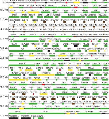
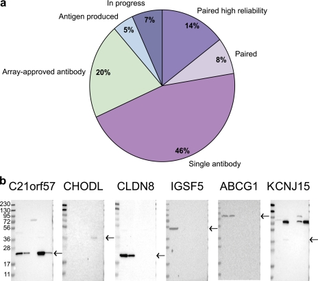
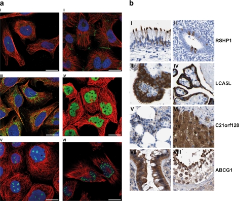
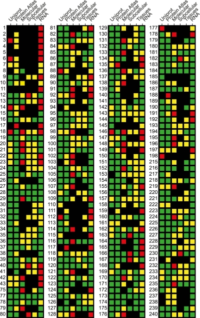
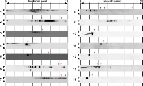
The Human Proteome Project has been proposed to create a knowledge-based resource based on a systematical mapping of all human proteins, chromosome by chromosome, in a gene-centric manner. With this background, we here describe the systematic analysis of chromosome 21 using an antibody-based approach for protein profiling using both confocal microscopy and immunohistochemistry, complemented with transcript profiling using next generation sequencing data. We also describe a new approach for protein isoform analysis using a combination of antibody-based probing and isoelectric focusing. The analysis has identified several genes on chromosome 21 with no previous evidence on the protein level, and the isoform analysis indicates that a large fraction of human proteins have multiple isoforms. A chromosome-wide matrix is presented with status for all chromosome 21 genes regarding subcellular localization, tissue distribution, and molecular characterization of the corresponding proteins. The path to generate a chromosome-specific resource, including integrated data from complementary assay platforms, such as mass spectrometry and gene tagging analysis, is discussed.


Figures

Fig. 1.
Fig. 1.
Overview and evidence level of the putative protein-coding genes on human chromosome 21. The 240 protein-coding genes annotated in Ensembl release 59 have been color coded according to the annotation of the corresponding protein in the UniProt database. The size of each box corresponds to the number of amino acids of the largest splice variant of the corresponding gene, but the distances between genes are not drawn in scale; instead 10 genes are shown across each horizontal line (except for the last two lines). The chromosomal position from the left arm of the chromosome is shown to the left. The color codes are as follows: green, UniProt category 1 (evidence at protein level); yellow, UniProt category 2 (evidence at transcript level); red, UniProt category 4 and 5 (predicted and uncertain); black, no reviewed data available in UniProt; brown, keratin-associated genes.
Fig. 2.
Fig. 2.
Status and molecular characterization of the chromosome 21 encoded proteins. a, the pie chart shows the status of the chromosome 21 genes as the fraction of genes having two published antibodies for which knowledge-based annotation show high reliability (Paired high reliability), having two published antibodies for which knowledge-based annotation is not conclusive (Paired), having one published antibody (Single antibody), having an antibody specifically recognizing its target on a protein array (Array-approved antibody), having an MS-verified antigen produced (Antigen produced), and being in an earlier stage in the workflow (In progress). b, the first five Western blots from the left show proteins that were detected as a single major band of predicted size and had no previous evidence at the protein level. The right-most Western blot is an example where there are strong indications that glycosylation of the protein is responsible for the shift of the band to a larger size than predicted.
Fig. 3.
Fig. 3.
Antibody-based profiling to determine subcellular localization and tissue distribution. a, examples of proteins localized to different subcellular compartments. The protein of interest is shown in green, the nucleus in blue, and microtubules are in red. The scale bar indicates 20 μm. Image I, image of the centrosomal protein PCNT, in U-2 OS cells using the antibody HPA016820. Image II, image of the receptor CXAR, localized to cell junctions in U-251 MG cells using the antibody HPA030411. Image III, image of the mitochondrial protein ATP5J in U-2 OS cells, using the antibody HPA031069. Image IV, image of the transcription factor BACH1 shown to localize to the nucleus but not to the nucleoli in A-431 cells using the antibody HPA003175. Image V, image of the ribosomal protein RRP1 localized to the nucleoli in A-431 cells using the antibody HPA018166. Image VI, image of the DNA-binding protein SON that localizes to the nucleus with a speckled pattern in U-251 MG cells using the antibody HPA023535. b, examples of immunohistochemistry stained tissue sections from various normal human tissues and cancer tissues. The previously unknown RSPH1 (Radial spoke head 1 homolog) protein was found expressed in ciliated cells in various tissues. The pattern of expression included a distinct positivity in cilia within the respiratory mucosa (image I). In hormonally active corpus mucosa, a small subset of normal glandular cells shows clear cytoplasmic expression of RSPH1 (image II). The unknown LCA5L (Leber congenital amaurosis 5-like) protein was found to be highly cell type-specific and in normal tissues expressed at high levels in only placental trophoblasts from both early (image III) and late stage placenta (image IV). The C21orf128 was found to be expressed in a selective pattern. Although two different antibodies showed a partly different pattern of immunohistochemical positivity, both antibodies showed a similar expression pattern in a subset of hematopoietic cells in normal bone marrow (image V) and widespread cytoplasmic and nuclear expression in hepatocytes from normal liver tissue (image VI). The ATP-binding cassette, subfamily G (WHITE), member 1 (ABCG1), belonging to the UniProt class of potential transmembrane proteins with no evidence at protein level, showed ubiquitous expression in epithelial cell types, whereas cells within the central nervous system and hematopoietic system were essentially negative. In normal colonic mucosa, ABCG1 expression was accentuated in surface epithelium and differentiating glandular cells lining upper parts of the colonic crypts (image VII). ABCG1 was also expressed in germinal cells from normal testis, with an apparent gradient, so that the most mature spermatocytes and spermatids showed a relative higher level of expression as compared with more basal layers in the seminiferous duct (image VIII).
Fig. 4.
Fig. 4.
RNA-seq to determine transcript abundance in three cell lines. a, the number of genes present in the three categories as defined by transcript expression levels in the three cell lines: supportive (detected at medium or high level in one or more cell lines), uncertain (detected at no more than low level in any cell line), and not supportive (not detected in any cell line). b, read coverage of RNA-seq reads mapping to the protein coding transcript of the gene AP000295.9 (ENSG00000249624). c, read coverage of RNA-seq reads mapping to the protein coding transcript of the gene AP001055.7 (ENSG00000248354). d, read coverage of RNA-seq reads mapping to the protein coding transcripts of the gene TTC3 (ENSG00000182670). The reads align almost exclusively to the exons of the genes.
Fig. 5.
Fig. 5.
Overview of data for proteins on chromosome 21. A status matrix is shown where, for each gene, the evidence levels according to UniProt (first columns), the current status in the Human Protein Atlas (second columns), the result of molecular characterization using Western blot (third columns), the reliability of the results from subcellular location analysis (fourth columns), and tissue profiling (fifth columns), and the evidence for existence on transcriptional level in cell lines (sixth columns) are represented using four colors as follows. First columns: green, protein evidence; yellow, transcript evidence; red, uncertain; black, not reviewed. Second columns: green, at least one antibody approved by the Human Protein Atlas; yellow, MS-verified antigen generated; red, failed or not started; black, in progress. Third through sixth columns: green, supportive; yellow, uncertain; red, nonsupportive; black, not done.
Fig. 6.
Fig. 6.
Analysis of protein isoforms using isoelectric focusing. Protein lysates for 14 chromosome 21 genes were separated according to isoelectric point on a pH gradient gel and subsequently reacted with corresponding antibodies. Red triangles indicate the theoretically calculated isoelectric point(s) for each protein, including known splice variants. To verify antibody specificity, each antibody is analyzed against one lysate from cells transfected with a chromosome 21 gene (rows P) and one lysate from cells infected with control vector (rows N). Pairs of protein and antibody are as follows: pair 1, C21orf59/HPA019055; pair 2, C21orf56/HPA018979; pair 3, CHAF1B/HPA016698; pair 4, CRYZL1/HPA019120; pair 5, DSCR4/HPA018460; pair 6, FAM3B/HPA015885; pair 7, NDUFV3/HPA020463; pair 8, PIGP/HPA026921; pair 9, RRP1/HPA018166; pair 10, RSPH1/HPA016816; pair 11, RWDD2B/HPA018316; pair 12, S100B/HPA015768; pair 13, SAMSN1/HPA017055; pair 14, TFF1/HPA003425.

Similar articles

Cited by

References

    1. (2010) A gene-centric human proteome project: HUPO–the Human Proteome Organisation. Mol. Cell. Proteomics 9, 427–429 - PMC - PubMed
    1. (2010) The call of the human proteome. Nat. Methods 7, 661. - PubMed
    1. Hochstrasser D. (2008) Should the human proteome project be gene- or protein-centric? J. Proteome Res. 7, 5071. - PubMed
    1. Legrain P., Aebersold R., Archakov A., Bairoch A., Bala K., Beretta L., Bergeron J., Borchers C. H., Corthals G. L., Costello C. E., Deutsch E. W., Domon B., Hancock W., He F., Hochstrasser D., Marko-Varga G., Salekdeh G. H., Sechi S., Snyder M., Srivastava S., Uhlen M., Wu C. H., Yamamoto T., Paik Y. K., Omenn G. S. (2011) The human proteome project: Current state and future direction. Mol. Cell. Proteomics 10, 10.1074/mcp.M111.009993 - DOI - PMC - PubMed
    1. Agaton C., Galli J., Höidén Guthenberg I., Janzon L., Hansson M., Asplund A., Brundell E., Lindberg S., Ruthberg I., Wester K., Wurtz D., Höög C., Lundeberg J., Ståhl S., Pontén F., Uhlén M. (2003) Affinity proteomics for systematic protein profiling of chromosome 21 gene products in human tissues. Mol. Cell. Proteomics 2, 405–414 - PubMed

Publication types

MeSH terms