

Inhibition of high-mobility group box 1 expression by siRNA in rat hepatic stellate cells
- PMID: 22039322
- PMCID: PMC3203359
- DOI: 10.3748/wjg.v17.i36.4090
Inhibition of high-mobility group box 1 expression by siRNA in rat hepatic stellate cells
Abstract
Aim: To explore the role of high-mobility group box 1 (HMGB1) protein during liver fibrogenesis and investigate the functional effects of HMGB1 gene silencing in hepatic stellate cells (HSCs) using siRNA.
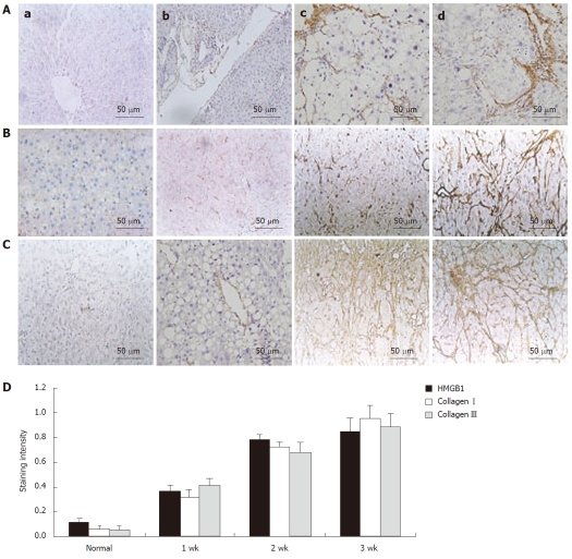
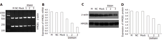
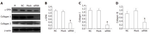
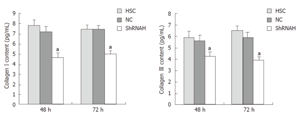
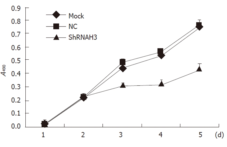
Methods: Hepatic fibrosis in rats was induced throu-gh serial subcutaneous injections of dimethylnitrosamine, and expression of HMGB1 was detected by immunohistochemistry. HMGB1 siRNAs were developed and transiently transfected into HSC-T6 cells using Lipofectamine 2000. HMGB1 expression was evaluated by real-time polymerase chain reaction (PCR) and Western blotting analysis. Expression of α-smooth muscle actin (α-SMA) and collagen types I and III was evaluated by real-time PCR. Cell proliferation and the cell cycle were determined using the methyl thiazolyl tetrazolium method. Finally, collagen content in HSC supernatant was evaluated by an enzyme-linked immunosorbent assay.
Results: The results showed that HMGB1 was upregulated during liver fibrosis and that its expression was closely correlated with the deposition of collagen. siRNA molecules were successfully transfected into HSCs and induced inhibition of HMGB1 expression in a time-dependent manner. Moreover, HMGB1 siRNA treatment inhibited synthesis of α-SMA and collagen types I and III in transfected HSCs.
Conclusion: This study suggests a significant fun-ctional role for HMGB1 in the development of liver fibrosis. It also demonstrates that downregulation of HMGB1 expression might be a potential strategy to treat liver fibrosis.
Keywords: Hepatic fibrosis; Hepatic stellate cells; High-mobility group box 1; RNA interference.
Figures
Similar articles
-
Inhibition of plasminogen activator inhibitor-1 expression by siRNA in rat hepatic stellate cells.J Gastroenterol Hepatol. 2008 Dec;23(12):1917-25. doi: 10.1111/j.1440-1746.2008.05485.x. Epub 2008 Aug 28. J Gastroenterol Hepatol. 2008. PMID: 18761555
-
[Effect of high fat on fibrosis in rat hepatic stellate cells].Zhonghua Gan Zang Bing Za Zhi. 2016 Mar 20;24(3):191-5. doi: 10.3760/cma.j.issn.1007-3418.2016.03.007. Zhonghua Gan Zang Bing Za Zhi. 2016. PMID: 27095762 Chinese.
-
Involvement of the nuclear high mobility group B1 peptides released from injured hepatocytes in murine hepatic fibrogenesis.Biochim Biophys Acta. 2014 Sep;1842(9):1720-32. doi: 10.1016/j.bbadis.2014.06.017. Epub 2014 Jun 23. Biochim Biophys Acta. 2014. PMID: 24970745
-
Emerging role of HMGB1 in fibrotic diseases.J Cell Mol Med. 2014 Dec;18(12):2331-9. doi: 10.1111/jcmm.12419. Epub 2014 Oct 6. J Cell Mol Med. 2014. PMID: 25284457 Free PMC article. Review.
-
From Inflammation to Fibrosis: Novel Insights into the Roles of High Mobility Group Protein Box 1 in Schistosome-Induced Liver Damage.Pathogens. 2022 Feb 24;11(3):289. doi: 10.3390/pathogens11030289. Pathogens. 2022. PMID: 35335612 Free PMC article. Review.
Cited by
-
Is serum high-mobility group box 1 (HMGB-1) level correlated with liver fibrosis in chronic hepatitis B?Medicine (Baltimore). 2017 Sep;96(36):e7547. doi: 10.1097/MD.0000000000007547. Medicine (Baltimore). 2017. PMID: 28885322 Free PMC article. Clinical Trial.
-
High-Mobility Group Box-1 and Liver Disease.Hepatol Commun. 2018 Sep 7;2(9):1005-1020. doi: 10.1002/hep4.1223. eCollection 2018 Sep. Hepatol Commun. 2018. PMID: 30202816 Free PMC article. Review.
-
Carnosic Acid Alleviates BDL-Induced Liver Fibrosis through miR-29b-3p-Mediated Inhibition of the High-Mobility Group Box 1/Toll-Like Receptor 4 Signaling Pathway in Rats.Front Pharmacol. 2018 Jan 19;8:976. doi: 10.3389/fphar.2017.00976. eCollection 2017. Front Pharmacol. 2018. PMID: 29403377 Free PMC article.
-
HMGB1 in health and disease.Mol Aspects Med. 2014 Dec;40:1-116. doi: 10.1016/j.mam.2014.05.001. Epub 2014 Jul 8. Mol Aspects Med. 2014. PMID: 25010388 Free PMC article. Review.
-
Artemisia annua Leaf Extract Attenuates Hepatic Steatosis and Inflammation in High-Fat Diet-Fed Mice.J Med Food. 2016 Mar;19(3):290-9. doi: 10.1089/jmf.2015.3527. Epub 2016 Jan 7. J Med Food. 2016. PMID: 26741655 Free PMC article.
References
-
- Moreira RK. Hepatic stellate cells and liver fibrosis. Arch Pathol Lab Med. 2007;131:1728–1734. - PubMed
-
- Wang H, Bloom O, Zhang M, Vishnubhakat JM, Ombrellino M, Che J, Frazier A, Yang H, Ivanova S, Borovikova L, et al. HMG-1 as a late mediator of endotoxin lethality in mice. Science. 1999;285:248–251. - PubMed
-
- Hamada N, Maeyama T, Kawaguchi T, Yoshimi M, Fukumoto J, Yamada M, Yamada S, Kuwano K, Nakanishi Y. The role of high mobility group box1 in pulmonary fibrosis. Am J Respir Cell Mol Biol. 2008;39:440–447. - PubMed
-
- Ilmakunnas M, Tukiainen EM, Rouhiainen A, Rauvala H, Arola J, Nordin A, Mäkisalo H, Höckerstedt K, Isoniemi H. High mobility group box 1 protein as a marker of hepatocellular injury in human liver transplantation. Liver Transpl. 2008;14:1517–1525. - PubMed
Publication types
MeSH terms
Substances
LinkOut - more resources
Full Text Sources
Other Literature Sources
Medical
