TGF-β drives epithelial-mesenchymal transition through δEF1-mediated downregulation of ESRP
- PMID: 22037216
- PMCID: PMC3391666
- DOI: 10.1038/onc.2011.493
TGF-β drives epithelial-mesenchymal transition through δEF1-mediated downregulation of ESRP
Abstract
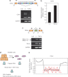
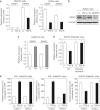
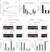
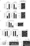
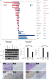
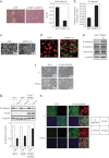
Epithelial-mesenchymal transition (EMT) is a crucial event in wound healing, tissue repair and cancer progression in adult tissues. We have recently shown that transforming growth factor (TGF)-β-induced EMT involves isoform switching of fibroblast growth factor receptors by alternative splicing. We performed a microarray-based analysis at single exon level to elucidate changes in splicing variants generated during TGF-β-induced EMT, and found that TGF-β induces broad alteration of splicing patterns by downregulating epithelial splicing regulatory proteins (ESRPs). This was achieved by TGF-β-mediated upregulation of δEF1 family proteins, δEF1 and SIP1. δEF1 and SIP1 each remarkably repressed ESRP2 transcription through binding to the ESRP2 promoter in NMuMG cells. Silencing of both δEF1 and SIP1, but not either alone, abolished the TGF-β-induced ESRP repression. The expression profiles of ESRPs were inversely related to those of δEF1 and SIP in human breast cancer cell lines and primary tumor specimens. Further, overexpression of ESRPs in TGF-β-treated cells resulted in restoration of the epithelial splicing profiles as well as attenuation of certain phenotypes of EMT. Therefore, δEF1 family proteins repress the expression of ESRPs to regulate alternative splicing during TGF-β-induced EMT and the progression of breast cancers.
Figures
Similar articles
-
Differential regulation of epithelial and mesenchymal markers by deltaEF1 proteins in epithelial mesenchymal transition induced by TGF-beta.Mol Biol Cell. 2007 Sep;18(9):3533-44. doi: 10.1091/mbc.e07-03-0249. Epub 2007 Jul 5. Mol Biol Cell. 2007. PMID: 17615296 Free PMC article.
-
TGF-β regulates isoform switching of FGF receptors and epithelial-mesenchymal transition.EMBO J. 2011 Feb 16;30(4):783-95. doi: 10.1038/emboj.2010.351. Epub 2011 Jan 11. EMBO J. 2011. PMID: 21224849 Free PMC article.
-
δEF1 associates with DNMT1 and maintains DNA methylation of the E-cadherin promoter in breast cancer cells.Cancer Med. 2015 Jan;4(1):125-35. doi: 10.1002/cam4.347. Epub 2014 Oct 15. Cancer Med. 2015. PMID: 25315069 Free PMC article.
-
Roles and Regulation of Epithelial Splicing Regulatory Proteins 1 and 2 in Epithelial-Mesenchymal Transition.Int Rev Cell Mol Biol. 2016;327:163-194. doi: 10.1016/bs.ircmb.2016.06.003. Epub 2016 Jul 30. Int Rev Cell Mol Biol. 2016. PMID: 27692175 Review.
-
Oncogenic functions of the EMT-related transcription factor ZEB1 in breast cancer.J Transl Med. 2020 Feb 3;18(1):51. doi: 10.1186/s12967-020-02240-z. J Transl Med. 2020. PMID: 32014049 Free PMC article. Review.
Cited by
-
Androgen-regulated transcription of ESRP2 drives alternative splicing patterns in prostate cancer.Elife. 2019 Sep 3;8:e47678. doi: 10.7554/eLife.47678. Elife. 2019. PMID: 31478829 Free PMC article.
-
Epithelial-Mesenchymal Transition and Metastasis under the Control of Transforming Growth Factor β.Int J Mol Sci. 2018 Nov 20;19(11):3672. doi: 10.3390/ijms19113672. Int J Mol Sci. 2018. PMID: 30463358 Free PMC article. Review.
-
Nanoscale Porphyrin Metal-Organic Frameworks Deliver siRNA for Alleviating Early Pulmonary Fibrosis in Acute Lung Injury.Front Bioeng Biotechnol. 2022 Jul 18;10:939312. doi: 10.3389/fbioe.2022.939312. eCollection 2022. Front Bioeng Biotechnol. 2022. PMID: 35923570 Free PMC article.
-
Epithelial splicing regulatory protein 2-mediated alternative splicing reprograms hepatocytes in severe alcoholic hepatitis.J Clin Invest. 2020 Apr 1;130(4):2129-2145. doi: 10.1172/JCI132691. J Clin Invest. 2020. PMID: 31945016 Free PMC article.
-
ESRP1 is overexpressed in ovarian cancer and promotes switching from mesenchymal to epithelial phenotype in ovarian cancer cells.Oncogenesis. 2017 Oct 9;6(10):e389. doi: 10.1038/oncsis.2017.87. Oncogenesis. 2017. PMID: 28991261 Free PMC article.
References
-
- Blencowe BJ. Alternative splicing: new insights from global analyses. Cell. 2006;126:37–47. - PubMed
-
- Chaffer CL, Dopheide B, Savagner P, Thompson EW, Williams ED. Aberrant fibroblast growth factor receptor signaling in bladder and other cancers. Differentiation. 2007;75:831–842. - PubMed
-
- Charafe-Jauffret E, Ginestier C, Monville F, Finetti P, Adelaide J, Cervera N, et al. Gene expression profiling of breast cell lines identifies potential new basal markers. Oncogene. 2006;25:2273–2284. - PubMed
-
- Coumoul X, Deng CX. Roles of FGF receptors in mammalian development and congenital diseases. Birth Defects Res C Embryo Today. 2003;69:286–304. - PubMed
-
- Dutertre M, Lacroix-Triki M, Driouch K, de la Grange P, Gratadou L, Beck S, et al. Exon-based clustering of murine breast tumor transcriptomes reveals alternative exons whose expression is associated with metastasis. Cancer Res. 2010;70:896–905. - PubMed
Publication types
MeSH terms
Substances
LinkOut - more resources
Full Text Sources
Other Literature Sources
Molecular Biology Databases
Research Materials
Miscellaneous

