

Dietary vitamin D3 supplements reduce demyelination in the cuprizone model
- PMID: 22028844
- PMCID: PMC3197632
- DOI: 10.1371/journal.pone.0026262
Dietary vitamin D3 supplements reduce demyelination in the cuprizone model
Abstract
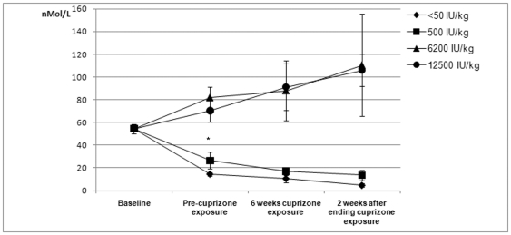
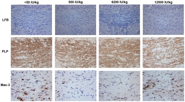
Vitamin D is emerging as a probably important environmental risk factor in multiple sclerosis, affecting both susceptibility and disease progression. It is not known to what extent this effect is due to a modulation of peripheral lymphocyte function, or to intrathecal effects of vitamin D. We investigated the effect of dietary vitamin D3 content on de/remyelination in the cuprizone model, which is a well established toxic model of demyelination, with no associated lymphocyte infiltration. The mice received diets either deficient of (<50 IU/kg), or supplemented with low (500 IU/kg), high (6200 IU/kg) or very high (12500 IU/kg) amounts of vit D3. Cuprizone (0.2%) was added to the diet for six weeks, starting two weeks after onset of the experimental diets. Mouse brain tissue was histopathologically evaluated for myelin and oligodendrocyte loss, microglia/macrophage activation, and lymphocyte infiltration after six weeks of cuprizone exposure, and two weeks after discontinuation of cuprizone exposure. High and very high doses of vitamin D3 significantly reduced the extent of white matter demyelination (p = 0.004) and attenuated microglia activation (p = 0.001). No differences in the density of oligodendrocytes were observed between the diet groups. Two weeks after discontinuation of cuprizone exposure, remyelination was only detectable in the white matter of mice receiving diets deficient of or with low vitamin D3 content. In conclusion, high dietary doses of vitamin D3 reduce the extent of demyelination, and attenuate microglia activation and macrophage infiltration in a toxic model of demyelination, independent of lymphocyte infiltration.
Conflict of interest statement
Figures


Similar articles
-
Effect of high-dose 1.25 dihydroxyvitamin D3 on remyelination in the cuprizone model.APMIS. 2014 Dec;122(12):1178-86. doi: 10.1111/apm.12281. Epub 2014 May 26. APMIS. 2014. PMID: 24862867
-
The cuprizone model: regional heterogeneity of pathology.APMIS. 2012 Aug;120(8):648-57. doi: 10.1111/j.1600-0463.2012.02882.x. Epub 2012 Feb 24. APMIS. 2012. PMID: 22779688
-
Time-dependent changes in the brain arachidonic acid cascade during cuprizone-induced demyelination and remyelination.Prostaglandins Leukot Essent Fatty Acids. 2011 Jul;85(1):29-35. doi: 10.1016/j.plefa.2011.04.001. Epub 2011 May 6. Prostaglandins Leukot Essent Fatty Acids. 2011. PMID: 21530210 Free PMC article.
-
De- and remyelination in the CNS white and grey matter induced by cuprizone: the old, the new, and the unexpected.Histol Histopathol. 2011 Dec;26(12):1585-97. doi: 10.14670/HH-26.1585. Histol Histopathol. 2011. PMID: 21972097 Review.
-
Oligodendrocyte death and myelin loss in the cuprizone model: an updated overview of the intrinsic and extrinsic causes of cuprizone demyelination.Mol Neurodegener. 2022 May 7;17(1):34. doi: 10.1186/s13024-022-00538-8. Mol Neurodegener. 2022. PMID: 35526004 Free PMC article. Review.
Cited by
-
Vitamin D₃ and monomethyl fumarate enhance natural killer cell lysis of dendritic cells and ameliorate the clinical score in mice suffering from experimental autoimmune encephalomyelitis.Toxins (Basel). 2015 Nov 13;7(11):4730-44. doi: 10.3390/toxins7114730. Toxins (Basel). 2015. PMID: 26580651 Free PMC article.
-
1,25-Dihydroxyvitamin D3 suppressed experimental autoimmune encephalomyelitis through both immunomodulation and oligodendrocyte maturation.Exp Mol Pathol. 2017 Jun;102(3):515-521. doi: 10.1016/j.yexmp.2017.05.015. Epub 2017 May 25. Exp Mol Pathol. 2017. PMID: 28552332 Free PMC article.
-
Efficacy of high-dose vitamin D3 supplementation in vitamin D deficient pregnant women with multiple sclerosis: Preliminary findings of a randomized-controlled trial.Iran J Neurol. 2015 Apr 4;14(2):67-73. Iran J Neurol. 2015. PMID: 26056550 Free PMC article.
-
Environmental Influencers, MicroRNA, and Multiple Sclerosis.J Cent Nerv Syst Dis. 2020 Jan 20;12:1179573519894955. doi: 10.1177/1179573519894955. eCollection 2020. J Cent Nerv Syst Dis. 2020. PMID: 32009827 Free PMC article. Review.
-
The benefits and detriments of macrophages/microglia in models of multiple sclerosis.Clin Dev Immunol. 2013;2013:948976. doi: 10.1155/2013/948976. Epub 2013 Jun 12. Clin Dev Immunol. 2013. PMID: 23840244 Free PMC article. Review.
References
-
- Grytten N, Glad SB, Aarseth JH, Nyland H, Midgard R, et al. A 50-year follow-up of the incidence of multiple sclerosis in Hordaland County, Norway. Neurology. 2006;66:182–186. - PubMed
-
- Compston A, Coles A. Multiple sclerosis. Lancet. 2008;372:1502–1517. S0140-6736(08)61620-7 [pii];10.1016/S0140-6736(08)61620-7 [doi] - PubMed
-
- Smolders J, Damoiseaux J, Menheere P, Hupperts R. Vitamin D as an immune modulator in multiple sclerosis, a review. J Neuroimmunol. 2008;194:7–17. - PubMed
-
- Myhr KM. Vitamin D treatment in multiple sclerosis. J Neurol Sci. 2009;286:104–108. S0022-510X(09)00586-3 [pii];10.1016/j.jns.2009.05.002 [doi] - PubMed
-
- Munger KL, Levin LI, Hollis BW, Howard NS, Ascherio A. Serum 25-hydroxyvitamin D levels and risk of multiple sclerosis. JAMA. 2006;296:2832–2838. - PubMed
Publication types
MeSH terms
Substances
LinkOut - more resources
Full Text Sources
Medical
