

Inflammatory cytokines in systemic lupus erythematosus
- PMID: 22028588
- PMCID: PMC3196871
- DOI: 10.1155/2011/432595
Inflammatory cytokines in systemic lupus erythematosus
Abstract
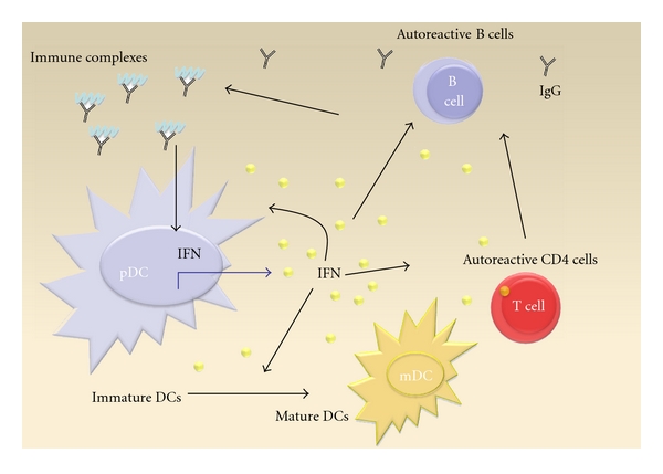
Systemic lupus erythematosus (SLE) is an autoimmune disease of unknown origin affecting virtually all organ systems. Beyond genetic and environmental factors, cytokine imbalances contribute to immune dysfunction, trigger inflammation, and induce organ damage. The key cytokine that is involved in SLE pathogenesis is interferon alpha. Interferon secretion is induced by immune complexes and leads to upregulation of several inflammatory proteins, which account for the so-called IFN signature that can be found in the majority of SLE PBMCs. Additionally IL-6 and IFN-y as well as T-cell-derived cytokines like IL-17, IL-21, and IL-2 are dysregulated in SLE. The latter induce a T-cell phenotype that is characterized by enhanced B-cell help and enhanced secretion of proinflammatory cytokines but reduced induction of suppressive T cells and activation-induced cell death. This paper will focus on these cytokines and highlights pathophysiological approaches and therapeutic potential.
Figures

Similar articles
-
IFN-λ1 with Th17 axis cytokines and IFN-α define different subsets in systemic lupus erythematosus (SLE).Arthritis Res Ther. 2017 Jun 15;19(1):139. doi: 10.1186/s13075-017-1344-7. Arthritis Res Ther. 2017. PMID: 28619037 Free PMC article.
-
Roles of IL-1 and IL-10 family cytokines in the progression of systemic lupus erythematosus: Friends or foes?IUBMB Life. 2022 Feb;74(2):143-156. doi: 10.1002/iub.2568. Epub 2021 Oct 19. IUBMB Life. 2022. PMID: 34668305 Review.
-
Cytokines as Biomarkers in Systemic Lupus Erythematosus: Value for Diagnosis and Drug Therapy.Int J Mol Sci. 2021 Oct 20;22(21):11327. doi: 10.3390/ijms222111327. Int J Mol Sci. 2021. PMID: 34768756 Free PMC article. Review.
-
Interferon-lambda1 induces peripheral blood mononuclear cell-derived chemokines secretion in patients with systemic lupus erythematosus: its correlation with disease activity.Arthritis Res Ther. 2011 Jun 16;13(3):R88. doi: 10.1186/ar3363. Arthritis Res Ther. 2011. PMID: 21679442 Free PMC article.
-
BAFF-R and TACI expression on CD3+ T cells: Interplay among BAFF, APRIL and T helper cytokines profile in systemic lupus erythematosus.Cytokine. 2019 Feb;114:115-127. doi: 10.1016/j.cyto.2018.11.008. Epub 2018 Nov 19. Cytokine. 2019. PMID: 30467093
Cited by
-
Intestinal homeostasis in the gut-lung-kidney axis: a prospective therapeutic target in immune-related chronic kidney diseases.Front Immunol. 2023 Nov 1;14:1266792. doi: 10.3389/fimmu.2023.1266792. eCollection 2023. Front Immunol. 2023. PMID: 38022571 Free PMC article. Review.
-
Normalization of CD4+ T cell metabolism reverses lupus.Sci Transl Med. 2015 Feb 11;7(274):274ra18. doi: 10.1126/scitranslmed.aaa0835. Sci Transl Med. 2015. PMID: 25673763 Free PMC article.
-
Pathophysiology of cutaneous lupus erythematosus.Arthritis Res Ther. 2015 Aug 10;17(1):182. doi: 10.1186/s13075-015-0706-2. Arthritis Res Ther. 2015. PMID: 26257198 Free PMC article. Review.
-
Performance of cytokine models in predicting SLE activity.Arthritis Res Ther. 2019 Dec 16;21(1):287. doi: 10.1186/s13075-019-2029-1. Arthritis Res Ther. 2019. PMID: 31842967 Free PMC article.
-
Retinoic Acid, Leaky Gut, and Autoimmune Diseases.Nutrients. 2018 Aug 3;10(8):1016. doi: 10.3390/nu10081016. Nutrients. 2018. PMID: 30081517 Free PMC article. Review.
References
-
- Herrmann M, Voll RE, Kalden JR. Etiopathogenesis of systemic lupus erythematosus. Immunology Today. 2000;21(9):424–426. - PubMed
-
- Siegal FP, Kadowaki N, Shodell M, et al. The nature of the principal type 1 interferon-producing cells in human blood. Science. 1999;284(5421):1835–1837. - PubMed
-
- Liu YJ. IPC: professional type 1 interferon-producing cells and plasmacytoid dendritic cell precursors. Annual Review of Immunology. 2005;23:275–306. - PubMed
Publication types
MeSH terms
Substances
LinkOut - more resources
Full Text Sources
Other Literature Sources
Medical
