. 2012 Mar;46(3):355-64.
doi: 10.1165/rcmb.2010-0155OC. Epub 2011 Oct 20.

The lysophosphatidic acid receptor LPA1 promotes epithelial cell apoptosis after lung injury

Affiliations

The lysophosphatidic acid receptor LPA1 promotes epithelial cell apoptosis after lung injury

Manuela Funke et al. Am J Respir Cell Mol Biol. 2012 Mar.

Abstract

Increased epithelial cell apoptosis in response to lung injury has been implicated in the development of idiopathic pulmonary fibrosis (IPF), but the molecular pathways promoting epithelial cell apoptosis in this disease have yet to be fully identified. Lysophosphatidic acid (LPA), which we have previously demonstrated to mediate bleomycin lung injury-induced fibroblast recruitment and vascular leak in mice and fibroblast recruitment in patients with IPF, is an important regulator of survival and apoptosis in many cell types. We now show that LPA signaling through its receptor LPA(1) promotes epithelial cell apoptosis induced by bleomycin injury. The number of apoptotic cells present in the alveolar and bronchial epithelia of LPA(1)-deficient mice was significantly reduced compared with wild-type mice at Day 3 after bleomycin challenge, as was lung caspase-3 activity. Consistent with these in vivo results, we found that LPA signaling through LPA(1) induced apoptosis in normal human bronchial epithelial cells in culture. LPA-LPA(1) signaling appeared to specifically mediate anoikis, the apoptosis of anchorage-dependent cells induced by their detachment. Similarly, LPA negatively regulated attachment of R3/1 rat alveolar epithelial cell line cells. In contrast, LPA signaling through LPA(1) promoted the resistance of lung fibroblasts to apoptosis, which has also been implicated in IPF. The ability of LPA-LPA(1) signaling to promote epithelial cell apoptosis and fibroblast resistance to apoptosis may therefore contribute to the capacity of this signaling pathway to regulate the development of pulmonary fibrosis after lung injury.


Figures

Figure 1.
Figure 1.
Bleomycin-induced alveolar epithelial apoptosis was attenuated in the lysophosphatidic acid receptor (LPA1) knockout (KO) mice. (A–C) The increase in terminal deoxynucleotidyl transferase dUTP nick end labeling (TUNEL)+ cells induced by bleomycin in the alveolar epithelium was attenuated in LPA1 KO mice. Representative TUNEL/peroxidase-stained sections of (A) wild-type (WT) and (B) LPA1 KO mouse lungs 3 days after bleomycin challenge. Scale bars = 50 μm for all images. (C) Mean numbers of TUNEL+ cells per high-power field ± SEM in lung sections of WT and LPA1 KO mice at Days 0, 1, 3, and 5 after bleomycin challenge. Data were pooled from two independent experiments; the combined numbers of samples were n = 3 (Day 0) or 6 (Days 1, 3, and 5) for WT and LPA1 KO mice. **P < 0.01, WT versus LPA1 KO mice at Day 3 after bleomycin challenge; significance for this comparison, and for all subsequent comparisons performed for experiments with only two experimental groups, was determined by two-tailed Student's t test. (D–F) The increase in p53+ cells induced by bleomycin in the alveolar epithelium was attenuated in LPA1 KO mice. Representative p53/peroxidase-stained sections of (D) WT and (E) LPA1 KO mouse lungs 3 days after bleomycin challenge. (F) Mean numbers of p53+ cells per high-power field ± SEM in WT and LPA1 KO lung sections at Day 0 and Day 3 after bleomycin challenge. n = 3 (Day 0) or 7 (Day 3) for each genotype. **P = 0.01, WT versus LPA1 KO mice at Day 3. (GI) The increase in p21+ cells induced by bleomycin in the alveolar epithelium was attenuated in LPA1 KO mice. Representative images of p21/peroxidase-stained sections of (G) WT and (H) LPA1 KO mouse lungs 3 days after bleomycin challenge. (I) Mean numbers of p21+ cells per high-power field ± SEM in WT and LPA1 KO lung sections at Day 0 and Day3 after bleomycin challenge. n = 3 (Day 0) or 7 (Day 3) for each genotype. #P < 0.001, WT versus LPA1 KO mice at Day 3. (J) Mean caspase 3 activity/whole lung set ± SEM measured in WT and LPA1 KO lung homogenates at Day 0 and Day 3 after bleomycin challenge. n = 7 to 10 for each genotype at each time point. **P = 0.01, WT versus LPA1 KO mice at Day 3.
Figure 2.
Figure 2.
Bleomycin-induced bronchial epithelial apoptosis was attenuated in LPA1 KO mice. The increase in TUNEL+ cells induced by bleomycin in the bronchial epithelium was attenuated in LPA1 KO mice. Representative TUNEL/peroxidase-stained sections of WT and LPA1 KO mouse lungs at Day 0 (A, B), Day 1 (C, D), and Day 3 (E, F) after bleomycin challenge. Scale bars = 50 μm for all images. (G) Mean numbers of TUNEL+ cells per high-power field ± SEM in lung sections of WT and LPA1 KO mice at Day 0, 1, and 3 after bleomycin challenge. Data were pooled from two independent experiments; the combined numbers of samples were n = 3 (Day 0) or 6 (Days 1 and 3) for WT and LPA1 KO mice. #P < 0.001, WT versus LPA1 KO mice at Day 3 after bleomycin challenge.
Figure 3.
Figure 3.
Bleomycin increased LPA levels early after injury. (A) Mean concentrations of six LPA species (16:0, 18:0, 18:1, 18:2, 20:4, and 22:6 LPA) and (B) mean total LPA concentrations in BAL of C57Bl/6 mice at Day 0, 1, and 3 after bleomycin challenge. Three independent experiments were performed. Data represent the means of the values produced in the individual experiments ± SEM; the values produced in the individual experiments themselves were the means of n = 5 mice per time point. Significant differences are indicated as follows: *P < 0.05, **P < 0.01, and ##P < 0.0001. Significance for these comparisons, and for all subsequent comparisons performed for experiments with more than two experimental groups, was determined by pairwise comparisons made with Bonferroni post-test corrections after one-way ANOVA rejected the hypothesis of equality of the group means.
Figure 4.
Figure 4.
LPA signaling through LPA1 promoted lung epithelial cell apoptosis. (A) LPA (18:1) induced the apoptosis of normal human bronchial epithelial (NHBE) cells grown on matrigel. Apoptotic cells were identified by flow cytometry after staining with annexin V and propidium iodide (24) as annexin V-positive, PI-negative cells. Data from one of three independent experiments are presented as mean percentages of annexin V(+) PI(−) cells ± SEM. n = 3 cultures per treatment condition in each experiment. **P < 0.01, LPA-treated versus untreated NHBE cells. (B) LPA (18:1) dose-dependently induced the apoptosis of NHBE cells grown on low-attachment polystyrene. Four independent experiments were performed. Data are presented as mean fold increases in percentages of apoptotic [annexin V(+), PI(−)] cells in LPA-treated NHBE cells compared with untreated cells from the four individual experiments ± SEM; the values produced in the individual experiments themselves were the means of n = 3 cultures per treatment condition. *P < 0.05, #P < 0.001, and ##P < 0.0001, NHBE cells treated with 1, 10, or 20 μM LPA, respectively, versus untreated cells. (C) LPA (18:0) also induced NHBE cell apoptosis, and this effect was abrogated by AM095, a selective LPA1 receptor antagonist. Data are from a representative experiment with n = 3 cultures per treatment condition. ##P < 0.0001, LPA-treated versus untreated NHBE cells and AM095-treated LPA-treated NHBE cells versus LPA-treated NHBE cells.
Figure 5.
Figure 5.
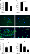
LPA promoted detachment and limited attachment of lung epithelial cells. (A) LPA promoted detachment of NHBE cells from low–attachment, untreated polystyrene. Six independent experiments were performed. Data represent the means of the adherence indices produced in the individual experiments ± SEM; the values produced in the individual experiments themselves were the means of n = 9 cultures per treatment condition. *P < 0.05, LPA-treated versus untreated NHBE cells. (B) LPA limited attachment of NHBE cells to high-attachment tissue culture–treated polystyrene. Four independent experiments were performed. Data represent the means of the percentages of spread cells produced in the individual experiments ± SEM; the values produced in the individual experiments themselves were the means of n = 3 cultures per treatment condition. Spread cells were identified visually by phase contrast microscopy as having flattened from their initial rounded shape such that the nucleus and cytoplasm could be differentiated. #P < 0.001, LPA-treated versus untreated NHBE cells. (C, D) LPA limited NHBE cell formation of focal adhesions. Representative NHBE cells labeled with anti–vinculin-fluorescein isothiocyanate 3 hours after transfer onto high-attachment tissue culture–treated permanox slides (C) without LPA treatment and (D) with LPA treatment. (E, F) LPA limited NHBE cell formation of actin stress fibers. Representative NHBE cells labeled with fluorescein-phalloidin 3 hours after transfer onto high-attachment permanox (E) without LPA treatment and (F) with LPA treatment. (G) LPA did not promote the apoptosis of NHBE cells transferred to ultra–low attachment polystyrene plates. Three independent experiments were performed. Data represent the means of the percentages of annexin V(+) PI(−) cells produced in the individual experiments ± SEM; the values produced in the individual experiments themselves were the means of n = 3 cultures per treatment condition. Differences between LPA-treated and untreated NHBE cells were not significant. (H) LPA limited attachment of R3/1 rat alveolar epithelial cell line cells to high-attachment tissue culture–treated polystyrene. Data represent the mean percentages of spread cells produced in a representative experiment ± SEM, with n = 3 cultures per treatment condition. Spread cells were identified as in (B). *P < 0.05 and ##P < 0.0001, untreated R3/1 cells versus R3/1 cells treated with 1 and 20 μM LPA, respectively.
Figure 6.
Figure 6.
LPA signaling through LPA1 promoted lung fibroblast resistance to apoptosis. (A) LPA promoted primary mouse lung fibroblast (PMLF) resistance to apoptosis induced by serum deprivation when these cells were grown on high-attachment tissue culture–treated polystyrene. Apoptotic cells were identified by flow cytometry after staining with annexin V and propidium iodide (8) as annexin V(+), PI(−). Three independent experiments were performed. In all panels of this figure, data represent the means of the percentages of annexin V(+) PI(−) cells produced in the individual experiments ± SEM; the values produced in the individual experiments themselves were the means of n = 3 cultures per treatment condition. ##P ≥ 0.0001, serum-deprived PMLFs versus PMLFs in serum and serum-deprived PMLFs treated with 1, 10, or 20 μM LPA versus untreated serum-deprived cells. (B) LPA-induced resistance to apoptosis of PMLFs grown on high-attachment polystyrene was abrogated by Ki16425. Three independent experiments were performed. **P < 0.01, serum-deprived PMLFs versus PMLFs in serum; serum-deprived, LPA-treated PMLFs versus untreated serum-deprived cells; and serum-deprived, LPA-treated PMLFs that were also treated with Ki16425 versus serum-deprived, LPA-treated PMLFs. (C) LPA did not promote resistance to apoptosis of PMLFs isolated from LPA1 KO mice that was induced by serum deprivation when these cells were grown on high-attachment polystyrene. Three independent experiments were performed. *P < 0.05, serum-deprived LPA1 KO PMLFs versus LPA1 KO PMLFs in serum. Differences between LPA-treated and untreated serum-deprived LPA1 KO PMLFs were not significant. (D) LPA did not promote resistance to apoptosis of PMLFs transferred to ultra–low attachment polystyrene. Three independent experiments were performed. Differences between LPA-treated and untreated serum-deprived PMLFs were not significant.

Similar articles

Cited by

References

    1. Selman M, King TE, Pardo A. Idiopathic pulmonary fibrosis: Prevailing and evolving hypotheses about its pathogenesis and implications for therapy. Ann Intern Med 2001;134:136–151 - PubMed
    1. Tager AM, LaCamera P, Shea BS, Campanella GS, Selman M, Zhao Z, Polosukhin V, Wain J, Karimi-Shah BA, Kim ND, et al. The lysophosphatidic acid receptor LPA1 links pulmonary fibrosis to lung injury by mediating fibroblast recruitment and vascular leak. Nat Med 2008;14:45–54 - PubMed
    1. Choi JW, Herr DR, Noguchi K, Yung YC, Lee CW, Mutoh T, Lin ME, Teo ST, Park KE, Mosley AN, et al. LPA receptors: subtypes and biological actions. Annu Rev Pharmacol Toxicol 2010;50:157–186 - PubMed
    1. Ye X, Ishii I, Kingsbury MA, Chun J. Lysophosphatidic acid as a novel cell survival/apoptotic factor. Biochim Biophys Acta 2002;1585:108–113 - PubMed
    1. Kuwano K, Kunitake R, Kawasaki M, Nomoto Y, Hagimoto N, Nakanishi Y, Hara N. P21Waf1/Cip1/Sdi1 and p53 expression in association with DNA strand breaks in idiopathic pulmonary fibrosis. Am J Respir Crit Care Med 1996;154:477–483 - PubMed

Publication types

MeSH terms