. 2011 Oct;23(10):3711-26.
doi: 10.1105/tpc.111.090670. Epub 2011 Oct 18.

Arabidopsis actin depolymerizing factor4 modulates the stochastic dynamic behavior of actin filaments in the cortical array of epidermal cells

Affiliations

Arabidopsis actin depolymerizing factor4 modulates the stochastic dynamic behavior of actin filaments in the cortical array of epidermal cells

Jessica L Henty et al. Plant Cell. 2011 Oct.

Abstract

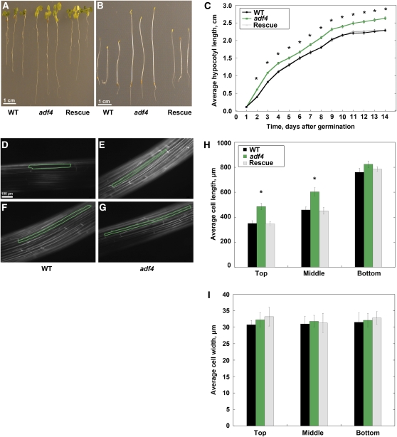
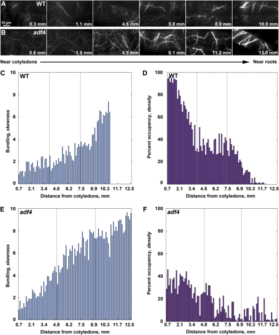
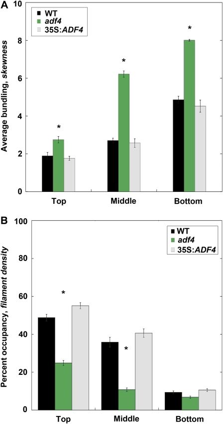
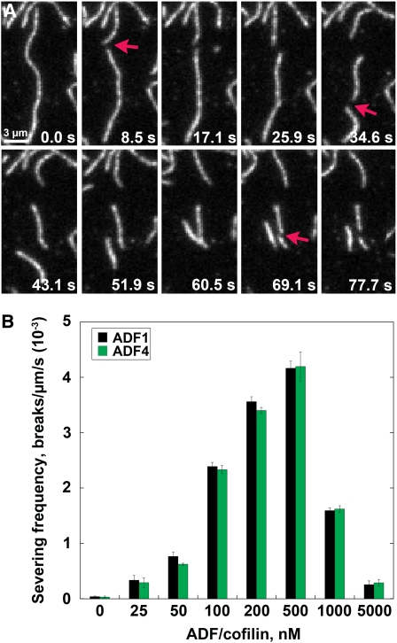
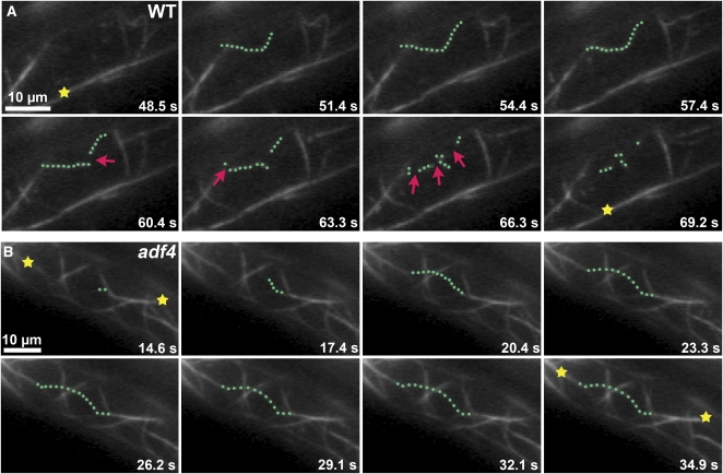
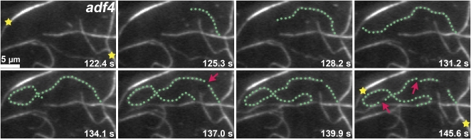
Actin filament arrays are constantly remodeled as the needs of cells change as well as during responses to biotic and abiotic stimuli. Previous studies demonstrate that many single actin filaments in the cortical array of living Arabidopsis thaliana epidermal cells undergo stochastic dynamics, a combination of rapid growth balanced by disassembly from prolific severing activity. Filament turnover and dynamics are well understood from in vitro biochemical analyses and simple reconstituted systems. However, the identification in living cells of the molecular players involved in controlling actin dynamics awaits the use of model systems, especially ones where the power of genetics can be combined with imaging of individual actin filaments at high spatial and temporal resolution. Here, we test the hypothesis that actin depolymerizing factor (ADF)/cofilin contributes to stochastic filament severing and facilitates actin turnover. A knockout mutant for Arabidopsis ADF4 has longer hypocotyls and epidermal cells when compared with wild-type seedlings. This correlates with a change in actin filament architecture; cytoskeletal arrays in adf4 cells are significantly more bundled and less dense than in wild-type cells. Several parameters of single actin filament turnover are also altered. Notably, adf4 mutant cells have a 2.5-fold reduced severing frequency as well as significantly increased actin filament lengths and lifetimes. Thus, we provide evidence that ADF4 contributes to the stochastic dynamic turnover of actin filaments in plant cells.


Figures

Figure 1.
Figure 1.
Homozygous adf4 Mutant Seedlings Exhibit Several Growth Phenotypes. (A) Light-grown adf4 seedlings have longer roots compared with controls; however, no hypocotyl phenotype was apparent. Three light-grown seedlings, 10 d after germination, per genotype are shown. Bar = 1 cm. WT, wild type. (B) Etiolated hypocotyls and roots from homozygous adf4 seedlings are longer than those from wild-type and 35S:ADF4;adf4 (Rescue) lines. Three representative dark-grown seedlings, 10 d after germination, per genotype are shown. Bar = 1 cm. (C) Etiolated adf4 mutant hypocotyls are significantly longer than wild-type and 35S:ADF4;adf4 rescue seedlings from day 2 onward (denoted by asterisks, P = 0.0001, ANOVA); however, no significant difference in length is present between wild-type and 35S:ADF4;adf4 rescue (P = 0.65 for day 2, t test). Measurements were taken daily from at least 100 seedlings per genotype per day. Values are means ± se. (D) to (G) Representative images of hypocotyl epidermal cells from wild-type ([D] and [F]) and adf4 mutant ([E] and [G]) seedlings 5 d after germination. Epidermal cells were stained with FM4-64 dye to visualize cell boundaries and imaged with epifluorescence microscopy. Bar = 100 μm. (D) An axially elongating wild-type epidermal cell located in the top third of the hypocotyl (outlined in green) is shown. (E) A cell from an adf4 mutant seedling located in the same region as (D) is longer along the axis of expansion than the wild-type cell. (F) A cell located in the bottom third of the wild-type hypocotyl has nearly completed axial growth. (G) A cell from an adf4 seedling imaged in the bottom third of the hypocotyl is not different from a wild-type cell imaged in the same region shown in (F). (H) Epidermal cells from adf4 hypocotyls are significantly longer in the axial dimension than wild-type and 35S:ADF4;adf4 controls (denoted by asterisk, P = 0.0166, ANOVA) during expansion. Knockout adf4 seedlings have significantly longer average epidermal cell lengths in the top (P = 0.0013, ANOVA) and middle (P = 0.0007, ANOVA) thirds of the dark-grown seedling when compared with wild-type and 35S:ADF4;adf4 rescue lines. Cell length values are the mean ± se from n > 100 cells and at least 10 hypocotyls per genotype. (I) Epidermal cell width is not altered in adf4 mutant hypocotyls. Epidermal cell width values are the mean ± se from n > 100 cells and least 10 hypocotyls per genotype with the wild type shown in black, adf4 shown in green, and 35S:ADF4;adf4 shown in gray.
Figure 2.
Figure 2.
Architecture of the Actin Cytoskeleton Is Altered in adf4 Hypocotyl Epidermal Cells. (A) VAEM micrographs demonstrate increased actin filament bundling and decreased filament density in the cortical array of dark-grown wild-type (WT) epidermal cells. A montage of images from a single representative hypocotyl is displayed, with cells near the cotyledons at the left and cells near the root at the right. Values shown correspond to the distance of each cell from the cotyledons. Bar = 10 μm. (B) VAEM micrographs of adf4 hypocotyl epidermal cells showing that actin filaments are more bundled and less dense than the wild type (A) in cells located in the lower two-thirds of the hypocotyl. Additionally, bundled actin filaments in cells located near the root in adf4 appear much brighter than bundled filaments in the wild type. (C) Bundling (skewness) quantitatively increases along the gradient of axial cell expansion in a representative hypocotyl. Skewness values were measured from micrographs for every cell of the hypocotyl shown in (A) and plotted as a function of distance from the cotyledons. (D) Quantitative analysis of actin filament density along the gradient of axial cell expansion in a representative hypocotyl. Actin filament density decreases in the cortical array of wild-type epidermal cells in the region spanning from cotyledons to root for the hypocotyl shown in (A). Moreover, filament density and filament bundling appear inversely correlated in vivo. (E) Bundling analysis of the adf4 hypocotyl shown in (B) reveals increased bundling in the cortical actin cytoskeleton. Like the wild type, bundling increases with distance from the cotyledons; however, bundling analysis of adf4 exhibits increased skewness values, indicating more pronounced bundling in the two-thirds of the hypocotyl epidermal cells closest to the roots. (F) Density analysis of adf4 reveals decreased filament density in the cortical actin cytoskeleton. The decrease in filament density in the adf4 mutant is more pronounced with lower values in the two-thirds of the hypocotyl cells closest to the roots when compared with the wild type. [See online article for color version of this figure.]
Figure 3.
Figure 3.
Actin Arrays in Actively Growing adf4 Cells Are More Bundled and Less Dense Than the Wild Type. (A) Average bundling was measured and binned into three regions corresponding to the dotted lines in Figures 2C to 2F and Supplemental Figures 4C to 4F online. Knockout adf4 seedlings have significantly elevated average bundling in the top (P = 0.0013, t test), middle (P = 0.0001, t test), and bottom (P = 0.0011, t test) thirds of the dark-grown seedling when compared with wild-type controls. Average bundling values for the 35S:ADF4;adf4 rescue line were not significantly different from the wild type (WT). Values given are means ± se (n = 300 images per region; n = 150 cells per region; n = 10 hypocotyls). Asterisks denote statistical difference by t test. (B) Average filament density was measured and binned for the same regions as in (A). Knockout adf4 seedlings have significantly decreased filament density in the top (P = 0.0003, t test) and middle (P = 0.0164, t test) thirds of dark-grown seedlings when compared by t test with wild-type controls. Average filament density values for the 35S:ADF4;adf4 rescue line were not significantly different from the wild type. Binned density results in vivo appear inversely related to the binned bundling results in (A). Values given are means ± se (n = 300 images per region; n = 150 cells per region; n = 10 hypocotyls). [See online article for color version of this figure.]
Figure 4.
Figure 4.
Recombinant ADF4 Severs Actin Filaments in Vitro. (A) Time-lapse TIRFM of 25 nM prepolymerized rhodamine-actin filaments attached to the cover slip of a perfusion chamber. At t = 0 s, 50 nM recombinant ADF4 was perfused into the chamber and imaged at 1.5-s intervals, with every other frame shown in the montage. Time points indicate elapsed time from the start of the experiment. Filaments became fragmented (arrows) over time (see Supplemental Movie 1 online). Bar = 3 μm. (B) Quantitative analysis of ADF severing frequency. Various concentrations of ADF1 or ADF4 were perfused into chambers containing 25 nM rhodamine-actin filaments. Time-lapse images were recorded at ~1.5- to 3-s intervals with TIRFM. Severing frequency was calculated as the maximum filament length divided by the number of breaks per filament over time (breaks/μm/s). The severing frequency for 50 filaments per concentration was calculated from three independent batches of each protein, with n = 3 replicates per concentration. Means ± sd are shown. No significant differences between ADF1 and ADF4 severing rates were found at any concentration tested (P > 0.05, t test).
Figure 5.
Figure 5.
Time-Lapse Imaging of Cortical Actin Filaments in Arabidopsis Epidermal Cells Shows Differences in the Dynamic Behavior between the Wild Type and adf4. (A) Time-lapse VAEM was used to image the cortical actin cytoskeleton in a dark-grown, 5-d-old wild-type hypocotyl epidermal cell expressing GFP-fABD2, as described previously (Staiger et al., 2009). A representative single actin filament is highlighted (green dots); it grows rapidly and is dismantled by prolific severing (arrows). By contrast, a representative actin filament cable (yellow star) remains relatively stationary throughout the ~20-s elapsed time (see Supplemental Movie 2 online). Bar = 10 μm. (B) Time-lapse VAEM of a dark-grown, 5-d-old adf4 homozygous seedling expressing GFP-fABD2 shows altered dynamics of individual actin filaments. A representative single actin filament is highlighted (green dots) that grows rapidly but persists throughout the ~20-s elapsed time. More actin filament cables (yellow stars) are present in the adf4 cell than in the wild type (A) (see Supplemental Movie 3 online). Bar = 10 μm.
Figure 6.
Figure 6.
Time-Lapse Imaging of Cortical Actin Filaments in Arabidopsis adf4 Mutant Epidermal Cells Show Enhanced Growth Rate and Maximum Filament Length. Time-lapse VAEM of an 11-d-old nonelongating adf4 homozygous mutant epidermal cell shows alterations in single filament dynamics. The representative filament highlighted (green dots) has an average growth rate of 3.4 μm/s and maximum filament length of 50 μm. Few instances of severing (arrows) are apparent and several actin cables remain stationary throughout the montage (stars). Every other consecutive frame is shown (see Supplemental Movie 4 online). Bar = 5 μm.

Comment in

Similar articles

Cited by

References

    1. Andrianantoandro E., Pollard T.D. (2006). Mechanism of actin filament turnover by severing and nucleation at different concentrations of ADF/cofilin. Mol. Cell 24: 13–23 - PubMed
    1. Augustine R.C., Vidali L., Kleinman K.P., Bezanilla M. (2008). Actin depolymerizing factor is essential for viability in plants, and its phosphoregulation is important for tip growth. Plant J. 54: 863–875 - PubMed
    1. Baskin T.I. (2005). Anisotropic expansion of the plant cell wall. Annu. Rev. Cell Dev. Biol. 21: 203–222 - PubMed
    1. Bernstein B.W., Bamburg J.R. (2010). ADF/cofilin: A functional node in cell biology. Trends Cell Biol. 20: 187–195 - PMC - PubMed
    1. Berro J., Sirotkin V., Pollard T.D. (2010). Mathematical modeling of endocytic actin patch kinetics in fission yeast: Disassembly requires release of actin filament fragments. Mol. Biol. Cell 21: 2905–2915 - PMC - PubMed

Publication types

MeSH terms