Huntington's Disease and Striatal Signaling
- PMID: 22007160
- PMCID: PMC3188786
- DOI: 10.3389/fnana.2011.00055
Huntington's Disease and Striatal Signaling
Abstract
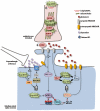
Huntington's Disease (HD) is the most frequent neurodegenerative disease caused by an expansion of polyglutamines (CAG). The main clinical manifestations of HD are chorea, cognitive impairment, and psychiatric disorders. The transmission of HD is autosomal dominant with a complete penetrance. HD has a single genetic cause, a well-defined neuropathology, and informative pre-manifest genetic testing of the disease is available. Striatal atrophy begins as early as 15 years before disease onset and continues throughout the period of manifest illness. Therefore, patients could theoretically benefit from therapy at early stages of the disease. One important characteristic of HD is the striatal vulnerability to neurodegeneration, despite similar expression of the protein in other brain areas. Aggregation of the mutated Huntingtin (HTT), impaired axonal transport, excitotoxicity, transcriptional dysregulation as well as mitochondrial dysfunction, and energy deficits, are all part of the cellular events that underlie neuronal dysfunction and striatal death. Among these non-exclusive mechanisms, an alteration of striatal signaling is thought to orchestrate the downstream events involved in the cascade of striatal dysfunction.
Keywords: Huntingtin; excitotoxicity; mitochondrial dysfunctions; polyglutamine; transcriptional deregulation.
Figures

Similar articles
-
Multiple Aspects of Gene Dysregulation in Huntington's Disease.Front Neurol. 2013 Oct 23;4:127. doi: 10.3389/fneur.2013.00127. Front Neurol. 2013. PMID: 24167500 Free PMC article. Review.
-
Huntington's disease.Adv Exp Med Biol. 2010;685:45-63. Adv Exp Med Biol. 2010. PMID: 20687494 Review.
-
Selective neuronal degeneration in Huntington's disease.Curr Top Dev Biol. 2006;75:25-71. doi: 10.1016/S0070-2153(06)75002-5. Curr Top Dev Biol. 2006. PMID: 16984809 Review.
-
Deterioration of neuroregenerative plasticity in association with testicular atrophy and dysregulation of the hypothalamic-pituitary-gonadal (HPG) axis in Huntington's disease: A putative role of the huntingtin gene in steroidogenesis.J Steroid Biochem Mol Biol. 2020 Mar;197:105526. doi: 10.1016/j.jsbmb.2019.105526. Epub 2019 Nov 9. J Steroid Biochem Mol Biol. 2020. PMID: 31715317 Review.
-
Striatal Vulnerability in Huntington's Disease: Neuroprotection Versus Neurotoxicity.Brain Sci. 2017 Jun 7;7(6):63. doi: 10.3390/brainsci7060063. Brain Sci. 2017. PMID: 28590448 Free PMC article. Review.
Cited by
-
Impaired TrkB Signaling Underlies Reduced BDNF-Mediated Trophic Support of Striatal Neurons in the R6/2 Mouse Model of Huntington's Disease.Front Cell Neurosci. 2016 Mar 9;10:37. doi: 10.3389/fncel.2016.00037. eCollection 2016. Front Cell Neurosci. 2016. PMID: 27013968 Free PMC article.
-
Bupropion for the treatment of apathy in Huntington's disease: A multicenter, randomised, double-blind, placebo-controlled, prospective crossover trial.PLoS One. 2017 Mar 21;12(3):e0173872. doi: 10.1371/journal.pone.0173872. eCollection 2017. PLoS One. 2017. PMID: 28323838 Free PMC article. Clinical Trial.
-
Mild cognitive impairment in Huntington's disease: challenges and outlooks.J Neural Transm (Vienna). 2024 Apr;131(4):289-304. doi: 10.1007/s00702-024-02744-8. Epub 2024 Jan 24. J Neural Transm (Vienna). 2024. PMID: 38265518 Review.
-
Multiple Aspects of Gene Dysregulation in Huntington's Disease.Front Neurol. 2013 Oct 23;4:127. doi: 10.3389/fneur.2013.00127. Front Neurol. 2013. PMID: 24167500 Free PMC article. Review.
-
Hypertension, antihypertensive drugs, and age at onset of Huntington's disease.Orphanet J Rare Dis. 2023 May 24;18(1):125. doi: 10.1186/s13023-023-02734-1. Orphanet J Rare Dis. 2023. PMID: 37226269 Free PMC article.
References
-
- Alford R. L., Ashizawa T., Jankovic J., Caskey C. T., Richards C. S. (1996). Molecular detection of new mutations, resolution of ambiguous results and complex genetic counseling issues in Huntington disease. Am. J. Med. Genet. 66, 281–28610.1002/(SICI)1096-8628(19961218)66:3<281::AID-AJMG9>3.0.CO;2-S - DOI - PubMed
-
- Andresen J. M., Gayan J., Cherny S. S., Brocklebank D., Alkorta-Aranburu G., Addis E. A., Cardon L. R., Housman D. E., Wexler N. S. (2007). Replication of twelve association studies for Huntington’s disease residual age of onset in large Venezuelan kindreds. J. Med. Genet. 44, 44–5010.1136/jmg.2006.045153 - DOI - PMC - PubMed
-
- Andrew S. E., Goldberg Y. P., Kremer B., Telenius H., Theilmann J., Adam S., Starr E., Squitieri F., Lin B., Kalchman M. A., Graham R. K., Hayden M. R. (1993). The relationship between trinucleotide (CAG) repeat length and clinical features of Huntington’s disease. Nat. Genet. 4, 398–40310.1038/ng0893-398 - DOI - PubMed
-
- Apostol B. L., Simmons D. A., Zuccato C., Illes K., Pallos J., Casale M., Conforti P., Ramos C., Roarke M., Kathuria S., Cattaneo E., Marsh J. L., Thompson L. M. (2008). CEP-1347 reduces mutant huntingtin-associated neurotoxicity and restores BDNF levels in R6/2 mice. Mol. Cell. Neurosci. 39, 8–2010.1016/j.mcn.2008.04.007 - DOI - PubMed
LinkOut - more resources
Full Text Sources

