Transgenic insulin-like growth factor-1 stimulates activation of COX-2 signaling in mammary glands
- PMID: 22006370
- PMCID: PMC3790642
- DOI: 10.1002/mc.20868
Transgenic insulin-like growth factor-1 stimulates activation of COX-2 signaling in mammary glands
Abstract
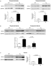
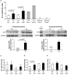
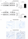
Studies show that elevated insulin-like growth factor-1 (IGF-1) levels are associated with an increased risk of breast cancer; however, mechanisms through which IGF-1 promotes mammary tumorigenesis in vivo have not been fully elucidated. To assess the possible involvement of COX-2 signaling in the pro-tumorigenic effects of IGF-1 in mammary glands, we used the unique BK5.IGF-1 mouse model in which transgenic (Tg) mice have significantly increased incidence of spontaneous and DMBA-induced mammary cancer compared to wild type (WT) littermates. Studies revealed that COX-2 expression was significantly increased in Tg mammary glands and tumors, compared to age-matched WTs. Consistent with this, PGE(2) levels were also increased in Tg mammary glands. Analysis of expression of the EP receptors that mediate the effects of PGE(2) showed that among the four G-protein-coupled receptors, EP3 expression was elevated in Tg glands. Up-regulation of the COX-2/PGE(2) /EP3 pathway was accompanied by increased expression of VEGF and a striking enhancement of angiogenesis in IGF-1 Tg mammary glands. Treatment with celecoxib, a selective COX-2 inhibitor, caused a 45% reduction in mammary PGE(2) levels, attenuated the influx of mast cells and reduced vascularization in Tg glands. These findings indicate that the COX-2/PGE(2) /EP3 signaling pathway is involved in IGF-1-stimulated mammary tumorigenesis and that COX-2-selective inhibitors may be useful in the prevention or treatment of breast cancer associated with elevated IGF-1 levels in humans. © 2011 Wiley Periodicals, Inc.
Copyright © 2011 Wiley Periodicals, Inc.
Figures


Similar articles
-
HER-2/neu status is a determinant of mammary aromatase activity in vivo: evidence for a cyclooxygenase-2-dependent mechanism.Cancer Res. 2006 May 15;66(10):5504-11. doi: 10.1158/0008-5472.CAN-05-4076. Cancer Res. 2006. PMID: 16707480
-
Developmental stage determines estrogen receptor alpha expression and non-genomic mechanisms that control IGF-1 signaling and mammary proliferation in mice.J Clin Invest. 2012 Jan;122(1):192-204. doi: 10.1172/JCI42204. Epub 2011 Dec 19. J Clin Invest. 2012. PMID: 22182837 Free PMC article.
-
Host prostaglandin EP3 receptor signaling relevant to tumor-associated lymphangiogenesis.Biomed Pharmacother. 2010 Feb;64(2):101-6. doi: 10.1016/j.biopha.2009.04.039. Epub 2009 Oct 20. Biomed Pharmacother. 2010. PMID: 20034758
-
IGF and insulin action in the mammary gland: lessons from transgenic and knockout models.J Mammary Gland Biol Neoplasia. 2000 Jan;5(1):19-30. doi: 10.1023/a:1009559014703. J Mammary Gland Biol Neoplasia. 2000. PMID: 10791765 Review.
-
The role of COX-2 inhibition in breast cancer treatment and prevention.Semin Oncol. 2004 Apr;31(2 Suppl 7):22-9. doi: 10.1053/j.seminoncol.2004.03.042. Semin Oncol. 2004. PMID: 15179621 Review.
Cited by
-
Urinary prostaglandin E2 metabolite and breast cancer risk.Cancer Epidemiol Biomarkers Prev. 2014 Dec;23(12):2866-73. doi: 10.1158/1055-9965.EPI-14-0685. Epub 2014 Sep 11. Cancer Epidemiol Biomarkers Prev. 2014. PMID: 25214156 Free PMC article.
-
Decoding the Role of Insulin-like Growth Factor 1 and Its Isoforms in Breast Cancer.Int J Mol Sci. 2024 Aug 27;25(17):9302. doi: 10.3390/ijms25179302. Int J Mol Sci. 2024. PMID: 39273251 Free PMC article. Review.
-
Prospective study of urinary prostaglandin E2 metabolite and pancreatic cancer risk.Int J Cancer. 2017 Dec 15;141(12):2423-2429. doi: 10.1002/ijc.31007. Epub 2017 Aug 29. Int J Cancer. 2017. PMID: 28815606 Free PMC article.
-
Resistance and escape from antiangiogenesis therapy: clinical implications and future strategies.J Clin Oncol. 2012 Nov 10;30(32):4026-34. doi: 10.1200/JCO.2012.41.9242. Epub 2012 Sep 24. J Clin Oncol. 2012. PMID: 23008289 Free PMC article. Review.
-
IGF-1-Overexpressing Mesenchymal Stem/Stromal Cells Promote Immunomodulatory and Proregenerative Effects in Chronic Experimental Chagas Disease.Stem Cells Int. 2018 Jul 24;2018:9108681. doi: 10.1155/2018/9108681. eCollection 2018. Stem Cells Int. 2018. PMID: 30140292 Free PMC article.
References
-
- Ruan W, Kleinberg DL. Insulin-like growth factor I is essential for terminal end bud formation and ductal morphogenesis during mammary development. Endocrinology. 1999;140(11):5075–5081. - PubMed
-
- Hankinson SE, Willett WC, Colditz GA, et al. Circulating concentrations of insulin-like growth factor-I and risk of breast cancer. Lancet. 1998;351(9113):1393–1396. - PubMed
-
- Lann D, LeRoith D. The role of endocrine insulin-like growth factor-I and insulin in breast cancer. J Mammary Gland Biol Neoplasia. 2008;13(4):371–379. - PubMed
-
- Yee D. The insulin-like growth factors and breast cancer--revisited. Breast Cancer Res Treat. 1998;47(3):197–199. - PubMed
-
- Yu H, Rohan T. Role of the insulin-like growth factor family in cancer development and progression. J Natl Cancer Inst. 2000;92(18):1472–1489. - PubMed
Publication types
MeSH terms
Substances
Grants and funding
LinkOut - more resources
Full Text Sources
Molecular Biology Databases
Research Materials
Miscellaneous

