. 2011 Dec;193(24):6852-63.
doi: 10.1128/JB.06190-11. Epub 2011 Oct 7.

The same periplasmic ExbD residues mediate in vivo interactions between ExbD homodimers and ExbD-TonB heterodimers

Affiliations

The same periplasmic ExbD residues mediate in vivo interactions between ExbD homodimers and ExbD-TonB heterodimers

Anne A Ollis et al. J Bacteriol. 2011 Dec.

Abstract

The TonB system couples cytoplasmic membrane proton motive force to TonB-gated outer membrane transporters for active transport of nutrients into the periplasm. In Escherichia coli, cytoplasmic membrane proteins ExbB and ExbD promote conformational changes in TonB, which transmits this energy to the transporters. The only known energy-dependent interaction occurs between the periplasmic domains of TonB and ExbD. This study identified sites of in vivo homodimeric interactions within ExbD periplasmic domain residues 92 to 121. ExbD was active as a homodimer (ExbD(2)) but not through all Cys substitution sites, suggesting the existence of conformationally dynamic regions in the ExbD periplasmic domain. A subset of homodimeric interactions could not be modeled on the nuclear magnetic resonance (NMR) structure without significant distortion. Most importantly, the majority of ExbD Cys substitutions that mediated homodimer formation also mediated ExbD-TonB heterodimer formation with TonB A150C. Consistent with the implied competition, ExbD homodimer formation increased in the absence of TonB. Although ExbD D25 was not required for their formation, ExbD dimers interacted in vivo with ExbB. ExbD-TonB interactions required ExbD transmembrane domain residue D25. These results suggested a model where ExbD(2) assembled with ExbB undergoes a transmembrane domain-dependent transition and exchanges partners in localized homodimeric interfaces to form an ExbD(2)-TonB heterotrimer. The findings here were also consistent with our previous hypothesis that ExbD guides the conformation of the TonB periplasmic domain, which itself is conformationally dynamic.


Figures

Fig. 1.
Fig. 1.
Cysteine substitutions in the ExbD periplasmic domain form spontaneous disulfide-linked dimers in vivo. TCA-precipitated proteins from strains expressing chromosomally encoded wild-type ExbD (W3110) or a ΔexbD ΔtolQR strain (RA1045) expressing plasmid-encoded ExbD variants near native ExbD levels (see Table S1 in the supplemental material for induction levels) were resolved on nonreducing or reducing 15% SDS-polyacrylamide gels and immunoblotted with ExbD-specific polyclonal antibodies. Reduced and nonreduced samples came from the same culture. d indicates the position of the homodimer, and m indicates the position of the monomer. The positions of nonreducing molecular mass standards are indicated on the left.
Fig. 2.
Fig. 2.
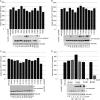
ExbD residues 92 through 121 are tolerant of replacement with cysteine, but disulfide-linked dimer formation can inhibit activity. A strain expressing chromosomally encoded wild-type ExbD (W3110) or a ΔexbD ΔtolQR strain (RA1045) expressing plasmid-encoded wild-type ExbD (pExbD) or ExbD Cys substitutions near native ExbD levels (see Table S1 in the supplemental material, 1× M9, for induction levels) was assayed for the ability to support transport of iron-loaded ferrichrome (Fe-Fc) as described in Materials and Methods. The results presented are representative data where activity was within 5% across at least two sets of triplicate assays. The y axis indicates the initial rate of transport. The ExbD variants assayed are indicated along the x axis. TCA-precipitated samples harvested just prior to each assay were resolved on 15% nonreducing or reducing SDS-polyacrylamide gels and immunoblotted with ExbD-specific polyclonal antibody. Δ indicates the ΔexbD ΔtolQR strain (RA1045), w indicates wild-type strain W3110, and pE indicates plasmid-encoded wild-type ExbD. The residue numbers indicate the positions of the ExbD Cys substitution. On the right, d indicates the position of the homodimer, and m indicates the position of the monomer. (A, B, and C) Sets of activity assays performed on the same days. (D) Activity assays for select ExbD homodimers catalyzed by CuoP treatment. − and + indicate buffer only and 0.03 mM CuoP treatment, respectively, prior to assay. The gray bars highlight CuoP-treated strains. The error bars indicate standard errors of the means.
Fig. 3.
Fig. 3.
Addition of an oxidizing agent increases the number of ExbD Cys substitutions trapped in disulfide-linked homodimers. (Nonreduced) ΔexbD ΔtolQR strains (RA1045) expressing plasmid-encoded ExbD variants near native ExbD levels (see Table S1 in the supplemental material, 1× M9, for induction levels) were treated with buffer only (−) or 0.03 mM CuoP (+) for 5 min at 37°C, with aeration. TCA-precipitated samples were resolved on nonreducing 15% SDS-polyacrylamide gels and immunoblotted with ExbD-specific polyclonal antibodies. The positions of nonreducing molecular mass standards are indicated on the left. d indicates the position of the homodimer, and m indicates the position of the monomer. (Reduced) TCA-precipitated samples taken from the same cultures prior to treatment were resolved on reducing 15% SDS-polyacrylamide gels and immunoblotted with ExbD-specific polyclonal antibodies.
Fig. 4.
Fig. 4.
ExbD disulfide-linked homodimers assemble with ExbB. Strains expressing chromosomally encoded wild-type ExbD (W3110) in the absence of TonB (KP1344) or a ΔexbD ΔtolQR strain (RA1045) or a ΔexbD ΔtolQR ΔtonB (KP1509) strain expressing plasmid-encoded wild-type ExbD (pExbD) or ExbD Cys substitutions near native ExbD levels (see Table S1 in the supplemental material for induction levels) were treated with CuoP, washed, and then cross-linked with formaldehyde as described in Materials and Methods. (A) Samples were resolved on 13% nonreducing or reducing SDS-polyacrylamide gels and immunoblotted with ExbD-specific polyclonal antibodies. The positions of monomeric ExbD and ExbD-specific cross-linked complexes are indicated on the left. The positions of molecular mass standards are indicated between the immunoblots. (B) Long exposures of the immunoblots in panel A showing only the strains expressing wild-type ExbD. NR indicates nonreduced. Red indicates reduced. (C) Short exposures of the immunoblots in panel A, cropped to the region of the ExbB-ExbD heterodimer, are shown for better comparison of relative levels of the complex under nonreducing or reducing conditions.
Fig. 5.
Fig. 5.
ExbD disulfide-linked dimers increase in the absence of TonB. TCA-precipitated samples of wild-type chromosomally encoded ExbD (W3110) and a ΔexbD ΔtolQR strain (RA1045) or a ΔexbD ΔtolQR ΔtonB strain (KP1509) expressing plasmid-encoded ExbD Cys substitutions near native ExbD levels (see Table S1 in the supplemental material for induction levels) were resolved on nonreducing or reducing 15% SDS-polyacrylamide gels and immunoblotted with ExbD-specific polyclonal antibodies. Reduced and nonreduced samples came from the same culture. + indicates the presence (RA1045), and − indicates the absence (KP1509) of TonB. ExbD Cys substitutions are indicated across the top. The positions of nonreducing molecular mass standards are indicated on the left. d indicates the position of the homodimer, and m indicates the position of the monomer. The middle set of immunoblots is a shorter exposure of the top set showing the monomer band only.
Fig. 6.
Fig. 6.
ExbD cysteine substitutions share common interfaces between homodimeric and heterodimeric interactions. TCA-precipitated samples of strains expressing chromosomally encoded wild-type ExbD and TonB (W3110) or a ΔexbD ΔtolQR ΔtonB strain (KP1509) coexpressing plasmid-encoded ExbD Cys substitutions and TonB(C18G, A150C) near native levels were resolved on nonreducing or reducing 13% and 11% SDS-polyacrylamide gels and immunoblotted with ExbD-specific polyclonal antibodies or TonB-specific monoclonal antibodies. Reduced and nonreduced samples came from the same culture. The positions of the monomeric proteins or disulfide-cross-linked complexes are indicated on the left. L93C, T94C, D107C, D111C, and D120C ExbD-TonB heterodimers were detected on longer exposures (data not shown). The positions of nonreducing molecular mass standards are indicated between the immunoblots. The strain designations across the top apply to both immunoblots below. All samples were processed on the same day.
Fig. 7.
Fig. 7.
ExbD D25 is important for ExbD-TonB disulfide-linked heterodimer formation. TCA-precipitated samples of strains expressing chromosomally encoded wild-type ExbD and TonB (W3110) or a ΔexbD ΔtolQR ΔtonB strain (KP1509) coexpressing plasmid-encoded TonB(C18G, A150C) and ExbD Cys substitutions without (−) or with (+) a D25N TMD substitution near native levels were resolved on nonreducing or reducing 13% and 11% SDS-polyacrylamide gels and immunoblotted with ExbD-specific polyclonal antibodies or TonB-specific monoclonal antibodies. Reduced and nonreduced samples came from the same culture. The positions of the monomeric proteins or disulfide-cross-linked complexes are indicated on the left. The positions of nonreducing molecular mass standards are indicated between the immunoblots. The strain designations across the top apply to both immunoblots below. All samples were processed on the same day.
Fig. 8.
Fig. 8.
Sites of disulfide-forming Cys substitutions map to opposite ends of the ExbD periplasmic domain solution structure. Side chains of residues where Cys substitutions were trapped in disulfide-linked homodimers in vivo are mapped on the ExbD periplasmic domain NMR structure, Protein Data Bank (PDB) code 2pfu. The image was generated using Swiss-PdbViewer (14). The darker gray ribbon represents the Cys-scanned region examined in this study. The black side chains indicate significant spontaneous ExbD homodimer formation. The gray side chains indicate weak spontaneous homodimer formation. With the exception of I102, V110, and M116, the side chains pictured showed significant ExbD heterodimer formation with TonB A150C formation. C and N indicate the carboxy and amino termini of the domain, respectively.

Similar articles

Cited by

References

    1. Braun V. 2009. FhuA (TonA), the career of a protein. J. Bacteriol. 191:3431–3436 - PMC - PubMed
    1. Braun V., et al. 1996. Energy-coupled transport across the outer membrane of Escherichia coli: ExbB binds ExbD and TonB in vitro, and leucine 132 in the periplasmic region and aspartate 25 in the transmembrane region are important for ExbD activity. J. Bacteriol. 178:2836–2845 - PMC - PubMed
    1. Braun V., Herrmann C. 1993. Evolutionary relationship of uptake systems for biopolymers in Escherichia coli: cross-complementation between the TonB-ExbB-ExbD and the TolA-TolQ-TolR proteins. Mol. Microbiol. 8:261–268 - PubMed
    1. Braun V., Herrmann C. 2004. Point mutations in transmembrane helices 2 and 3 of ExbB and TolQ affect their activities in Escherichia coli K-12. J. Bacteriol. 186:4402–4406 - PMC - PubMed
    1. Brinkman K. K., Larsen R. A. 2008. Interactions of the energy transducer TonB with noncognate energy-harvesting complexes. J. Bacteriol. 190:421–427 - PMC - PubMed

Publication types

MeSH terms

LinkOut - more resources