

Stachybotrys chartarum-induced hypersensitivity pneumonitis is TLR9 dependent
- PMID: 21982832
- PMCID: PMC3260863
- DOI: 10.1016/j.ajpath.2011.08.019
Stachybotrys chartarum-induced hypersensitivity pneumonitis is TLR9 dependent
Abstract
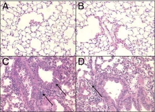
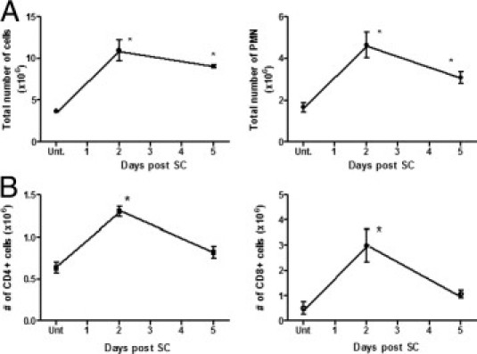
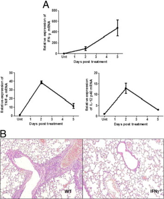
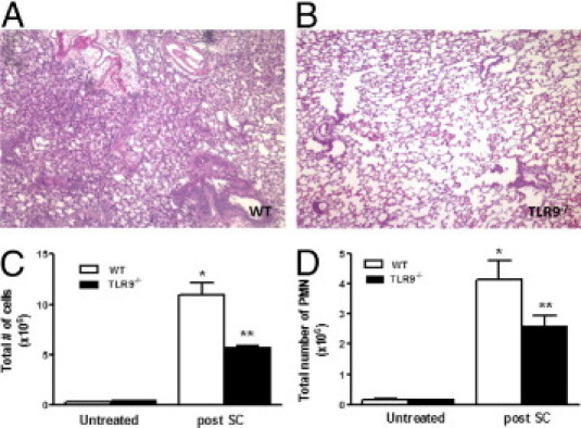
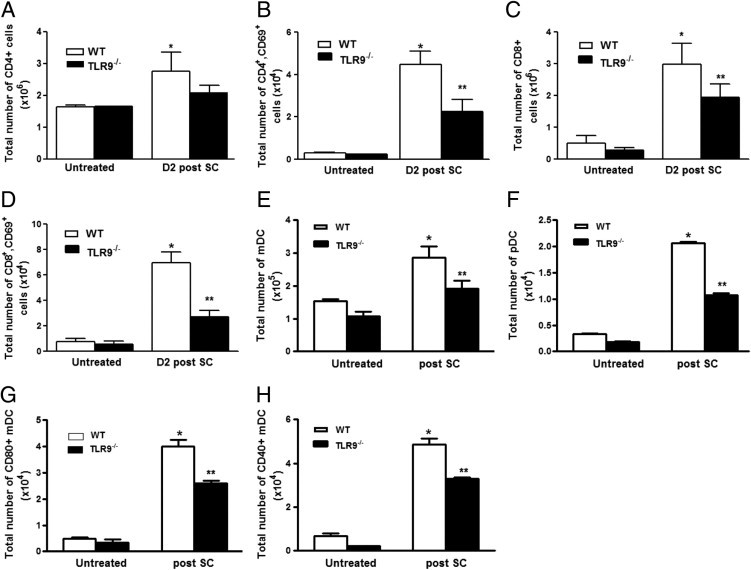
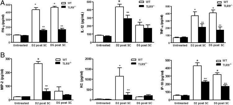
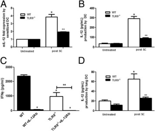
Hypersensitivity pneumonitis (HP), an inflammatory lung disease, develops after repeated exposure to inhaled particulate antigen and is characterized by a vigorous T helper type 1-mediated immune response, resulting in the release of IL-12 and interferon (IFN)-γ. These T helper type 1 cytokines may participate in the pathogenesis of HP. Stachybotrys chartarum (SC) is a dimorphic fungus implicated in a number of respiratory illnesses, including HP. Here, we have developed a murine model of SC-induced HP that reproduces pathology observed in human HP and hypothesized that toll receptor-like 9 (TLR9)-mediated dendritic cell responses are required for the generation of granulomatous inflammation induced by inhaled SC. Mice sensitized and challenged with 10(6) SC spores develop granulomatous inflammation with multinucleate giant cells, accompanied by increased accumulation of neutrophils and CD4(+) and CD8(+) T cells. SC sensitization and challenge resulted in robust pulmonary expression of tumor necrosis factor-α, IL-12, and IFN-γ. SC-mediated granulomatous inflammation required IFN-γ and was TLR9 dependent, because TLR9(-/-) mice displayed reduced peribronchial inflammation, decreased accumulation and/or activation of polymorphonuclear (PMN) and CD4(+) and CD8(+) T cells, and reduced lung expression of type 1 cytokines and chemokines. T-cell production of IFN-γ was IL-12 dependent. Our studies suggest that TLR9 is critical for dendritic cell-mediated development of a type 1 granulomatous inflammation in the lung in response to SC.
Copyright © 2011 American Society for Investigative Pathology. Published by Elsevier Inc. All rights reserved.
Figures
Similar articles
-
TLR9-dependent IL-23/IL-17 is required for the generation of Stachybotrys chartarum-induced hypersensitivity pneumonitis.J Immunol. 2013 Jan 1;190(1):349-56. doi: 10.4049/jimmunol.1202225. Epub 2012 Nov 23. J Immunol. 2013. PMID: 23180821 Free PMC article.
-
Mechanisms accounting for granulomatous responses in hypersensitivity pneumonitis.Sarcoidosis Vasc Diffuse Lung Dis. 1997 Sep;14(2):131-8. Sarcoidosis Vasc Diffuse Lung Dis. 1997. PMID: 9306503 Review.
-
Intranasal exposure to Stachybotrys chartarum enhances airway inflammation in allergic mice.Am J Respir Crit Care Med. 2006 Mar 1;173(5):512-8. doi: 10.1164/rccm.200503-466OC. Epub 2005 Dec 1. Am J Respir Crit Care Med. 2006. PMID: 16322647
-
TLR2 regulates neutrophil recruitment and cytokine production with minor contributions from TLR9 during hypersensitivity pneumonitis.PLoS One. 2013 Aug 30;8(8):e73143. doi: 10.1371/journal.pone.0073143. eCollection 2013. PLoS One. 2013. PMID: 24023674 Free PMC article.
-
Hypersensitivity pneumonitis and alpha-chemokines.Clin Ter. 2017 Mar-Apr;168(2):e140-e145. doi: 10.7417/CT.2017.1996. Clin Ter. 2017. PMID: 28383627 Review.
Cited by
-
A critical role of Dectin-1 in hypersensitivity pneumonitis.Inflamm Res. 2016 Mar;65(3):235-44. doi: 10.1007/s00011-015-0910-1. Epub 2015 Dec 7. Inflamm Res. 2016. PMID: 26644324
-
Protein kinase D1 in myeloid lineage cells contributes to the accumulation of CXCR3+CCR6+ nonconventional Th1 cells in the lungs and potentiates hypersensitivity pneumonitis caused by S. rectivirgula.Front Immunol. 2024 Oct 11;15:1403155. doi: 10.3389/fimmu.2024.1403155. eCollection 2024. Front Immunol. 2024. PMID: 39464896 Free PMC article.
-
Management of hypersensivity pneumonitis.Clin Transl Allergy. 2013 Feb 4;3(1):5. doi: 10.1186/2045-7022-3-5. Clin Transl Allergy. 2013. PMID: 23374544 Free PMC article.
-
The Roles of Autoimmunity and Biotoxicosis in Sick Building Syndrome as a "Starting Point" for Irreversible Dampness and Mold Hypersensitivity Syndrome.Antibodies (Basel). 2020 Jun 22;9(2):26. doi: 10.3390/antib9020026. Antibodies (Basel). 2020. PMID: 32580407 Free PMC article.
-
TLR9-dependent IL-23/IL-17 is required for the generation of Stachybotrys chartarum-induced hypersensitivity pneumonitis.J Immunol. 2013 Jan 1;190(1):349-56. doi: 10.4049/jimmunol.1202225. Epub 2012 Nov 23. J Immunol. 2013. PMID: 23180821 Free PMC article.
References
-
- Girard M., Israel-Assayag E., Cormier Y. Pathogenesis of hypersensitivity pneumonitis. Curr Opin Allergy Clin Immunol. 2004;4:93–98. - PubMed
-
- Suga M., Yamasaki H., Nakagawa K., Kohrogi H., Ando M. Mechanisms accounting for granulomatous responses in hypersensitivity pneumonitis. Sarcoidosis Vasc Diffuse Lung Dis. 1997;14:131–138. - PubMed
-
- Denis M. Proinflammatory cytokines in hypersensitivity pneumonitis. Am J Respir Crit Care Med. 1995;151:164–169. - PubMed
-
- Denis M., Cormier Y., Laviolette M., Ghadirian E. T cells in hypersensitivity pneumonitis: effects of in vivo depletion of T cells in a mouse model. Am J Respir Cell Mol Biol. 1992;6:183–189. - PubMed
-
- Israel-Assayag E., Fournier M., Cormier Y. Blockade of T cell costimulation by CTLA4-Ig inhibits lung inflammation in murine hypersensitivity pneumonitis. J Immunol. 1999;163:6794–6799. - PubMed
Publication types
MeSH terms
Substances
Grants and funding
LinkOut - more resources
Full Text Sources
Molecular Biology Databases
Research Materials
Miscellaneous
