. 2011 Oct 4;9(4):317-29.
doi: 10.1016/j.stem.2011.09.001.

Sox2(+) adult stem and progenitor cells are important for tissue regeneration and survival of mice

Affiliations

Sox2(+) adult stem and progenitor cells are important for tissue regeneration and survival of mice

Katrin Arnold et al. Cell Stem Cell. .

Abstract

The transcription factor Sox2 maintains the pluripotency of early embryonic cells and regulates the formation of several epithelia during fetal development. Whether Sox2 continues to play a role in adult tissues remains largely unknown. We show here that Sox2 marks adult cells in several epithelial tissues where its expression has not previously been characterized, including the stomach, cervix, anus, testes, lens, and multiple glands. Genetic lineage tracing and transplantation experiments demonstrate that Sox2-expressing cells continuously give rise to mature cell types within these tissues, documenting their self-renewal and differentiation potentials. Consistent with these findings, ablation of Sox2(+) cells in mice results in a disruption of epithelial tissue homeostasis and lethality. Developmental fate mapping reveals that Sox2(+) adult stem cells originate from fetal Sox2(+) tissue progenitors. Thus, our results identify Sox2 expression in numerous adult endodermal and ectodermal stem cell compartments, which are critical for normal tissue regeneration and survival.


Figures

Figure 1
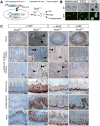
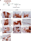
Figure 1. Widespread Sox2-GFP expression in epithelial tissues of mice
(A) Experimental outline for the generation and analysis of Sox2GFP reporter mice. IHC, immunohistochemistry; FACS, fluorescence-activated cell sorting. (B) Sox2-GFP expression in targeted embryonic stem cells (ESCs), the inner cell mass of blastocysts (asterisk marks GFP-negative control blastocyst), and neonatal neural progenitor cells (NPCs). BF, brightfield. (C) IHC for GFP, Sox2 and the basal cell marker p63 on paraffin-embedded sections of adult testis, pylorus and corpus of the glandular stomach, esophagus, forestomach, and anus of wild type (Sox2WT) and Sox2GFP mice. Inserts also show cross-sections of corpus and pylorus. Asterisk marks unspecific antibody staining of blood vessels. Dashed lines outline gastric units (glandular stomach) or the basement membrane (testes, esophagus, forestomach, anus). Original magnifications as indicated. See also Suppl. Figure 1 and 2.
Figure 2
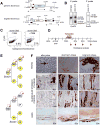
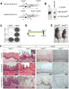
Figure 2. Genetic lineage tracing identifies Sox2+ stem cells in testes, glandular stomach and lens
(A) Targeting strategy for generating Sox2-CreER ESCs and mice. Restriction sites, southern blot analysis probes and expected restriction fragment lengths are indicated. (B) Southern blot analyses with 5′ and 3′ probes, as indicated in (A), to verify correct targeting. (C) ESCs carrying the Sox2-CreER and ROSA26-lsl-EYFP alleles were analyzed by FACS for EYFP expression 2 weeks after exposure to 4-OHT. (D) Experimental outline for pulse-chase experiment. Tamoxifen (TAM) was given intraperitoneally on 4 consecutive days to adult Sox2-CreER, ROSA26-lsl-EYFP mice (“pulse”). Animals were sacrificed at the indicated time points and tissues analyzed by FACS and IHC for EYFP. E, embryonic day; wks, weeks; mo, months. (E) Possible outcomes of lineage tracing assay. If Sox2-expressing cells are stem cells (SC), they should activate the EYFP reporter upon TAM administration and give rise to permanently labeled Sox2+ stem cells as well as differentiated progeny (top image). In contrast, Sox2+ progenitors (P) and derivative differentiated cells (D) would only be transiently labeled (indicated by dashed circle, bottom image). (F) IHC for EYFP on lineage tracing samples. Note individual cells or small patches of EYFP+ cells immediately after the pulse, which gradually expand over the subsequent months to span the entire epithelium. Short-term chase reflects 1mo (testes) and 2mo (stomach, lens), respectively. The long-term chase periods shown are 6mo for testes and lens, and 15mo for stomach. Original magnifications as indicated. See also Suppl. Figure 3.
Figure 3
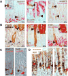
Figure 3. Self-renewal and multipotency of Sox2+ stomach stem cells
(A) Co-staining for Sox2-GFP and gastric markers on pylorus sections. Sox2-GFP+ cells (arrowheads) do not express differentiated cell markers gastrin (enteroendocrine cells), MUC5AC (mucus cells) or H-K-ATPase (parietal cells). (B) Co-staining of lineage tracing sections of Sox2-CreER; ROSA26-lsl-EYFP mice with differentiation markers in glandular stomach. Co-staining (arrowhead) was seen between EYFP and Chromogranin A (enteroendocrine cells) and MUC5AC (mucus cells) in the pylorus and with H-K-ATPase (parietal cells) in the corpus-pylorus transition zone. (C) Co-localization analysis for Sox2 and Lgr5-EGFP in pylorus. Sox2 (arrowhead) and Lgr5-EGFP (pink) IHC signals appear to mark different cells in pyloric glands. (D) Co-localization analysis between proliferation marker Ki67 and Sox2-GFP. Black arrowheads depict Ki67+ cells, red arrowheads depict Ki67+Sox2-GFP+ cells. Original magnifications as indicated. See also Suppl. Figure 4.
Figure 4
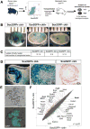
Figure 4. Sox2-GFP+ testis cells reconstitute spermatogenesis in infertile mice
(A) Experimental outline. Sox2GFP animals were crossed with ROSA26-lacZ animals to enable tracking of transplanted donor cells. Sox2GFP+ckit cells (1), Sox2GFP+ckit+ cells (2), and Sox2GFPckit cells (3) from the testes of 2-week-old animals were transplanted into the vas deferens of germ cell-depleted, infertile ckitW/Wv recipient mice and testes analyzed for beta-gal activity after 6 months. (B) X-gal stain of transplanted ckitW/Wv testis 6 months after transplantation with the indicated cell populations. Light blue signal (asterisk) reflects background staining of the interstitium. (C) Table summarizing repopulation experiments. (D) Whole mount X-gal staining (left image) and paraffin sections of repopulated seminiferous tubules counterstained with neutral red (center image). Note the presence of immature spermatogonia at periphery and mature sperm in center of tubules transplanted with Sox2GFP+ckit cells and lack of spermatogenesis upon transplantation of Sox2GFPckit cells (right image). (E) Scatter plot of gene expression profiles comparing Sox2GFP+ckit and Sox2GFP+ckit+ testis cells. Green dots depict genes with 2-fold and higher expression in Sox2GFP+ckit+ cells, red dots depict genes 2-fold and higher expression in Sox2GFP+ckit cells. (pairwise analysis, two-fold change, t-test P=0.05, Benjamini and Hochberg correction.) Previously characterized spermatogonial stem cell genes are shown in bold. (F) Phase contrast and GFP images of a spermatogonial stem cell line derived from Sox2-GFP mice (passage 10). See also Suppl. Figure 5.
Figure 5
Figure 5. Fetal Sox2+ progenitors give rise to adult Sox2+ stem cells
(A) Experimental strategy for embryonic lineage tracing. (B) IHC for EYFP in Sox2-CreER; ROSA26-lsl-EYFP mice that received one pulse of tamoxifen at E14.5 and were analyzed after 22 months. Labeled cells were detected in corpus and pylorus sections of the glandular stomach, forestomach esophagus, tongue, lens, ducts (arrow) and acini (arrowhead) of salivary glands, duodenum and tracheal glands. Original magnifications as indicated. See also Suppl. Figure 6.
Figure 6
Figure 6. Ablation of Sox2+ cells disrupts tissue homeostasis and causes lethality
(A) Targeting strategy to generate Sox2TK mice. Restriction sites, Southern blot analysis probes and expected restriction fragment lengths are indicated. (B) Southern blot analysis to confirm correct integration of Sox2TK allele in ESCs. (C) Crystal violet stain of Sox2TK and control (V6.5) ESCs after 4 days of gancyclovir (GCV) treatment. (D) Experimental outline. ALZET pumps, releasing a constant dose of GCV over 2 weeks, were implanted into Sox2TK mice and control Sox2WT littermates. Animals were then sacrificed and tissues analyzed histologically. (E) Morbid Sox2TK mouse next to Sox2WT littermate after 13 days of GCV treatment. (F) H&E staining of paraffin-embedded sections from tongue, esophagus, and forestomach of GCV-treated Sox2TK mice shows loss of basal layer (dashed line) in tongue, esophagus and forestomach (second column) compared with control mice (left column). Note the appearance of atypical cells (arrows) in all tissues and severe inflammation as well as edema formation in the forestomach. GCV-treated WT animals remained unaffected (left column). IHC for Sox2 confirmed almost complete absence of Sox2+ cells in Sox2TK mice with very rare Sox2+ cells remaining (right column). Original magnifications as indicated.
Figure 7
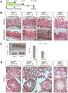
Figure 7. Rescue of morbidity upon withdrawal of gancyclovir
(A) Experimental strategy. GCV was given for 7 consecutive days to 3- or 10-week old mice. Treated Sox2TK mice and Sox2WT controls were then allowed to recover. Tissues were analyzed 7 days (7d) and 3 months (3mo) after removal of GCV pumps. (B) Histological analysis of Sox2TK tongue and forestomach after 7 days of GCV treatment followed by 7 days (second column) or 3 months (third column) of recovery. GCV-treated WT mice and 7 month-old untreated Sox2TK mice showed no abnormalities in tissue architecture (first and last columns, respectively). (C) 3 months after GCV treatment, testes from Sox2TK mice exhibited a significant reduction in size compared with treated control littermates. (D) Histological analysis of Sox2TK testes after 7d of GCV treatment followed by 7d or 3mo, respectively, of recovery. After 7d of recovery, the seminiferous tubules of Sox2TK testes showed a mild phenotype with less sperm (second column) compared to GCV-treated Sox2WT animals (left column). This phenotype was exacerbated after 3mo when entire atrophic tubules were seen (third column). No phenotypic abnormalities were seen in testes of age-matched untreated Sox2TK mice (right column). Original magnifications as indicated. See also Suppl. Figure 7.

Comment in

Similar articles

Cited by

References

    1. Avilion AA, Nicolis SK, Pevny LH, Perez L, Vivian N, Lovell-Badge R. Multipotent cell lineages in early mouse development depend on SOX2 function. Genes & development. 2003;17:126–140. - PMC - PubMed
    1. Barker N, Bartfeld S, Clevers H. Tissue-resident adult stem cell populations of rapidly self-renewing organs. Cell stem cell. 7:656–670. - PubMed
    1. Barker N, Huch M, Kujala P, van den Wetering M, Snippert HJ, van Es JH, Sato T, Stange DE, Begthel H, van den Born M, et al. Lgr5(+ve) stem cells drive self-renewal in the stomach and build long-lived gastric units in vitro. Cell Stem Cell. 6:25–36. - PubMed
    1. Barker N, van Es JH, Kuipers J, Kujala P, van den Born M, Cozijnsen M, Haegebarth A, Korving J, Begthel H, Peters PJ, et al. Identification of stem cells in small intestine and colon by marker gene Lgr5. Nature. 2007;449:1003–1007. - PubMed
    1. Bass AJ, Watanabe H, Mermel CH, Yu S, Perner S, Verhaak RG, Kim SY, Wardwell L, Tamayo P, Gat-Viks I, et al. SOX2 is an amplified lineage-survival oncogene in lung and esophageal squamous cell carcinomas. Nature genetics. 2009;41:1238–1242. - PMC - PubMed

Publication types

MeSH terms

LinkOut - more resources