. 2011 Dec;85(24):13260-70.
doi: 10.1128/JVI.06005-11. Epub 2011 Oct 5.

The cytomegaloviral protein pUL138 acts as potentiator of tumor necrosis factor (TNF) receptor 1 surface density to enhance ULb'-encoded modulation of TNF-α signaling

Affiliations

The cytomegaloviral protein pUL138 acts as potentiator of tumor necrosis factor (TNF) receptor 1 surface density to enhance ULb'-encoded modulation of TNF-α signaling

Vu Thuy Khanh Le et al. J Virol. 2011 Dec.

Abstract

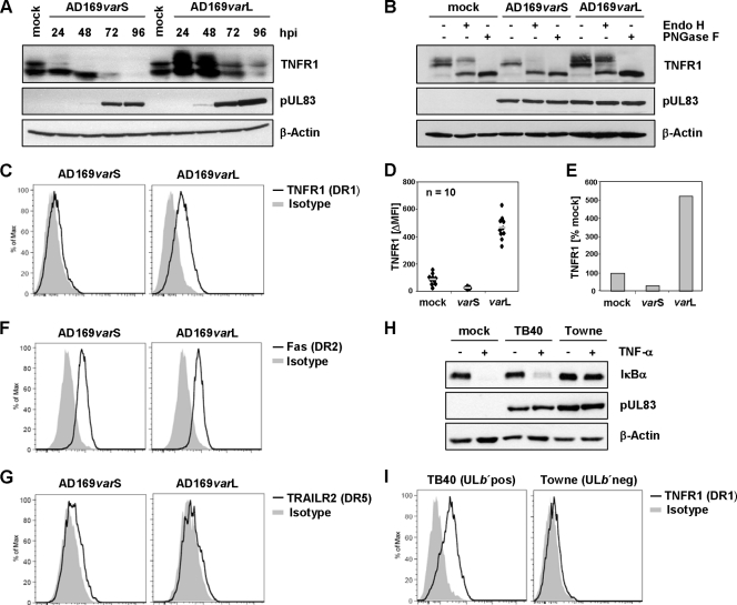
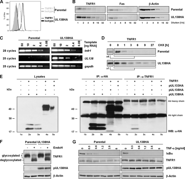
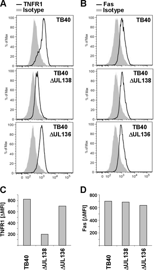
Human cytomegalovirus is a ubiquitous herpesvirus that establishes lifelong latent infection. Changes in immune homeostasis induce the reactivation of lytic infection, which is mostly inapparent in healthy individuals but often causes overt disease in immunocompromised hosts. Based on discrepant tumor necrosis factor receptor 1 surface disposition between human cytomegalovirus AD169 variants differing in the ULb' region, we identified the latency-associated gene product pUL138, which also is expressed during productive infection, as a selective potentiator of tumor necrosis factor receptor 1, one of the key receptors of innate immunity. Ectopically expressed pUL138 coprecipitated with tumor necrosis factor receptor 1, extended the protein half-life, and enhanced its signaling responses, thus leading to tumor necrosis factor receptor 1 hyperresponsiveness. Conversely, the targeted deletion of UL138 from the human cytomegaloviral genome strongly reduced tumor necrosis factor receptor 1 surface densities of infected cells. Remarkably, the comparison of UL138 deficiency to ULb' deficiency revealed the presence of further positive modulators of tumor necrosis factor alpha signal transduction encoded within the human cytomegalovirus ULb' region, identifying this region as a hub for multilayered tumor necrosis factor alpha signaling regulation.


Figures

Fig. 1.
Fig. 1.
HCMV-AD169 variants differ in their modulation of TNF-α signaling. (A) Viral DNA of AD169varATCC and AD169varX (genomically undefined) was digested with HindIII and separated on an agarose gel. (B) Scheme of HCMV genome organization. UL, unique long; US, unique short; TRL, terminal repeat long; IRS, internal repeat short; TRS, terminal repeat short; IRL, internal repeat long; RL, repeat long. (C to E) MRC-5 fibroblasts were mock treated or infected for 72 h (MOI, 5) with AD169varS or AD169varL. (C and D) Cells treated for 20 min with 20 ng/ml TNF-α (C) or 2 ng/ml IL-1β (D) were lysed for the immunoblot analysis of IκBα protein. pUL83 and β-actin served as infection and loading controls, respectively. (E) Cells were treated with 20 ng/ml TNF-α for 3 h, and total RNA was prepared for Northern blot analysis of the indicated transcripts.
Fig. 2.
Fig. 2.
ULb′ gene product(s) selectively modulate TNFR1. (A and B) MRC-5 fibroblasts were mock treated or infected (MOI, 5) with AD169varS or AD169varL. (A) Cells were lysed at the indicated time points postinfection for the detection of the indicated proteins. (B) Lysates of cells infected for 24 h were treated with endoglycosidase H (EndoH) or N-glycosidase F (PNGase F). The immunoblot analysis of the indicated proteins was performed. (C to G) MRC-5 fibroblasts were infected (MOI, 5) with AD169varS or AD169varL. Cells were stained at 72 hpi with anti-TNFR1 (C), anti-Fas (F), or anti-TRAILR2 (G) and the respective isotype control antibody and were analyzed by flow cytometry. Histograms of DAPI-negative cells are shown. (D) To quantify the upregulation of TNFR1 surface densities by AD169 infection, 10 independent experiments were performed, and the arithmetic means were calculated (gray bar). (E) Changes of TNFR1 surface densities (calculated based on arithmetic mean values). (H) MRC-5 fibroblasts were mock treated or infected (MOI, 5) with TB40 (ULb′pos) or Towne (ULb′neg). Cells were treated at 72 hpi with 20 ng/ml TNF-α for 20 min before being lysed for the immunoblot analysis of the indicated proteins. (I) Cells infected for 72 h with TB40 or Towne were stained with anti-TNFR1 or isotype control antibody and analyzed by flow cytometry. Histograms of DAPI-negative cells are shown.
Fig. 3.
Fig. 3.
Identification of pUL138 as an enhancer of TNFR1 surface density. (A) Scheme of AD169-ULb′ ORFs. Candidate ORFs for TNFR1 upregulation (present in AD169varL and absent from Towne) are depicted in gray. (B) Subcellular localization of ectopically expressed HA-tagged ULb′ candidate proteins. HeLa cells were transfected with the respective expression plasmid. Twenty-four h posttransfection, cells were analyzed by immunofluorescence staining (anti-HA). (C) pUL138 mediates the upregulation of TNFR1 surface expression. HeLa cells were transfected with pIRES-UL138HA-EGFP. Twenty-four h posttransfection, cells were harvested for analysis by flow cytometry. DAPI-negative cells of GFP-positive and GFP-negative gates were analyzed for TNFR1 surface density. TNF-α treatment (20 ng/ml for 30 min) before staining served as a specificity control of TNFR1 detection, since ligand binding competes with MAB225 binding. (D to G) MRC-5 fibroblasts were mock treated or infected with AD169varL (MOI, 5). (D) Cells were harvested for RNA preparation at the indicated times. Equal amounts of total RNA were separated on a MOPS gel, and UL138 transcripts were detected by Northern blotting. (E) Selective transcription of HCMV immediate-early genes was achieved by infection in the presence of cycloheximide (CHX; 50 μg/ml). (F) HCMV DNA replication was prevented by the use of ganciclovir (GCV; 50 μM) to determine GCV-sensitive transcripts. Infected cells were harvested for RNA preparation at the indicated times. Equal amounts of total RNA were separated on a MOPS gel, and UL138-spanning transcripts were detected by Northern blotting. (G) Cells were harvested at the indicated times for the analysis of TNFR1 surface expression by flow cytometry. Histograms of DAPI-negative cells are shown.
Fig. 4.
Fig. 4.
pUL138 interaction with TNFR1 leading to the stabilization of TNFR1, enhancement of TNFR1 surface disposition, and TNF-α hyperresponsiveness. (A) DAPI-negative parental HeLa and HeLa-UL138HA cells were gated and analyzed for TNFR1 surface density by flow cytometry. (B) Lysates of parental HeLa and HeLa-UL138HA were log2 diluted to analyze the abundance of the indicated proteins. (C) Increase of TNFR1 protein amount in HeLa-UL138HA cells is not due to the upregulation of tnfr1 transcription. Total RNA from parental and HeLa-UL138HA cells was prepared and treated with DNase I. Graded template amounts were used for the semiquantitative analysis of the indicated transcripts. (D) Cells were treated with 100 μg/ml cycloheximide (CHX), blocking protein neosynthesis for the indicated times. Protein amounts were adjusted to reach the same TNFR1 levels at 0 h to ensure fair comparisons. (E) HEK293T cells were transfected with the indicated expression constructs. Twenty-four h posttransfection, cells were lysed and immunoprecipitation (IP) was performed using TNFR1- and HA-specific antibodies. WB, Western blotting. (F) Lysates of parental HeLa and HeLa-UL138HA cells were treated with endoglycosidase H (EndoH) as indicated, and the immunoblot analysis of indicated proteins was performed. (G) Parental HeLa and HeLa-UL138HA cells were treated with the indicated concentrations of TNF-α for 20 min before being lysed and analyzed by immunoblotting.
Fig. 5.
Fig. 5.
Deletion of UL138 results in decreased TNFR1 surface density on HCMV-infected cells. (A to D) MRC-5 fibroblasts infected with the indicated virus (MOI, 5) were stained at 72 hpi with anti-TNFR1 (A), anti-Fas (B), or the respective isotype control antibody and analyzed by flow cytometry. Shown are histograms of DAPI-negative cells. The difference between the mean fluorescence intensity (ΔMFI) of TNFR1 (C) and Fas signal (D) and the respective isotype control signal was calculated for better comparison.
Fig. 6.
Fig. 6.
Comparison of UL138 with ULb′ deficiency defines the ULb′ region as a hub for TNF-α signaling regulation. (A to F) MRC-5 fibroblasts infected with BAC-derived AD169varL, AD169ΔUL138, AD169ΔUL136, or AD169varS HCMV (MOI, 5). Cells were stained at 72 hpi with anti-TNFR1 (A), anti-Fas (B), or the respective isotype control antibody and analyzed by flow cytometry. Histograms of DAPI-negative cells are shown. The difference between the mean fluorescence intensities (ΔMFI) of TNFR1 (C) or Fas signal (D) and the respective isotype control signal are indicated. (E and F) The TNFR1 surface phenotype of ΔUL138 is virus dose independent. MRC-5 fibroblasts infected with the indicated virus (MOI of 2.5, 5, and 10) were stained at 72 hpi with anti-TNFR1, anti-Fas, or the respective isotype control antibody. DAPI-negative cells were analyzed by flow cytometry to calculate the difference between the mean fluorescence intensity (ΔMFI) of TNFR1 (E) or Fas signal (F) and the respective isotype control signal. (G) MRC-5 fibroblasts were mock treated or infected with BAC-derived AD169varL, AD169ΔUL138, AD169ΔUL136, or AD169varS HCMV (MOI, 5). Cells were incubated at 72 hpi with 20 ng/ml TNF-α for 15 min before being lysed for the immunoblot detection of IκBα protein. pUL83 and β-actin served as infection and loading controls, respectively.

Similar articles

Cited by

References

    1. Atalay R., et al. 2002. Identification and expression of human cytomegalovirus transcription units coding for two distinct Fc gamma receptor homologs. J. Virol. 76:8596–8608 - PMC - PubMed
    1. Baillie J., Sahlender D. A., Sinclair J. H. 2003. Human cytomegalovirus infection inhibits tumor necrosis factor alpha (TNF-alpha) signaling by targeting the 55-kilodalton TNF-alpha receptor. J. Virol. 77:7007–7016 - PMC - PubMed
    1. Bego M., Maciejewski J., Khaiboullina S., Pari G., St. Jeor S. 2005. Characterization of an antisense transcript spanning the UL81-82 locus of human cytomegalovirus. J. Virol. 79:11022–11034 - PMC - PubMed
    1. Benedict C. A., et al. 2001. Three adenovirus E3 proteins cooperate to evade apoptosis by tumor necrosis factor-related apoptosis-inducing ligand receptor-1 and -2. J. Biol. Chem. 276:3270–3278 - PubMed
    1. Borst E. M., Hahn G., Koszinowski U. H., Messerle M. 1999. Cloning of the human cytomegalovirus (HCMV) genome as an infectious bacterial artificial chromosome in Escherichia coli: a new approach for construction of HCMV mutants. J. Virol. 73:8320–8329 - PMC - PubMed

Publication types

MeSH terms

Substances

LinkOut - more resources