. 2011;6(9):e24962.
doi: 10.1371/journal.pone.0024962. Epub 2011 Sep 19.

A functional interface at the rDNA connects rRNA synthesis, pre-rRNA processing and nucleolar surveillance in budding yeast

Affiliations

A functional interface at the rDNA connects rRNA synthesis, pre-rRNA processing and nucleolar surveillance in budding yeast

Nathalie Leporé et al. PLoS One. 2011.

Abstract

Ribogenesis is a multistep error-prone process that is actively monitored by quality control mechanisms. How ribosomal RNA synthesis, pre-rRNA processing and nucleolar surveillance are integrated is unclear. Nor is it understood how defective ribosomes are recognized. We report in budding yeast that, in vivo, the interaction between the transcription elongation factor Spt5 and Rpa190, the largest subunit of RNA polymerase (Pol) I, requires the Spt5 C-terminal region (CTR), a conserved and highly repetitive domain that is reminiscent of the RNA Pol II C-terminal domain (CTD). We show that this sequence is also required for the interaction between Spt5 and Nrd1, an RNA specific binding protein, and an exosome cofactor. Both the Spt4-Spt5, and the Nrd1-Nab3 complexes interact functionally with Rrp6, and colocalize at the rDNA. Mutations in the RNA binding domain of Nrd1, but not in its RNA Pol II CTD-interacting domain, and mutations in the RRM of Nab3 led to the accumulation of normal and aberrant polyadenylated pre-rRNAs. Altogether these results indicate that Nrd1-Nab3 contributes to recruiting the nucleolar surveillance to elongating polymerases to survey nascent rRNA transcripts.


Conflict of interest statement

Competing Interests: The authors have declared that no competing interests exist.

Figures

Figure 1
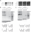
Figure 1. Involvement of the RRM and CID domains of Nrd1 in nucleolar surveillance.
A, (left) Domain mapping of Nrd1. All three nrd1 alleles used in this work (provided by Prof. Jeff Corden, Johns Hopkins University School of Medicine) are HA-epitope tagged at the carboxyl terminal end of the protein, which allowed us to establish that neither mutation affects protein stability (see Fig S2B and C). Nrd1 consists of an amino terminal RNA Pol II CTD interacting domain (CID), followed by a Nab3-interacting domain (Nab3), a central RE/RS-rich domain (RE/RS), an RNA binding domain (RRM), and a carboxyl terminal P/Q-rich-domain. The amino acid substitutions responsible for the thermosensitive phenotypes for growth in the conditional alleles, nrd1-101 and nrd1-102, are indicated. (right) Ten-fold serial dilutions of yeast cultures. Cells were grown on solid rich medium at the indicated temperatures for 2 d (30°C and 37°C) or 3 d (25°C). The Nrd1-HA-tagged wild-type control construct was fully functional. B, Analysis of the RRM mutation. (top) Northern-blot analysis of total and purified poly(A)+ RNAs in different mutant background at the indicated temperatures. Poly(A)+ RNA was purified on poly-dT-coated magnetic beads (see [6]). Total RNA and poly(A)+ RNAs were loaded in a 1∶50 ratio, separated on 1.2% agarose/6% formaldehyde gel, transferred to nylon membranes and hybridized. Cells were grown to mid-log phase at 25°C and then transferred to the non-permissive temperature for 2 h. The probe used to detect the pre-rRNAs was LD471 (panel I, see Fig S3A). Total RNA was normalized to SCR1. Poly(A)+ RNA was normalized to the PGK1 mRNA. (bottom) Phosphor Imager quantitation. Histograms represent the relative distribution of specific RNA normalized to the PGK1 mRNA in different mutant background at different temperatures. C, Analysis of the CID mutation. Legend as in panel B. See also Fig S2.
Figure 2
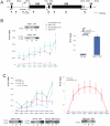
Figure 2. Involvement of the RRM domain of Nab3 in nucleolar surveillance.
A, (left) Domain mapping of Nab3. Nab3 consists of an amino terminal D/E-rich, a central RRM and a carboxyl terminal P/Q-rich domain. The two amino acid substitutions in the RRM domain responsible for the thermosensitive phenotypes in the nab3-11 are indicated (provided by Prof. Jeff Corden, Johns Hopkins University School of Medicine). (right) Ten-fold serial dilutions of yeast cultures. Cells were grown on solid rich medium at the indicated temperatures for 2 d (30°C and 37°C) or 3 d (25°C). B, Analysis of the Nab3 RRM mutation. (left) Northern-blot analysis of total and purified poly(A)+ RNAs in different mutant background at the indicated temperatures (see legend to Fig 1). (right) Phosphor Imager quantitation. Histograms represent the relative distribution of specific RNA normalized to the PGK1 mRNA in different mutant background at different temperatures.
Figure 3
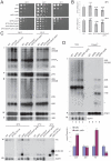
Figure 3. The Nrd1-Nab3 and Spt4-Spt5 complexes colocalize at the rDNA with a particular enrichment towards the 3′-end of the gene.
A, Yeast rDNA unit and amplicons used in the ChIP analysis. The RNA Pol I transcript extends from the promoter to the terminator regions of the rDNA and encodes the 18S, 5.8S, and 25S rRNAs. Mature rRNAs are interspersed with external (5′- and 3′-ETS) and internal (ITS1 and ITS2) transcribed spacers. The 5S gene is transcribed by RNA Pol III in the opposite direction and is embedded in nontranscribed regions 1 and 2 (NTS1 and NTS2). Amplicons used in the ChIP analysis are indicated (1, 3, 6, 5′-ETS, 7, 10, 11, 13, 17, and 18). Scale bar is 500 bp. B, The Nrd1-Nab3 complex interacts with the rDNA. Chromatin extracts prepared from yeast cells expressing a functional carboxyl terminal TAP-tagged version of Nrd1 (NRD1::TAP) or Nab3 (NAB3::TAP), and an otherwise isogenic wild-type strain (No tag), grown to mid-log phase at 30°C, were submitted to coprecipitation analysis with IgG-coated magnetic beads, and the copurifying DNA were analyzed by qPCR with oligonucleotide pairs specific to the entire stretch of the rDNA locus (see panel A). Enrichment at each position is indicated (n = 3). Importantly, chromatin was extracted from independent cultures and used in independent coprecipitation analysis (biological triplicate). Inset: Western-blot analysis of protein coprecipitation efficiency. Chromatin extracts from the total (T), supernatant (S), and pellet (P) fractions were loaded in a 1∶1∶10 ratio. Membranes were probed with an anti-ProtA antibody. Right panel: the interaction of Nrd1 at the snR33 locus was established as a control. The cartoon depicts the snRN33 locus with its promoter (arrow) and the amplicon (−87 to 106) used in the qPCR. For the left panel, p-values were calculated with a standard paired t-test, and are as follows: NRD1::TAP: 0.0039 (rDNA1), 0.012 (rDNA6), 0.0347 (5′-ETS), 0.0357 (rDNA7), 0.0348 (rDNA10), 0.0032 (rDNA11), 0.0119 (rDNA13), 0.0297 (rDNA18); for NAB3::TAP: 0.0475 (rDNA1), 0.0495 (rDNA6), 0.1256 (5′-ETS), 0.0408 (rDNA7), 0.0229 (rDNA10), 0.0109 (rDNA11), 0.0102 (rDNA13), 0.0372 (rDNA18). C, The Spt4-Spt5 RNA polymerase elongation complex interacts with the rDNA and the interaction of Spt5 with the rDNA is reduced in the absence of Spt4 (left). See legend to panel B for details. As a control, the interaction of a functional carboxyl terminal TAP-tagged version of the largest subunit of RNA Pol I (Rpa190) at the rDNA was established (right). n = 3, except for Rpa190 and Spt4 (n = 2). Lower panels: Western-blot analysis of protein coprecipitation efficiency. p-values for SPT5::TAP are 0.0231 (rDNA3), 0.0436 (rDNA6), 0.0258 (5′-ETS), 0.0163 (rDNA7), 0.0252 (rDNA10), 0.0134 (rDNA11), 0.0253 (rDNA13), 0.0239 (rDNA18); for SPT5::TAP spt4Δ: 0.013 (rDNA3), 0.0317 (rDNA6), 0.0187 (5′-ETS), 0.0068 (rDNA7), 0.0146 (rDNA10), 0.0074 (rDNA11), 0.01 (rDNA13), 0.0171 (rDNA18).
Figure 4
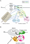
Figure 4. Spt5 interacts with Nrd1 and with the RNA Pol I through its conserved CTR.
A, Spt5 interacts in vivo with Nrd1 Cells expressing functional Spt5-TAP and Nrd1-HA fusions were grown to mid-log phase in complete medium at 30°C. Glass-bead lysates were used in affinity purification analysis with IgG-coated magnetic beads that specifically capture TAP-tagged constructs. Isogenic strains expressing Spt5-TAP or Nrd1-HA alone were used as controls. Fractions of the total (T), supernantant (S) and pellet (P) were loaded in a 1∶1∶20 ratio. Membranes were probed with peroxidase-anti-peroxidase raised in rabbit (recognizes the Protein A component of the TAP-tag fusions) or an anti-HA antibody. This experiment was repeated three times. B, Characterization of a SPT5ΔCTR::HA construct. The precise deletion of the CTR of Spt5 in the genome of haploid cells resulted in a cold-sensitive phenotype for growth (left, cells were incubated for 5 d at 16°C, 2 d at 30°C), and the expression of a protein reduced in length by the expected size (right). The presence of an HA-tag at the carboxyl end of the construct, which did not affect functionality of the full-length construct (see SPT5::HA at 30°C), allowed Western-blot detection with an anti-HA antibody. C, The interaction between Spt5 and Nrd1 requires the CTR domain of Spt5. Cells expressing functional Nrd1-TAP and either a full-length Spt5-HA fusion, or a Spt5-HA construct precisely truncated for its entire CTR, were used as described in panel A. This experiment was repeated two times. D, The interaction between Spt5 and RNA Pol I requires the CTR domain of Spt5. Cells expressing functional Rpa190-TAP and either a full-length Spt5-HA fusion, or a Spt5-HA construct truncated for its CTR were used as described in panel A. This experiment was repeated three times. To the left of each western blot panel, molecular weight markers (in kDa).
Figure 5
Figure 5. The Spt4-Spt5 complex interacts functionally with Rrp6, is required for efficient pre-rRNA processing, and is involved in nucleolar surveillance.
A, Spt4 and Spt5 interact functionally with Rrp6. Legend as in Fig 1. B, RTqPCR analysis of the mRNA level of SPT5 (normalized to ACT1) in SPT5 DAmP at different temperature in the presence, and in the absence of Spt4 (n = 3). C, Pre-rRNA processing analysis. Total RNA was extracted from different mutants grown at the indicated temperature, separated on denaturing gels. Probes used to detect pre-rRNAs and rRNAs were: LD359 (panel I), LD339 (panel II), LD471 (panel III), LD1099 (panel IV), LD871 (panel V), and LD906 (panel VI); they are depicted in Fig S3A. Panel VI, as a control for detection of 6S and 5.8S+30, total RNA was extracted from ngl2 and rrp6 mutants, respectively. These precursors are so abundant in these mutants, that 10x less RNA was loaded in these lanes; consequently, these lanes show no signal for SCR1 on this exposure. This experiment was repeated three times and a representative case is shown. D, Spt4 is involved in nucleolar surveillance. (top) Northern-blot analysis of total and purified poly(A)+ RNAs in different mutant background at 30°C (see legends to Figs 1 and 2). (bottom) Phosphor Imager quantitation (see legends to Figs 1 and 2).
Figure 6
Figure 6. A physico-functional interface at the rDNA that connects rRNA synthesis, rRNA processing, and rRNA surveillance.
A, The RNA Pol I, the Spt4–Spt5 elongation complex, the Nrd1-Nab3-Sen1, and the RNA exosome interact physically and are poised to interact functionally. We suggest that the Nrd1-Nab3-Sen1 complex contributes to recruiting 3′-to-5′ nucleolar surveillance to nascent pre-rRNA transcripts. Interactomics suggest that 5′-3′ nucleolar surveillance might also be recruited, but this has not addressed in this work. Rpa135-Rpa190, catalytic core of RNA Pol I; Rpa34.5-Rpa49, intrinsic elongation factor. Color-code: blue (physical interaction, affinity purification and in vitro pull-down), green (genetic interaction, limited to synthetic growth defect). Interaction dataset curated from BioGrid and from this work. B, Model for early recognition of defective pre-ribosomes: in conditions in which ribosome assembly is compromised (delayed assembly of ribosome synthesis factors or ribosomal proteins and/or misfolded RNA, lightning symbol), we speculate that cryptic Nrd1-Nab3 binding sites become available for binding. We suggest that the elongation factor Spt4–Spt5 recruits and deposits the Nrd1-Nab3 complex, which in turn attracts the RNA exosome to defective pre-rRNA transcripts.

Similar articles

Cited by

References

    1. Henras AK, Soudet J, Gerus M, Lebaron S, Caizergues-Ferrer M, et al. The post-transcriptional steps of eukaryotic ribosome biogenesis. Cell Mol Life Sci. 2008;65:2334–2359. - PMC - PubMed
    1. Lafontaine DLJ. A ‘garbage can’ for ribosomes: how eukaryotes degrade their ribosomes? Trends Biochem Sci. 2010;35:267–277. - PubMed
    1. Fang F, Phillips S, Butler JS. Rat1p and Rai1p function with the nuclear exosome in the processing and degradation of rRNA precursors. Rna. 2005;11:1571–1578. - PMC - PubMed
    1. Wang M, Pestov DG. 5′-end surveillance by Xrn2 acts as a shared mechanism for mammalian pre-rRNA maturation and decay. Nucleic Acids Res. 2010. pp. 1811–1822. - PMC - PubMed
    1. Dez C, Dlakic M, Tollervey D. Roles of the HEAT repeat proteins Utp10 and Utp20 in 40S ribosome maturation. Rna. 2007;13:1516–1527. - PMC - PubMed

Publication types

MeSH terms