. 2011 Sep 15;25(18):1943-54.
doi: 10.1101/gad.17257711.

Cdc15 integrates Tem1 GTPase-mediated spatial signals with Polo kinase-mediated temporal cues to activate mitotic exit

Affiliations

Cdc15 integrates Tem1 GTPase-mediated spatial signals with Polo kinase-mediated temporal cues to activate mitotic exit

Jeremy M Rock et al. Genes Dev. .

Abstract

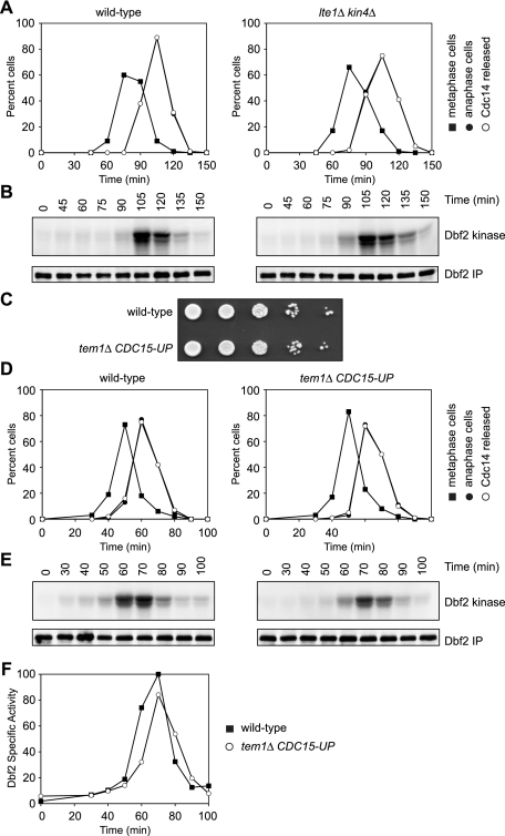
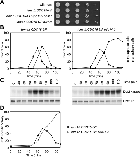
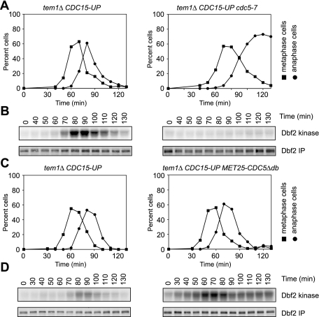
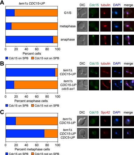
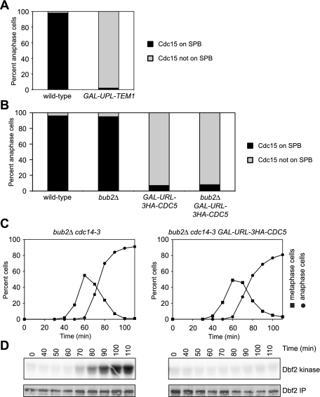
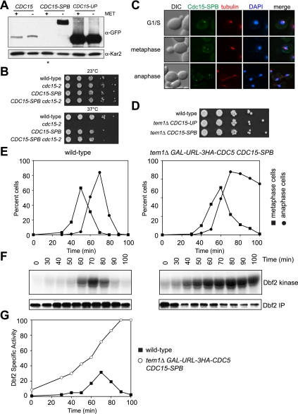
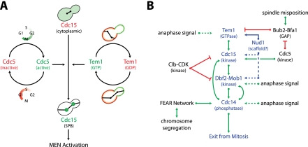
In budding yeast, a Ras-like GTPase signaling cascade known as the mitotic exit network (MEN) promotes exit from mitosis. To ensure the accurate execution of mitosis, MEN activity is coordinated with other cellular events and restricted to anaphase. The MEN GTPase Tem1 has been assumed to be the central switch in MEN regulation. We show here that during an unperturbed cell cycle, restricting MEN activity to anaphase can occur in a Tem1 GTPase-independent manner. We found that the anaphase-specific activation of the MEN in the absence of Tem1 is controlled by the Polo kinase Cdc5. We further show that both Tem1 and Cdc5 are required to recruit the MEN kinase Cdc15 to spindle pole bodies, which is both necessary and sufficient to induce MEN signaling. Thus, Cdc15 functions as a coincidence detector of two essential cell cycle oscillators: the Polo kinase Cdc5 synthesis/degradation cycle and the Tem1 G-protein cycle. The Cdc15-dependent integration of these temporal (Cdc5 and Tem1 activity) and spatial (Tem1 activity) signals ensures that exit from mitosis occurs only after proper genome partitioning.


Figures

Figure 1.
Figure 1.
Anaphase-specific activation of the MEN in the absence of TEM1. (A,B) Wild-type (A2747) and lte1Δ kin4Δ (A26379) cells containing 3HA-Cdc14 and 3MYC-Dbf2 fusion proteins were arrested in G1 with α-factor pheromone (5 μg/mL) in YEP medium containing glucose (YEPD). When the arrest was complete (after 150 min), cells were released into pheromone-free YEPD medium. After 80 min, α-factor pheromone (10 μg/mL) was re-added to prevent entry into the subsequent cell cycle. The percentage of cells with metaphase spindles (closed squares; A), anaphase spindles (closed circles; A), and 3HA-Cdc14 released from the nucleolus (open circles; A), and the amount of Dbf2-associated kinase activity (Dbf2 kinase; B) and immunoprecipitated 3MYC-Dbf2 (Dbf2 IP; B) were determined at the indicated times. (C) Wild-type (A2747) and tem1Δ CDC15-UP (A22670) cells containing 3HA-Cdc14 and 3MYC-Dbf2 fusion proteins were spotted on YEP plates containing raffinose and galactose (YEPRG) at 30°C. Approximately 3 × 10⁴ cells were deposited in the first spot, and each subsequent spot is a 10-fold serial dilution. The picture shown depicts 3 d of growth. (D,E) Wild-type (A2747) and tem1Δ CDC15-UP (A22670) cells containing 3HA-Cdc14 and 3MYC-Dbf2 fusion proteins were arrested in G1 with α-factor pheromone (5 μg/mL) in YEPRG medium. When the arrest was complete (after 2 h 50 min), cells were released into pheromone-free YEPRG medium. After 60 min, α-factor pheromone (10 μg/mL) was added to prevent entry into the subsequent cell cycle. The percentage of cells with metaphase spindles (closed squares; D), anaphase spindles (closed circles; D), and 3HA-Cdc14 released from the nucleolus (open circles; D), and the amount of Dbf2-associated kinase activity (Dbf2 kinase; E) and immunoprecipitated 3MYC-Dbf2 (Dbf2 IP; E) were determined at the indicated times. (F) The amount of Dbf2-associated kinase activity and immunoprecipitated 3MYC-Dbf2 from E was determined by quantitative autoradiography and quantitative Western blot, respectively. Shown is the specific Dbf2-associated kinase activity.
Figure 2.
Figure 2.
The FEAR network is not required for Dbf2 activity in tem1Δ CDC15-UP cells. (A) Wild-type (A2747), tem1Δ CDC15-UP (A22670), tem1Δ spo12Δ bns1Δ CDC15-UP (A23392), and tem1Δ slk19Δ CDC15-UP (A23387) cells containing 3HA-Cdc14 and 3MYC-Dbf2 fusion proteins were spotted on YEPRG plates, as in Figure 1C. (B,C) tem1Δ CDC15-UP (A23782) and tem1Δ cdc14-3 CDC15-UP (A23712) cells containing a 3MYC-Dbf2 fusion protein were arrested in G1 with α-factor pheromone (5 μg/mL) in YEPRG medium at room temperature. Thirty minutes prior to release, the cells were shifted to 35°C. When the arrest was complete (after 3 h 30 min), cells were released into pheromone-free YEPRG medium at 35°C. After 65 min, α-factor pheromone (10 μg/mL) was re-added to prevent entry into the subsequent cell cycle. The percentage of cells with metaphase spindles (closed squares, B) and anaphase spindles (closed circles, B) and the amount of Dbf2-associated kinase activity (Dbf2 kinase, C) and immunoprecipitated 3MYC-Dbf2 (Dbf2 IP, C) were determined at the indicated times. (D) The amount of Dbf2-associated kinase activity and immunoprecipitated 3MYC-Dbf2 from C was determined as in Figure 1F. Shown is the specific Dbf2-associated kinase activity. Note that the specific Dbf2-associated kinase activity continues to rise in the tem1Δ cdc14-3 CDC15-UP strain as a result of a prolonged anaphase arrest.
Figure 3.
Figure 3.
Polo-like kinase Cdc5 controls MEN activity in the absence of TEM1. (A,B) tem1Δ CDC15-UP (A22670) and tem1Δ cdc5-7 CDC15-UP (A24305) cells containing 3HA-Cdc14 and 3MYC-Dbf2 fusion proteins were arrested in G1 with α-factor pheromone (5 μg/mL) in YEPRG medium at 30°C. Thirty minutes prior to release, the cells were shifted to 37°C. When the arrest was complete (after 3 h), cells were released into pheromone-free YEPRG medium at 37°C. After 65 min, α-factor pheromone (10 μg/mL) was added to prevent entry into the subsequent cell cycle. The percentage of cells with metaphase spindles (closed squares; A) and anaphase spindles (closed circles; B) and the amount of Dbf2-associated kinase activity (Dbf2 kinase; B) and immunoprecipitated 3MYC-Dbf2 (Dbf2 IP; B) were determined at the indicated times. (C,D) tem1Δ CDC15-UP (A22670) and tem1Δ MET25-CDC5Δdb CDC15-UP (A25175) cells containing 3HA-Cdc14 and 3MYC-Dbf2 fusion proteins were arrested in G1 with α-factor pheromone (5 μg/mL) in YEPRG medium. Ninety minutes prior to release, the cells were transferred to −Met medium containing raffinose and galactose (−MetRG; to induce the expression of Cdc5Δdb) supplemented with α-factor pheromone (5 μg/mL). When the arrest was complete (after 3 h), cells were released into pheromone-free −MetRG medium. After 70 min, α-factor pheromone (10 μg/mL) was re-added to prevent entry into the subsequent cell cycle. The percentage of cells with metaphase spindles (closed squares; C) and anaphase spindles (closed circles; C) and the amount of Dbf2-associated kinase activity (Dbf2 kinase; D) and immunoprecipitated 3MYC-Dbf2 (Dbf2 IP; D) were determined at the indicated times.
Figure 4.
Figure 4.
Cdc5 promotes localization of Cdc15 to SPBs. (A) tem1Δ CDC15-eGFP-UP (A25630) cells containing a mCherry-Tub1 fusion protein were arrested in G1 with α-factor pheromone (5 μg/mL) in YEPRG medium. When the arrest was complete (after 2 h 50 min), cells were released into pheromone-free YEPRG medium and imaged after a brief paraformaldehyde fixation. Cell cycle stage was determined based on spindle morphology and correlated with Cdc15 localization at SPBs (n ≥ 100 cells for each cell cycle stage). Representative images of G1/S, metaphase, and anaphase cells are shown. Cdc15 is shown in green, microtubules are shown in red, and DNA is shown in blue. (B) tem1Δ CDC15-eGFP-UP (A25630) and tem1Δ CDC15-eGFP-UP cdc5-as1 (A25633) cells containing a mCherry-Tub1 fusion protein were arrested in G1 as in A. Cells were released into pheromone-free YEPRG medium supplemented with 5 μM CMK (cdc5-as1 inhibitor). Cells were scored as in A. Representative images of anaphase cells are shown. (C) tem1Δ CDC15-eGFP-UP (A25744) and tem1Δ CDC15-eGFP-UP MET25-CDC5ΔN70 (tem1Δ CDC15-eGFP-UP CDC5-UP; A25983) cells containing a Spc42-mCherry fusion protein were arrested in G1 with α-factor pheromone (5 μg/mL) in YEPRG medium supplemented with 8 mM methionine. Ninety minutes prior to release, the cells were transferred to −MetRG medium (to induce the expression of Cdc5ΔN70) supplemented with α-factor pheromone. When the arrest was complete (after 3 h), cells were released into pheromone-free −MetRG medium. Cells were imaged and scored as in A. Representative images of metaphase cells are shown. Cdc15 is shown in green, Spc42 is shown in red, and DNA is shown in blue.
Figure 5.
Figure 5.
Cdc15 functions as a coincidence detector of Tem1 and Cdc5 activity. (A) CDC15-eGFP (A26481) and CDC15-eGFP GAL-UPL-TEM1 (A27055) cells containing a mCherry-Tub1 fusion protein were arrested in G1 with α-factor pheromone (5 μg/mL) in YEPRG medium. Ubiquitin-proline-LacZ (UPL) acts as a destabilizing module that permits rapid degradation of appended proteins. One hour prior to release, glucose was added to a final concentration of 2% (to repress expression of GAL-UPL-TEM1). When the arrest was complete (after 2 h 40 min), cells were released into pheromone-free YEPD medium. Cells were imaged and scored as in Figure 4A. (B) CDC15-eGFP (A26481), CDC15-eGFP bub2Δ (A26480), CDC15-eGFP GAL-URL-3HA-CDC5 (A26556), and CDC15-eGFP bub2Δ GAL-URL-3HA-CDC5 (A26558) cells containing a mCherry-Tub1 fusion protein were arrested in G1 with α-factor pheromone (5 μg/mL) in YEPRG medium. Ubiquitin-arginine-LacZ (URL) acts as a destabilizing module that permits rapid degradation of appended proteins. Two hours prior to release, glucose was added to a final concentration of 2% (to repress expression of GAL-URL-3HA-CDC5). When the arrest was complete (after 2 h 45 min), cells were released into pheromone-free YEPD medium. Cells were imaged and scored as in Figure 4A. (C,D) bub2Δ cdc14-3 (A26844) and bub2Δ cdc14-3 GAL-URL-3HA-CDC5 (A26842) cells containing a 3MYC-Dbf2 fusion protein were arrested in G1 with α-factor pheromone (5 μg/mL) in YEPRG medium. Two hours prior to release, glucose was added to repress expression of GAL-URL-3HA-CDC5. When the arrest was complete (after 2 h 45 min), cells were released into pheromone-free YEPD medium. The percentage of cells with metaphase spindles (closed squares; C) and anaphase spindles (closed circles; C) and the amount of Dbf2-associated kinase activity (Dbf2 kinase; D) and immunoprecipitated 3MYC-Dbf2 (Dbf2 IP; D) were determined at the indicated times.
Figure 6.
Figure 6.
Targeting Cdc15 to SPBs bypasses the need for TEM1 and CDC5 in MEN activation. (A) CDC15-eGFP (CDC15; A20935), pMET3-CDC15-eGFP-CNM67 (CDC15-SPB; A26417), and CDC15-eGFP-UP (CDC15-UP; A25515) cells were grown to log phase in either YEPRG + methionine (+MET) or −Met medium to determine the amount of Cdc15-eGFP (α-GFP) in cells. Kar2 was used as a loading control in Western blots. (B) Wild-type (A2587), cdc15-2 (A2597), pMET3-CDC15-eGFP-CNM67 (CDC15-SPB; A26419), and pMET3-CDC15-eGFP-CNM67 cdc15-2 (CDC15-SPB cdc15-2; A26413) cells were spotted on YEPRG plates supplemented with 8 mM methionine as in Figure 1C. The picture shown depicts 2 d of growth at 37°C and 3 d of growth at 23°C. (C) pMET3-CDC15-eGFP-CNM67 (CDC15-SPB; A26486) cells containing a mCherry-Tub1 fusion protein were grown to log phase in YEPRG medium supplemented with 8 mM methionine and imaged after a brief paraformaldehyde fixation. Representative images of G1/S, metaphase, and anaphase cells are shown. (D) Wild-type (A2747), tem1Δ CDC15-UP (A22670), and tem1Δ pMET3-CDC15-eGFP-CNM67 (tem1Δ CDC15-SPB; A26396) cells containing 3HA-Cdc14 and 3MYC-Dbf2 fusion proteins were spotted on YEPRG plates supplemented with 8 mM methionine as in Figure 1C. The picture shown depicts 3 d of growth. (E,F) Wild-type (A2747) and tem1Δ GAL-URL-3HA-CDC5 pMET3-CDC15-eGFP-CNM67 (tem1Δ GAL-URL-3HA-CDC5 CDC15-SPB; A27051) cells containing 3HA-Cdc14 and 3MYC-Dbf2 fusion proteins were arrested in G1 with α-factor pheromone (5 μg/mL) in YEPRG medium supplemented with 8 mM methionine. Two hours prior to release, glucose was added (to repress expression of GAL-URL-3HA-CDC5). When the arrest was complete (after 2 h 50 min), cells were released into pheromone-free YEPD medium supplemented with 8 mM methionine. After 65 min, α-factor pheromone (10 μg/mL) was added to prevent entry into the subsequent cell cycle. The percentage of cells with metaphase spindles (closed squares; E) and anaphase spindles (closed circles; E) and the amount of Dbf2-associated kinase activity (Dbf2 kinase; F) and immunoprecipitated 3MYC-Dbf2 (Dbf2 IP; F) were determined at the indicated times. (G) The amount of Dbf2-associated kinase activity and immunoprecipitated 3MYC-Dbf2 from F was determined as in Figure 1F. Shown is the specific Dbf2-associated kinase activity.
Figure 7.
Figure 7.
A model for the coordination of exit from mitosis with spatial and temporal cues. (A) Cdc15 functions as a coincidence detector of Tem1 and Cdc5 activity, both of which are required for the association of Cdc15 with SPBs. See the text for details. (B) Multiple signals control MEN activity. The core MEN components are shown in blue, activators of the MEN are shown in green, and inhibitors of the MEN are shown in red. Experimentally validated interactions are shown with solid lines, and more speculative interactions are shown with dashed lines. See the text for details.

Similar articles

Cited by

References

    1. Asakawa K, Yoshida S, Otake F, Toh-e A 2001. A novel functional domain of Cdc15 kinase is required for its interaction with Tem1 GTPase in Saccharomyces cerevisiae. Genetics 157: 1437–1450 - PMC - PubMed
    1. Bachmair A, Finley D, Varshavsky A 1986. In vivo half-life of a protein is a function of its amino-terminal residue. Science 234: 179–186 - PubMed
    1. Bardin AJ, Visintin R, Amon A 2000. A mechanism for coupling exit from mitosis to partitioning of the nucleus. Cell 102: 21–31 - PubMed
    1. Bardin AJ, Boselli MG, Amon A 2003. Mitotic exit regulation through distinct domains within the protein kinase Cdc15. Mol Cell Biol 23: 5018–5030 - PMC - PubMed
    1. Bertazzi DT, Kurtulmus B, Pereira G 2011. The cortical protein Lte1 promotes mitotic exit by inhibiting the spindle position checkpoint kinase Kin4. J Cell Biol 193: 1033–1048 - PMC - PubMed

Publication types

MeSH terms

LinkOut - more resources