. 2011 Nov 25;286(47):40974-86.
doi: 10.1074/jbc.M111.290874. Epub 2011 Sep 21.

Binding site specificity and factor redundancy in activator protein-1-driven human papillomavirus chromatin-dependent transcription

Affiliations

Binding site specificity and factor redundancy in activator protein-1-driven human papillomavirus chromatin-dependent transcription

Wei-Ming Wang et al. J Biol Chem. .

Abstract

Activator protein-1 (AP-1) regulates diverse gene responses triggered by environmental cues and virus-induced cellular stress. Although many signaling events leading to AP-1 activation have been described, the fundamental features underlying binding site selection and factor recruitment of dimeric AP-1 complexes to their target genes remain mostly uncharacterized. Using recombinant full-length human AP-1 dimers formed between c-Jun and Fos family members (c-Fos, FosB, Fra-1, Fra-2) for DNA binding and transcriptional analysis, we found that each of these AP-1 complex exhibits differential activity for distinct non-consensus AP-1 sites present in human papillomavirus (HPV), and each AP-1 complex is capable of activating transcription from in vitro-reconstituted HPV chromatin in a p300- and acetyl-CoA-dependent manner. Transcription from HPV chromatin requires AP-1-dependent and contact-driven recruitment of p300. Acetylation of dimeric AP-1 complexes by p300 enhances AP-1 binding to DNA. Using a human C-33A cervical cancer-derived cell line harboring the episomal HPV type 11 genome, we illustrate binding site selectivity recognized by c-Jun, JunB, JunD, and various Fos family members in a combinatorial and unique pattern, highlighting the diversity and importance of non-canonical binding site recognition by various AP-1 family proteins.


Figures

FIGURE 1.
FIGURE 1.
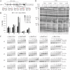
Recombinant full-length dimeric c-Jun-containing human AP-1 complexes are all active in binding the consensus TRE sequence found in the human cyclin D1 gene. A, shown is a schematic drawing of human c-Jun and Fos family proteins tagged at the N terminus with the FLAG (F:) or hexahistidine (6His) sequence. Numbers indicate the first and last amino acids of each protein. B, purification scheme of dimeric AP-1 complex is shown. C, purified AP-1 complexes are visualized by Coomassie Blue staining. Protein size markers (in kDa) are shown on the left. D, protein-DNA complexes formed on DNA fragments containing the TRE derived from the human cyclin D1 (hCyclin D1) gene are shown. The sequences of wild-type (WT) and mutated (Mut) TREs present in the non-radiolabeled cold competitors are shown on the right. Antibody supershift assay was performed with anti-6His (α-His) or α-c-Jun antibodies. E, each dimeric AP-1 complex binds the TRE in a dose-dependent manner. EMSA was performed as described under “Experimental Procedures” with the indicated amounts of protein complexes.
FIGURE 2.
FIGURE 2.
Non-canonical AP-1 binding sites found in HPV-11 are differentially recognized by different AP-1 complexes. A, the position and sequence of each AP-1 site in HPV-11 are shown, with nucleotides deviating from the consensus TRE indicated in red. B, affinity (1/Kd) of distinct AP-1 complexes for each AP-1 site in HPV-11. Kd (see Table 1) was calculated as described under “Experimental Procedures” according to the EMSA results shown in C. D, relative binding affinity to HPV-11 #3–#5 sites exhibited by different AP-1 complexes as resolved by DNase I footprinting. Protected regions around and between core AP-1 sequences are marked in solid and dashed lines, respectively, with hypersensitivity sites depicted by asterisks on the right. A schematic drawing of AP-1 sites is shown on the left.
FIGURE 3.
FIGURE 3.
Non-canonical AP-1 sites are well conserved among genital HPVs. The position of each non-canonical and consensus AP-1 site, whose sequence is shown in a unique color on the right, is indicated for eight prevalent genital HPV types. Four conserved E2 binding sites are also marked for position referencing. Accession numbers for the analyzed HPV sequences are listed in parentheses.
FIGURE 4.
FIGURE 4.
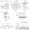
Distinct AP-1 complexes initiate transcription from HPV chromatin in a p300- and acetyl-CoA-dependent manner. A, G-less cassette DNA templates used for transcriptional analysis are shown. B, shown is a scheme for chromatin assembly with purified factors, including HeLa core histones, human NAP-1 (hNAP-1), and ACF, added at different time points. C, shown is the outline of the order-of-addition transcription experiment performed with HeLa nuclear extract (NE). D, in vitro transcription was performed with HPV chromatin or DNA with an internal DNA template (pMLΔ53) included for comparison. Transcription was set up as outlined in C in the presence (+) or absence (−) of AP-1, p300, and acetyl-CoA as indicated. Relative transcription (Rel Txn) is defined as the signal intensity, quantified by Typhoon 9200 PhosphorImager (GE Healthcare) from the HPV template relative to that performed in the presence of 60 ng of c-Jun/c-Fos, acetyl-CoA, and p300 (i.e. lane 4 for HPV chromatin and lane 21 for HPV DNA) after initial normalization with the signal derived from the internal pMLΔ53 control template.
FIGURE 5.
FIGURE 5.
p300 acetylates AP-1, enhancing its binding to DNA and stimulating HPV chromatin transcription at a step post-AP-1 entry. A, dimeric AP-1 complexes interact directly with full-length (FL) but not truncated p300 proteins. AP-1, immobilized on the beads via its hexahistidine tag (residing at the Fos partner) association with Ni2+-NTA, was incubated with p300 or its truncated mutant (HAT or ΔHAT, see Fig. 6A) input (InP) as described under “Experimental Procedures.” Bound proteins were then analyzed by Western blotting (WB) with anti-FLAG antibody, which detects FLAG-tagged p300 and FLAG-tagged c-Jun in AP-1 complexes. B–D, p300 acetylates c-Jun, c-Fos, FosB, Fra-1, Fra-2, JunB, and JunD in each dimeric AP-1 complex. HAT assay was performed in the absence (−) or presence (+) of AP-1 and p300 as described under “Experimental Procedures.” Tritium-labeled proteins were then separated by SDS-PAGE and visualized after fluorography and film exposure. Protein size markers (in kDa) are shown on the left. E, acetylation enhances dimeric AP-1 complex binding to DNA. EMSA was performed with dimeric AP-1 complexes, with (+) or without (-) prior incubation with p300 and acetyl-CoA (Ac-CoA) using a 32P-labeled DNA probe containing the HPV-11 #5 AP-1 site. F, AP-1 stimulates HPV chromatin transcription at a step before or concomitant with p300 entry. Transcription reactions were assembled as outlined with c-Jun/c-Fos (AP-1) added at a step either before, during, or after p300 addition. Transcription was then initiated upon the addition of [α-32P]CTP and unlabeled nucleoside triphosphates (NTPs). Relative transcription (Rel Txn) is defined in Fig. 4D, with the transcription signal from HPV chromatin in lane 4 set as 100.
FIGURE 6.
FIGURE 6.
p300-stimulated HPV chromatin transcription and acetylation of nucleosomal histones is an AP-1-dependent event requiring additional regions outside its HAT domain. A, Coomassie staining of purified full-length (FL) p300 and its truncated mutants containing the wild-type (HAT) or acetylase activity-impaired (ΔHAT) HAT domain is shown. A schematic representation of protein domains in p300, HAT, and ΔHAT is shown on the left. B, a HAT assay performed with free core histones as substrates in the absence (−) or presence of different amounts of p300 (FL), ΔHAT, and HAT as indicated is shown. Protein size markers (in kDa) are shown on the left. C, in vitro transcription was performed as described in Fig. 4D with c-Jun/c-Fos in the absence (−) or presence of p300 (FL), ΔHAT, or HAT as indicated. D, in vitro HAT assay was performed with HPV chromatin as the substrate in the absence (−) or presence (+) of c-Jun/c-Fos and p300 (FL), ΔHAT, or HAT as indicated.
FIGURE 7.
FIGURE 7.
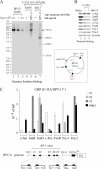
Differential recruitment of Jun and Fos family members to individual AP-1 sites in HPV-11 episomes. A, establishment of a C-33A-derived cell line harboring HPV-11 episomes is shown. Genomic Southern blotting was performed by digesting 7.5 μg of genomic DNA, isolated from C-33A cells (−) or C-33A/HPV-11+ cells (HPV-11) with either a single-cutter (BamHI) or a triple-cutter (ApaL1) of HPV-11 DNA (map shown in the box) and probed with 32P-labeled HPV-11 DNA as described under “Experimental Procedures.” BamHI-cleaved HPV-11 DNA representing different genomic copy numbers as indicated was mixed with 7.5 μg of C-33A genomic DNA and loaded as controls for quantification. B, detection of Jun and Fos family proteins in HeLa and C-33A cells with (+) or without (−) HPV-11 episomes by Western blotting with different anti-protein antibodies as indicated is shown. C, association of various Jun and Fos family members with different HPV-11 AP-1 sites as monitored by a ChIP assay performed with chromatin samples isolated from C-33A/HPV-11+ cells is shown. ChIP assay was performed with antibodies against different components of AP-1 family proteins as indicated. The locations of PCR-amplified DNA fragments containing specific AP-1 sites in HPV-11 are shown on the bottom.
FIGURE 8.
FIGURE 8.
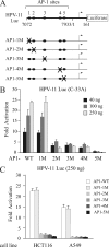
Non-canonical #2–#5 AP-1 binding sites are crucial for HPV transcription in different cell types. A, shown are reporter plasmids containing HPV-11 wild-type or mutated AP-1 sites used for luciferase assay. Mutated nucleotides (underlined) are #1: TACGTAA; #2: TACCGAA; #3: TACCGAA; #4: TACGTAA; #5: TACGTAA (see supplemental Table S1). B, #2–#5 AP-1 sites are crucial for HPV transcription in C-33A cervical cancer cells. Different amounts of reporter plasmids as indicated were transfected into C-33A cells, and luciferase activity was measured 24 h later as described under “Experimental Procedures.” C, #2–#5 AP-1 sites are important for HPV transcription in HCT116 colon cancer cells and A549 lung adenocarcinoma-derived cells. Transfection of HCT116 and A549 cells was conducted with 250 ng of each reporter plasmid. Luciferase activity was measured 24 h post-transfection.
FIGURE 9.
FIGURE 9.
A promoter-proximal AP-1 site is spatially conserved in genital HPVs. A, shown is a schematic drawing of the promoter-proximal AP-1 site relative to the #2 E2 binding site and the TATA box of the E6 promoter in representative low-risk and high-risk genital HPVs. B, distance from the promoter-proximal AP-1 site to the #2 E2 binding site and the E6 TATA box is highly conserved among genital HPVs. C, sequence alignment of the heptanucleotide core (in brown) and conserved flanking nucleotides (indicated in yellow) is shown.

Similar articles

Cited by

References

    1. Lee W., Mitchell P., Tjian R. (1987) Cell 49, 741–752 - PubMed
    1. Angel P., Karin M. (1991) Biochim. Biophys. Acta 1072, 129–157 - PubMed
    1. Jochum W., Passegué E., Wagner E. F. (2001) Oncogene 20, 2401–2412 - PubMed
    1. Mechta-Grigoriou F., Gerald D., Yaniv M. (2001) Oncogene 20, 2378–2389 - PubMed
    1. Shaulian E., Karin M. (2002) Nat. Cell Biol. 4, E131–E136 - PubMed

Publication types

MeSH terms