. 2011;6(9):e23926.
doi: 10.1371/journal.pone.0023926. Epub 2011 Sep 7.

Transgenic expression of human LAMA5 suppresses murine Lama5 mRNA and laminin α5 protein deposition

Affiliations

Transgenic expression of human LAMA5 suppresses murine Lama5 mRNA and laminin α5 protein deposition

Brooke M Steenhard et al. PLoS One. 2011.

Abstract

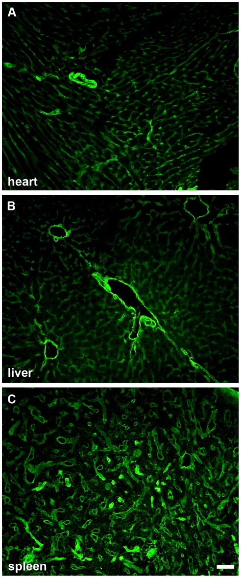
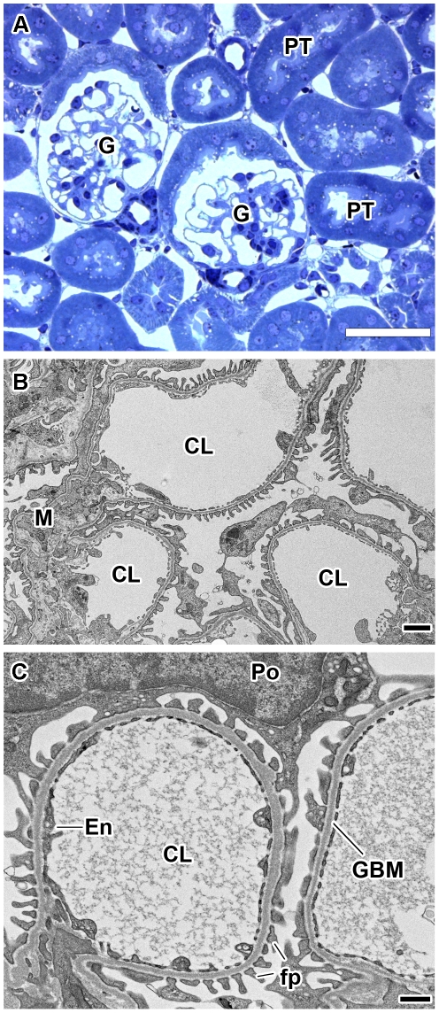
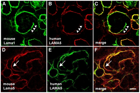
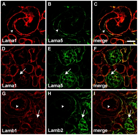
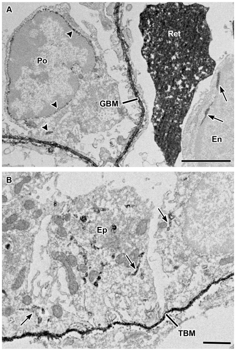
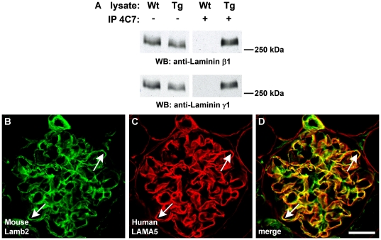
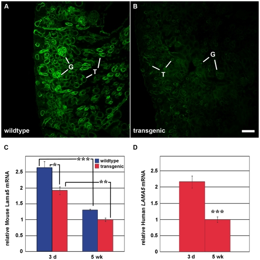
Laminin α5 is required for kidney glomerular basement membrane (GBM) assembly, and mice with targeted deletions of the Lama5 gene fail to form glomeruli. As a tool to begin to understand factors regulating the expression of the LAMA5 gene, we generated transgenic mice carrying the human LAMA5 locus in a bacterial artificial chromosome. These mice deposited human laminin α5 protein into basement membranes in heart, liver, spleen and kidney. Here, we characterized two lines of transgenics; Line 13 expressed ∼6 times more LAMA5 than Line 25. Mice from both lines were healthy, and kidney function and morphology were normal. Examination of developing glomeruli from fetal LAMA5 transgenics showed that the human transgene was expressed at the correct stage of glomerular development, and deposited into the nascent GBM simultaneously with mouse laminin α5. Expression of human LAMA5 did not affect the timing of the mouse laminin α1-α5 isoform switch, or that for mouse laminin β1-β2. Immunoelectron microscopy showed that human laminin α5 originated in both glomerular endothelial cells and podocytes, known to be origins for mouse laminin α5 normally. Notably, in neonatal transgenics expressing the highest levels of human LAMA5, there was a striking reduction of mouse laminin α5 protein in kidney basement membranes compared to wildtype, and significantly lower levels of mouse Lama5 mRNA. This suggests the presence in kidney of a laminin expression monitor, which may be important for regulating the overall production of basement membrane protein.


Conflict of interest statement

Competing Interests: The authors have declared that no competing interests exist.

Figures

Figure 1
Figure 1. Human LAMA5 BAC transgenic mice deposit human laminin α5 protein in kidney basement membranes.
A: Immunofluorescence micrograph of frozen kidney section from a transgenic mouse labeled with mouse anti-human laminin α5, followed by goat anti-mouse IgG2a conjugated to Alexa Fluor-594. Basement membranes within glomeruli (G) and surrounding tubules (T) immunolabel in bright linear patterns. B: Section from a wildtype littermate incubated with anti-human laminin α5 is negative. In both A and B, cell nuclei are stained blue by DAPI. Magnification: 200×; scale bar = 50 µm.
Figure 2
Figure 2. Human LAMA5 BAC transgenic mice deposit human laminin α5 protein in basement membranes.
Frozen sections of tissues from human LAMA5 BAC transgenics immunolabeled with mouse anti-human laminin α5 antibody and goat anti- mouse IgG2a conjugated to Alexa Fluor-488. Human laminin α5 is deposited widely in most or all basement membranes of heart (A), liver (B), and spleen (C). Magnification: 200×; scale bar = 50 µm.
Figure 3
Figure 3. Characterization of two different human LAMA5 BAC transgenic lines.
A–D: Fresh frozen kidney sections labeled with mouse anti-human laminin α5 antibody, followed by anti-mouse IgG conjugated to Alexa Fluor-594. Sections from Line 13 and Line 25 were imaged by routine immunofluorescence microscopy (A and B), and by scanning confocal microscopy (C and D), using the same exposure settings. Linear basement membrane labeling of anti-human laminin α5 is seen in both line 13 (A and C) and 25 (B and D), but fluorescent signal appears brighter in Line 13. Magnification (A and B): 200×; scale bar = 50 µm. Magnification (C and D): 630×; scale bar = 20 µm. E: Quantification of glomerular immunofluorescence intensities shows significantly higher expression of human laminin α5 in GBMs of Line 13 mice, *** p<0.0001. F: Relative transgene copy number estimates were made using cycle threshold from quantitative real time PCR of human LAMA5 genomic primers normalized to thresholds obtained with mouse hemoglobin A (Hba) primers. Compared to Line 25, Line 13 has more than 6 times as many copies of LAMA5, **p = 0.0013. G: Whole kidney total RNA from Line 13 or Line 25 (n = 3 samples per line) was amplified with human LAMA5 intron-spanning primers, relative to PPIA (cyclophilin). Considerably more LAMA5 mRNA is detected in Line 13, *** p<0.0001.
Figure 4
Figure 4. Normal kidney histology and glomerular capillary ultrastructure in Line 13 human LAMA5 BAC transgenics.
A: Semithin kidney section from an 8 week old transgenic stained with Toluidine Blue. Profiles of glomeruli (G) and proximal tubules (PT) are normal and there is no evidence of fibrosis or other defects. Magnification: 400×; scale bar = 50 µm. B: Electron micrograph of glomerular capillary loops from a human LAMA5 BAC transgenic mouse showing normal mesangial (M) architecture and open capillary lumens (CL). Magnification: 7,000×; scale bar = 500 nm. C: Higher power view of glomerular capillary loops showing fenestrated endothelium (En), and normal, interdigitating foot processes (fp). The glomerular basement membrane (GBM) also appears to be of normal density and width. CL: capillary lumen, Po: Podocyte cell body. Magnification: 13,500×; scale bar = 500 nm.
Figure 5
Figure 5. Human LAMA5 expression occurs temporally correctly and coordinately with expression of mouse Lama5.
A–C: Early GBM in vascular cleft of a comma-shaped nephric figure in neonatal human LAMA5 transgenic immunolabeled with anti-mouse laminin α1 (A, green) and anti-human laminin α5 (B, red). At this early stage of glomerular development, the nascent GBM within the vascular cleft (arrowheads) contains predominantly the laminin α1 isoform with only trace amounts of laminin α5 present. D–F: At a slightly later stage of glomerular development, linear deposition of both mouse and human laminin α5 is evident in the GBM (arrow). Digitally merged images in F show colocalization of mouse and human laminin α5 in the same GBM (arrow). Magnification: 250×; scale bar = 20 µm.
Figure 6
Figure 6. Expression of human LAMA5 does not alter timing of mouse laminin isoform substitution.
A–C: S-shaped nephric figure immunolabled for mouse laminin α1 (A, red) and mouse laminin α5 (B, green). Deposition of laminin α1 declines markedly (arrowhead) as laminin α5 appears. D–F: By the time glomeruli reach the capillary loop stage, laminin α1 is seen only in mesangial regions whereas laminin α5 occurs in loop GBMs (arrow). G–I: The normal laminin β1–β2 switch occurs somewhat slower than that for laminin α1–α5. G: Laminin β1 can be seen in vascular clefts (arrowhead) as well as in capillary loop stage GBMs (arrow). H: Trace amounts of laminin β2 are seen in vascular clefts and it becomes much more abundant in GBMs of capillary loop stage glomeruli (arrow). Magnification: 400×; scale bar = 20 µm.
Figure 7
Figure 7. Post-fixation immunoperoxidase electron microscopy of newborn LAMA5 transgenic kidney showing cellular origins of human laminin α5.
A: Lightly fixed kidney sections were sequentially incubated with mouse anti-human laminin α5 IgG and then anti-mouse IgG conjugated to horseradish peroxidase. Tissue was then processed for peroxidase histochemistry and electron microscopy as described in Materials and Methods. Peroxidase reaction product, signifying anti-human laminin α5 IgG, is observed within biosynthetic organelles of glomerular endothelial cell (En) (arrows) and podocyte (Po) (arrowheads), as well as in the GBM. Ret: reticulocyte. Magnification: 13,000×. Scale bar = 2 µm. B: Human laminin α5 is also observed within biosynthetic organelles of tubular epithelial cells (Ep) (arrows) and in the tubular basement membrane (TBM). Magnification: 7,000×; scale bar = 2 µm.
Figure 8
Figure 8. Human laminin α5 forms heterotrimers with mouse β1 and γ1 chains and co-localizes with mouse laminin β2 in GBMs.
A: Postnatal day 2 kidneys were harvested from wildtype (Wt) or human LAMA5 BAC transgenic littermates (Tg). Lysates were incubated with anti-human LAMA5 antibody 4C7, and recovered with protein G beads (+). Western blotting with chain-specific anti-laminin β1 (top blot) or anti-laminin γ1 (bottom blot) shows that both wildtype and transgenic lysates contain laminin β1 and laminin γ1. Lysates from wildtype kidneys immunoprecipitated with anti-human laminin α5 4C7 antibody do not contain mouse laminin β1 or γ1, but both chains are present in immunoprecipitates from transgenic kidney. B–D: Double label immunofluorescence microscopy of fully mature LAMA5 transgenic glomeruli shows widespread co-localization of mouse laminin β2 and human laminin α5 in GBMs (arrows). Magnification: 600×; scale bar = 20 µm.
Figure 9
Figure 9. Human LAMA5 BAC transgenics express less mouse laminin α5 protein and Lama5 mRNA.
A–B: Wildtype (A) and Line 13 LAMA5 BAC transgenic (B) littermate kidneys were harvested and frozen sections were labeled with anti-mouse laminin α5. Digital images were captured using same exposure parameters. Compared to wildtype (A), note the marked reduction in immunolabeling for mouse laminin α5 in the transgenics (B). G: glomeruli; T: tubules. Magnification: 80×; scale bar = 100 µm. C: Total kidney RNAs from 3 day- (3 d) and 5 week-old (5 wk) wildtype (blue) and Line 13 transgenic (red) mice were amplified using mouse Lama5 primers normalized to PPIA (cyclophilin). Significantly less mouse Lama5 mRNA expression was seen in 3-day old LAMA5 transgenic mice than in 3-day old wildtypes. Compared to 3-day olds, there was significantly less Lama5 expression at 5 weeks for both wildtypes and transgenics. ANOVA, * p<0.05, ** p<0.01, *** p<0.001. D: Total kidney RNA from 3 day- and 5 week-old Line 13 human LAMA5 BAC transgenics were amplified using human LAMA5 primers. Compared to the 3-day old time point, the fold reduction in expression of human LAMA5 mRNA at 5 weeks was similar to that seen for native mouse Lama5 (C).

Similar articles

Cited by

References

    1. Haraldsson B, Nystrom J, Deen WM. Properties of the glomerular barrier and mechanisms of proteinuria. Physiol Rev. 2008;88:451–487. - PubMed
    1. Haraldsson B, Jeansson M. Glomerular filtration barrier. Curr Opin Nephrol Hypertens. 2009;18:331–335. - PubMed
    1. Patrakka J, Tryggvason K. Molecular make-up of the glomerular filtration barrier. Biochem Biophys Res Commun. 2010;396:164–169. - PubMed
    1. Jeansson M, Haraldsson B. Morphological and functional evidence for an important role of the endothelial cell glycocalyx in the glomerular barrier. Am J Physiol Renal Physiol. 2006;290:F111–F116. - PubMed
    1. Eremina V, Sood J, Haigh J, Nagy A, Lajoie G, et al. Glomerular-specific alterations of VEGF-A expression lead to distinct congenital and acquired renal diseases. J Clin Invest. 2003;111:707–716. - PMC - PubMed

Publication types