. 2011 Sep 7;14(3):378-89.
doi: 10.1016/j.cmet.2011.06.015.

C2 domain-containing phosphoprotein CDP138 regulates GLUT4 insertion into the plasma membrane

Affiliations

C2 domain-containing phosphoprotein CDP138 regulates GLUT4 insertion into the plasma membrane

Xiangyang Xie et al. Cell Metab. .

Abstract

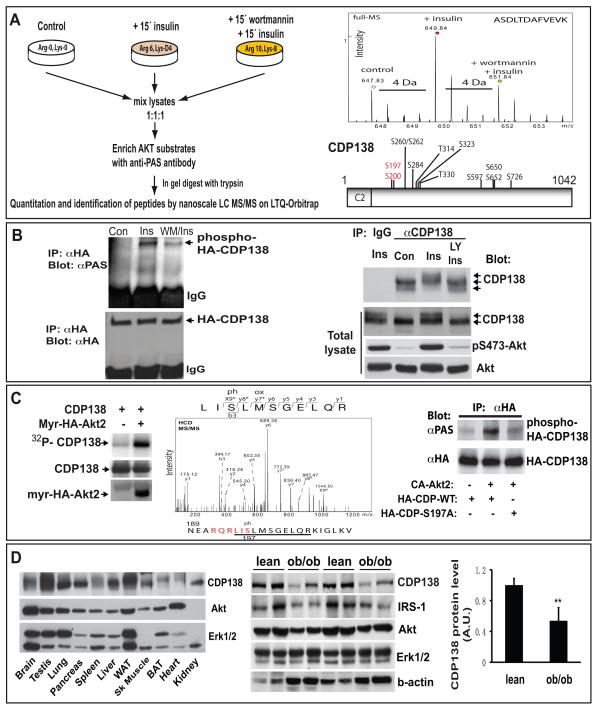
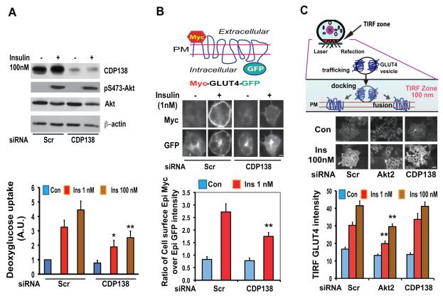
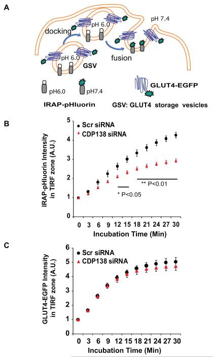
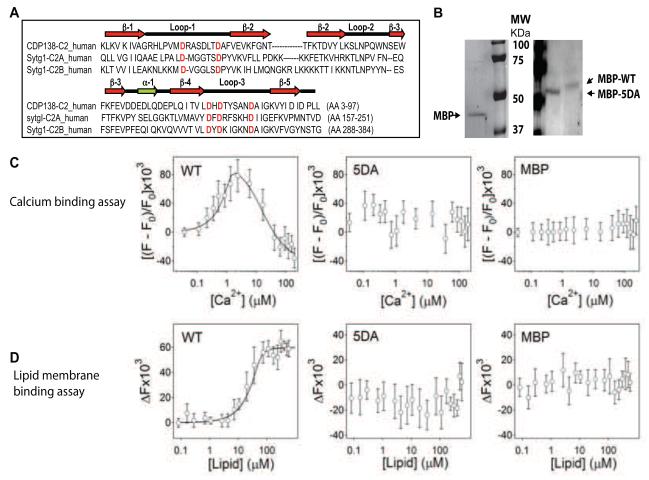
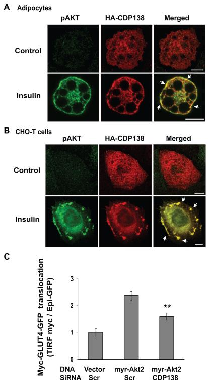
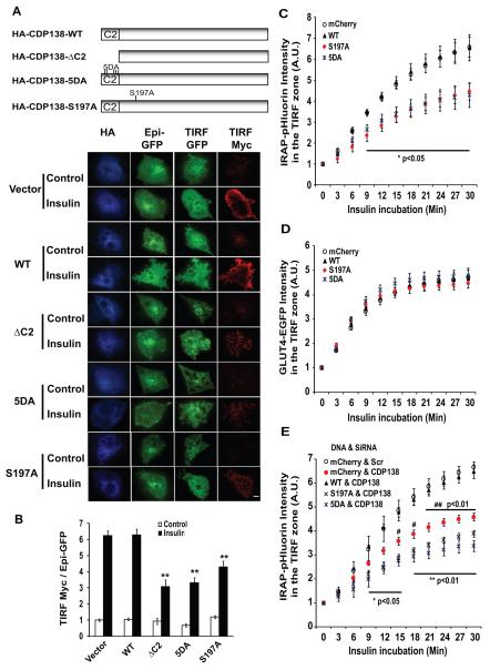
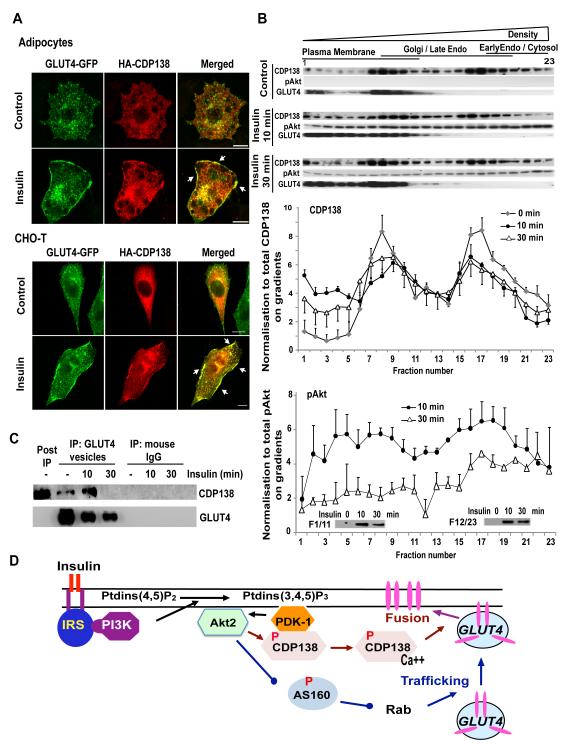
The protein kinase B(β) (Akt2) pathway is known to mediate insulin-stimulated glucose transport through increasing glucose transporter GLUT4 translocation from intracellular stores to the plasma membrane (PM). Combining quantitative phosphoproteomics with RNAi-based functional analyses, we show that a previously uncharacterized 138 kDa C2 domain-containing phosphoprotein (CDP138) is a substrate for Akt2, and is required for optimal insulin-stimulated glucose transport, GLUT4 translocation, and fusion of GLUT4 vesicles with the PM in live adipocytes. The purified C2 domain is capable of binding Ca(2+) and lipid membranes. CDP138 mutants lacking the Ca(2+)-binding sites in the C2 domain or Akt2 phosphorylation site S197 inhibit insulin-stimulated GLUT4 insertion into the PM, a rate-limiting step of GLUT4 translocation. Interestingly, CDP138 is dynamically associated with the PM and GLUT4-containing vesicles in response to insulin stimulation. Together, these results suggest that CDP138 is a key molecule linking the Akt2 pathway to the regulation of GLUT4 vesicle-PM fusion.


Figures

Figure 1
Figure 1. An uncharacterized C2 domain-containing protein encoded by 5730419I09Rik is a novel phosphoprotein identified in insulin-stimulated adipocytes using a SILAC phosphoproteomic approach
(A): Schematic procedure for SILAC quantitative proteomics used for identification and quantification of peptide from CDP138 (5730419I09Rik). Top right panel: quantification of CDP138 peptide from different groups of adipocytes. Lower right panel: Schematic diagram of CDP138 and the identified phosphorylation sites. (B): Confirmation of CDP138 phosphorylation induced by insulin. CHO-T cells expressing HA-CDP138, or differentiated 3T3-L1 adipocytes were treated with or without insulin (100 nM) for 15 min. A third sample was pretreated with wortmannin (100nM, WM) for 20 min or LY294002 (50 μM, LY) for 1 hour before insulin stimulation. Left panel: HA-CDP138 was immunoprecipitated with anti-HA Ab from CHO-T cells and blotted first with the phospho-Akt substrate PAS pAb (#9611, Lot 2, Cell Signaling), and then reblotted with an anti-HA Ab. Right panel: An anti-CDP138 peptide Ab was used for immunoprecipitation of CDP138 from cell lysates of adipocytes followed by immunoblotting with the same Ab. CDP138, pS473-Akt and Akt protein were also detected with total cell lysates. The arrows indicate the insulin-stimulated CDP138 mobility shift. (C): Constitutively active Akt2 (myr-HA-Akt2) directly phosphorylates HA-CDP138 in vitro kinase assays (left panel) and identification of Ser197 residue in CDP138 as the phosphorylation target of myr-HA-Akt2 with MS (middle panel) as described in S.I. Right panel: purified constitutively active Akt2 (Millipore) induces HA-CDP138-WT, but not HA-CDP-S197A, phosphorylation detected with PAS antibodies. (D): CDP138 protein expression in mouse tissues. Tissue protein extracts (25μg) from different tissues of lean C57BL/6J mice (left panel) or epididymal fat pads from ob/ob and lean male mice (24 weeks old, The Jackson Lab; middle panel) were analyzed by immunoblotting. Right panel: quantification of CDP138 protein levels in fat tissues from lean and obese mice. Data are mean ± SEM, ** P <0.01 lean vs ob/ob mice (n=4). WAT or BAT: white or brown adipose tissue. A, B & C are representatives of 2 to 3 independent experiments. See also Figure S1 and Table S1.
Figure 2
Figure 2. Knockdown of CDP138 in 3T3-L1 adipocytes inhibits insulin-stimulated glucose transport (A) and myc-GLUT4-GFP translocation (B), but not endogenous GLUT4 movement to the periphery detected in TIRF zone (C)
(A): Differentiated adipocytes at day 5 were transfected with siRNAs against mouse 5730419I09Rik or the scrambled siRNA (Scr) as described earlier (Jiang et al., 2003) for 60 hrs, then serum starved overnight. Cells were then treated with or without insulin (1 nM and 100nM) for 30 min for the glucose uptake assay, or 15 min for immunoblotting of CDP138, pAkt, Akt and β-actin. Glucose transport data are presented as mean ± SD of 4 independent experiments. * P < 0.05 vs Scr Insulin (1nM) group; ** P < 0.01 vs Scr Insulin (100nM) group. (B): Day 5 adipocytes were transfected with siRNAs and myc-GLUT4-GFP for 60 hrs and then serum starved overnight. Cells were then treated with insulin (1 nM) for 20 min. Cell surface myc-GLUT4-GFP was detected with anti-myc monoclonal Ab (9E10) and Alexa Fluor 568-labeled goat anti-mouse IgG in non-permeablized cells. The myc signal and GFP signal were quantified as previously described (Jiang et al., 2002). Data presented are representative microscopic images and mean ± SD of about 160 GFP-positive cells in each group from three independent experiments. ** P < 0.01 vs Scr Insulin (1 nM) group. (C): Endogenous GLUT4 accumulation in the TIRF zone in fixed adipocytes treated with or without insulin for 20 min. GLUT4 was detected with a goat anti-GLUT4 Ab and Alexa Fluor 488-conjugated donkey anti-goat Ab in permeabilized cells. Top and middle panels show 100nm TIRF zone and representative GLUT4 TIRFM images, respectively. Data are mean ± SD of 3 independent experiments with 300 plus cells in each group. ** P < 0.01 vs Scr Insulin groups. Scare Bars: 5 μm. See also Figure S2.
Figure 3
Figure 3. Knockdown of CDP138 in live 3T3-L1 adipocytes inhibits insulin-stimulated membrane fusion between GLUT4 storage vesicles (GSV) and the PM, but not GLUT4-EGFP trafficking to the TIRF zone
(A): Schematic illustration of the molecular probes and TIRF microcopy-based live cell assays for GLUT4 trafficking and GSV - PM fusion. (B) and (C): The effect of CDP138 knockdown on insulin-stimulated IRAP-pHluorin insertion into the PM (B), and accumulation of GLUT4-EGFP in the TIRF zone (C). Adipocytes (day 4) were transfected by electroporation with plasmid DNA encoding IRAP-pHluorin or GLUT4-EGFP, together with either the scrambled siRNA or smartPool siRNA against mouse 5730419I09Rik. Cells were reseeded for 72 hrs, serum starved for 2 hr then stimulated with 100 nM insulin for 30 min. Analyses were performed in a cell warmer adapted for a Nikon TiE with fully motorized combined dual laser (488 and 561nm). Images were acquired every 3 min immediately after addition of insulin and analyzed as described in the S.I.. Perfect focus system and multiple points capture program were used to acquire images from multiple positively transfected cells at each time point. Data are mean ± SEM of three independent experiments (n=3) with total 159 cells (43,46,70/exp, Scr siRNA) or 154 cells (43, 37, 74/exp, CDP138 siRNA) in the GLUT4-EGFP trafficking assay; and 125 cells (41, 48, 36/exp, Scr siRNA) or 120 cells (43, 46, 31/exp, CDP138 siRNA) in the GSV - PM fusion assay. P value: CDP138 siRNAs vs scrambled siRNA. Live cell movies for IRAP-pHluorin membrane fusion assay are provided in S.I. See also Figure S3, Movie S1 and Movie S2.
Figure 4
Figure 4. The purified C2 domain from CDP138 is capable of binding Ca2+ ions and lipid membranes
(A): Diagram and amino acid alignment of the C2 domains of CDP138 (CDP138-C2) and synaptotagmin-1 (Sytg1-C2A & Sytg-C2B). β: beta strands; α: alpha helix; loop: coil loop. The conserved potential Ca2+-binding aspartate residues are highlighted. (B): Gel images of purified MBP (42kDa), MBP-C2-WT domain, and MBP-C2-5DA mutant. (C): Calcium binds to the wild-type C2-domain but not to the 5DA or the MBP proteins. Change in tryptophan fluorescence intensity at 340 nm as a signal of calcium interaction with the MBP-C2-WT fusion protein, MBP-C2-5DA mutant fusion protein, and MBP measured at 37°C. The calcium-binding isotherm was constructed as described in S.I.. Calcium exerts a biphasic effect on tryptophan fluorescence of the wild-type protein, but has little effect on the 5DA or MBP proteins. The curve of calcium binding to MBP-C2-WT was constructed using two independent binding sites per wild-type C2 domain, with dissociation constants of KD,1 = 0.03±0.012 μM and KD,2 = 15.0±6 μM. (D): Fluorescence resonance energy transfer (RET) from protein tryptophan residues to Py-PE in membrane indicates membrane binding by the MBP-C2 WT fusion protein but not by the MBP-5DA-C2 mutant or MBP proteins. Change in tryptophan fluorescence intensity as a function of lipid concentration, corrected for the effect of membranes without energy acceptor Py-PE, measured at 37°C. The solid line describing membrane binding of MBP-C2-WT was simulated using a lipid-to-protein stoichiometry N = 20 and a dissociation constant KD = 0.06±0.015 μM. All data is presented as mean ± SD from three independent experiments. See also Figure S4.
Figure 5
Figure 5. CDP138 co-localizes with phospho-Akt (A & B), and is required for constitutively active myr-Akt2-induced GLUT4 translocation (C)
A & B: HA-CDP138-WT was transfected into adipocytes (A) and CHO-T cells (B) for 48 hrs before serum starvation overnight. Cells were then treated with or without insulin (100nM) for 10 min. Cells were fixed and permeabilized before immunostaining with mouse anti-HA and rabbit anti-phospho-Akt (S473) antibodies followed by goat anti-mouse (Alexa Fluor568) and goat anti-rabbit (Alexa Fluor488) secondary antibodies, respectively. The white arrow indicates co-localization of phospho-Akt and HA-CDP138. Scale bar: 10μm. C: Differentiated adipocytes were transfected by electroporation with the scrambled siRNA or CDP138 siRNAs together with plasmid DNAs encoding myc-GLUT4-GFP and myr-HA-Akt2 or HA-empty vector. Cells were reseeded for 60 hours before serum starvation overnight. Myc-GLUT4 translocation assays were carried out with TIRF microscopy as described in the Experimental Procedures. Data are mean ± SEM of four independent experiments. ** P< 0.01 myr-Akt2 / CDP138 siRNA vs myr-Akt2 / Scr siRNA.
Figure 6
Figure 6. The effects of CDP138-ΔC2, CDP138-5DA and CDP138-S197A mutants on myc-GLUT4-GFP translocation and GSV-PM fusion in 3T3-L1 adipocytes
Plasmid DNAs for pCMV5-HA, HA-CDP138-WT, HA-CDP138-ΔC2, HA-CDP138-5DA, or HA-CDP138-S197A were transfected by electroporation into adipocytes together with the myc-GLUT4-GFP expression vector. Cells were reseeded for 48 hrs and serum-starved overnight. Cells were then treated with or without insulin (100nM) for 30 min, before immunostaining with rabbit anti-myc and mouse anti-HA antibodies. (A): Schematic diagram for CDP138 (wild-type and mutants) and representative images of the myc-GLUT4-GFP translocation assay using TIRF microscopy. Scare Bars: 5 μm. (B): GLUT4 translocation to the cell surface of fixed adipocytes is shown as the ratio of surface TIRF myc signal to total Epi GFP. Data are presented as mean ± SEM of three independent experiments. ** P< 0.01 (n=3); ΔC2, 5DA, or S197A vs HA vector. (C) & (D): The effects of CDP138-mCherry constructs on insulin-induced GSV - PM fusion and GLUT4-EGFP trafficking in live adipocytes, respectively. Data are expressed in arbitrary units as the ratio of pHluorin or EGFP intensity to the basal intensity at time zero. Data are mean ± SEM of three independent experiments (total 65 to 137 cells each group). P value (n=3): S197A or 5DA vs mCherry vector. (E): Overexpression of human WT but not mutant CDP138-mCherry rescures siRNA-induced inhibition of GSV - PM fusion in live adipocytes. Adipocytes (day 4) were transfected with the scrambled siRNA or smartPool siRNA against mouse 5730419I09Rik together with plasmid DNAs encoding IRAP-pHluorin and mCherry alone, CDP138-WT-mCherry, CDP138-S197A-mCherry or CDP138-5DA-mCherry for 72 hrs before GSV – PM fusion assay. Data are mean ± SEM of three independent experiments (total 137 to 153 cells each group). P value (n=3): * and ** CDP138 siRNA plus S197A or 5DA vs Scr siRNA plus mCherry vector. # P < 0.05 or ## P < 0.01: CDP138 siRNA plus mCherry vector vs Scr siRNA plus mCherry vector. See also Figure S5.
Figure 7
Figure 7. CDP138 is colocalized with GLUT4 at the PM (A) and dynamically associated with the PM fractions (B) and GLUT4 vesicles (C)
HA-CDP138-WT plasmid DNA was transfected alone into a CHO-T cell line stably expressing myc-GLUT4-GFP, or into adipocytes together with the myc-GLUT4-GFP vector. Forty-eight hr later, serum starved cells were treated with or without insulin for 10 min before immunofluorescent staining with anti-HA Ab, as described for Figure 5. Scare Bars: 10 μm. (B): Post nuclear supernatants from 3T3-L1 adipocytes treated with or without insulin 100nM were fractionated with iodixanol gradients (10-20-30% Optiprep) as described in S.I. Equal volumes of each fraction were resolved in 4-20% SDS-PAGE followed by immunoblotting with antibodies against CDP138, p-Akt and GLUT4. For quantification, the results were normalized to the total level of the indicated protein. Data are mean ± SEM of 3 independent experiments. (C): GLUT4 vesicles were enriched by immunoabsorption with anti-GLUT4 Ab (1F8) from adipocytes after removal of nuclear fraction as described in S.I. Samples were then immunoblotted with anti-CDP138 and anti-GLUT4 antibodies. Images are representative of three independent experiments (A, B & C). (D): Akt substrate CDP138 is a potential link between Akt2 activation and GSV - PM fusion. See also Figure S6.

Similar articles

Cited by

References

    1. Alessi DR, Caudwell FB, Andjelkovic M, Hemmings BA, Cohen P. Molecular basis for the substrate specificity of protein kinase B; comparison with MAPKAP kinase-1 and p70 S6 kinase. FEBS Lett. 1996;399:333–338. - PubMed
    1. Alessi DR, James SR, Downes CP, Holmes AB, Gaffney PR, Reese CB, Cohen P. Characterization of a 3-phosphoinositide-dependent protein kinase which phosphorylates and activates protein kinase Balpha. Curr Biol. 1997;7:261–269. - PubMed
    1. Bai J, Chapman ER. The C2 domains of synaptotagmin--partners in exocytosis. Trends Biochem Sci. 2004;29:143–151. - PubMed
    1. Bai L, Wang Y, Fan J, Chen Y, Ji W, Qu A, Xu P, James DE, Xu T. Dissecting multiple steps of GLUT4 trafficking and identifying the sites of insulin action. Cell Metab. 2007;5:47–57. - PubMed
    1. Bryant NJ, Govers R, James DE. Regulated transport of the glucose transporter GLUT4. Nat Rev Mol Cell Biol. 2002;3:267–277. - PubMed

Publication types

MeSH terms