. 2011 Dec 30;286(52):44821-32.
doi: 10.1074/jbc.M111.249177. Epub 2011 Sep 6.

The vitamin K-dependent carboxylase generates γ-carboxylated glutamates by using CO2 to facilitate glutamate deprotonation in a concerted mechanism that drives catalysis

Affiliations

The vitamin K-dependent carboxylase generates γ-carboxylated glutamates by using CO2 to facilitate glutamate deprotonation in a concerted mechanism that drives catalysis

Mark A Rishavy et al. J Biol Chem. .

Abstract

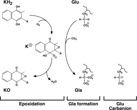
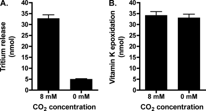
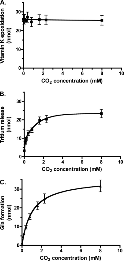
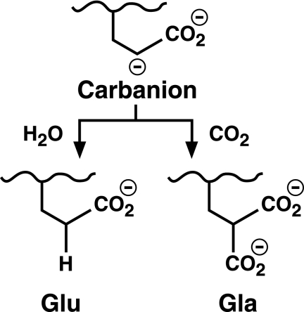
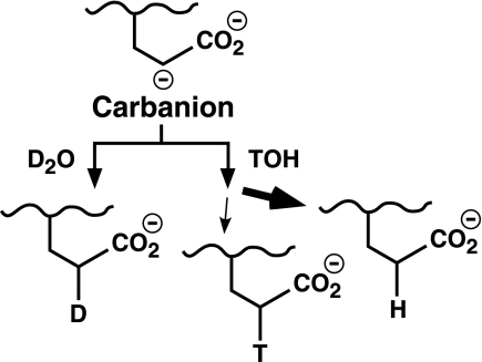
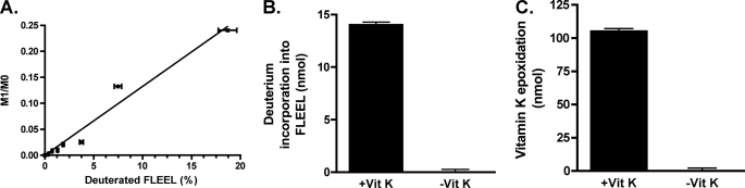
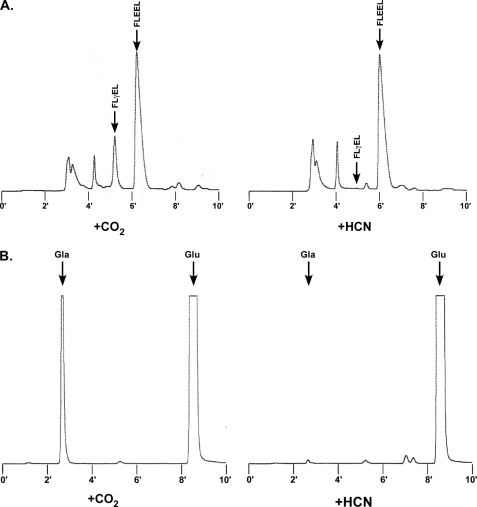
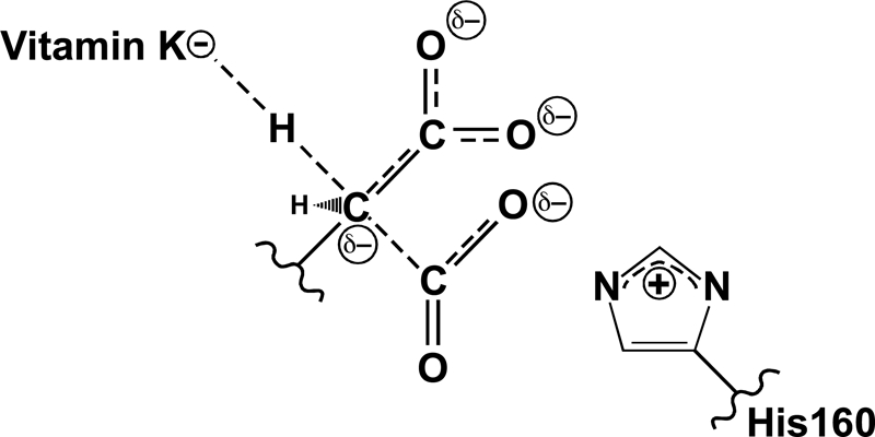
The γ-glutamyl carboxylase converts Glu to carboxylated Glu (Gla) to activate a large number of vitamin K-dependent proteins with diverse functions, and this broad physiological impact makes it critical to understand the mechanism of carboxylation. Gla formation is thought to occur in two independent steps (i.e. Glu deprotonation to form a carbanion that then reacts with CO(2)), based on previous studies showing unresponsiveness of Glu deprotonation to CO(2). However, our recent studies on the kinetic properties of a variant enzyme (H160A) showing impaired Glu deprotonation prompted a reevaluation of this model. Glu deprotonation monitored by tritium release from the glutamyl γ-carbon was dependent upon CO(2), and a proportional increase in both tritium release and Gla formation occurred over a range of CO(2) concentrations. This discrepancy with the earlier studies using microsomes is probably due to the known accessibility of microsomal carboxylase to water, which reprotonates the carbanion. In contrast, tritium incorporation experiments with purified carboxylase showed very little carbanion reprotonation and consequently revealed the dependence of Glu deprotonation on CO(2). Cyanide stimulated Glu deprotonation and carbanion reprotonation to the same extent in wild type enzyme but not in the H160A variant. Glu deprotonation that depends upon CO(2) but that also occurs when water or cyanide are present strongly suggests a concerted mechanism facilitated by His-160 in which an electrophile accepts the negative charge on the developing carbanion. This revised mechanism provides important insight into how the carboxylase catalyzes the reaction by avoiding the formation of a high energy discrete carbanion.


Figures

FIGURE 1.
FIGURE 1.
The carboxylase reaction. The carboxylase is a bifunctional enzyme that converts KH2 to vitamin K epoxide (KO) and Glu residues to Gla. A highly basic vitamin K intermediate (K) (e.g. the alkoxide that is shown) is proposed to abstract a hydrogen from the glutamyl γ-carbon to form a carbanion that reacts with CO2 to generate Gla. Previous studies (26) led to a model in which the formation of a discrete Glu carbanion (shown on the right) and the reaction of the carbanion with CO2 to form Gla are two independent steps. However, the results presented here show that Glu deprotonation depends upon CO2 and suggest that Gla formation occurs in a single step.
FIGURE 2.
FIGURE 2.
Analysis of purified carboxylase. Wild type carboxylase and an H160A mutant with C-terminal FLAG tags were affinity-purified using immobilized anti-FLAG antibody, and 1 μg of each protein was then subjected to SDS-PAGE and Coomassie staining.
FIGURE 3.
FIGURE 3.
Removal of endogenous CO2 from carboxylase reaction mixtures. A, Gla formation is usually quantitated by a radioactivity assay that measures [14C]CO2 incorporation into Glu-containing substrate; however, this assay can be confounded by the presence of endogenous CO2, and we therefore developed an alternative method for quantitating Gla. Reactions were performed and then chromatographed on P-2 columns to isolate peptide and remove interfering substances. HPLC was then used to separate the carboxylated product (FLγEL) from FLEEL substrate. The FLγEL peptide was hydrolyzed with base, and Gla was derivatized with o-phthalaldehyde and quantitated using HPLC and fluorescence detection. An example of the resolution of FLγEL and FLEEL by HPLC is shown. B, analysis of reaction mixtures without exogenously added CO2 revealed significant levels of carboxylation due to endogenous CO2 (lane 1). Gassing the samples with a 1:1 mixture of nitrogen and oxygen, either over the headspace in a sealed vial (lane 2) or directly in the sample (lane 3) reduced but did not eliminate Gla formation. When the samples were gassed in the sample at pH 5.5 and then adjusted to pH 6.9 and reacted, carboxylation was barely detectable (lane 4). Error bars, S.E.
FIGURE 4.
FIGURE 4.
CO2 impacts the level of Glu deprotonation. Wild type carboxylase was incubated in reaction mixtures containing a peptide substrate with Glu tritiated at the γ-carbon position (FL-[R,S-3H]EEL), either in the presence or absence of CO2, and the release of tritium to solvent was measured (A). Aliquots of each reaction were also withdrawn to determine the level of vitamin K epoxidation (B). Error bars, S.E.
FIGURE 5.
FIGURE 5.
Glu deprotonation and Gla formation show proportional increases over a range of CO2 concentrations. Reactions were performed using carboxylase, FL-[R,S-3H]EEL, KH2, and the indicated concentrations of CO2. At the end of the reaction, aliquots were withdrawn to determine the level of vitamin K epoxidation (A), and the remainder of the reaction mixtures was used to measure the release of tritium from peptide into solvent (B). To monitor Gla formation (C), a separate set of reactions was also performed in which FL-[R,S-3H]EEL was replaced by FLEEL. Vitamin K epoxidation was measured as above and gave values that were essentially the same as those shown in A. Peptide product (FLγEL) isolated from the remaining reaction mixture was base-hydrolyzed, and Gla was quantitated using HPLC and fluorescence detection. The lines in B and C were fit using GraphPad Prism (version 4.0), and the details for the assays are described under “Experimental Procedures.” Error bars, S.E.
SCHEME 1.
SCHEME 1.
Alternative consequences of Glu deprotonation.
FIGURE 6.
FIGURE 6.
FLEEL carboxylation is undetectable in the absence of CO2. Reactions containing T2O but no added CO2 were performed, and peptide was isolated using P-2 chromatography followed by HPLC, which showed that very little carboxylation and generation of FLγEL product occurred (e.g. compare with Fig. 3A). The FLEEL peptide was collected and subjected to scintillation counting to determine the amount of tritium incorporation due to Glu reprotonation. The elution time for the FLEEL peptide is different from that shown in Fig. 3A due to the use of different mobile phases.
SCHEME 2.
SCHEME 2.
Glu reprotonation in deuterium oxide or tritiated water.
FIGURE 7.
FIGURE 7.
Measuring Glu reprotonation in a deuterium-containing reaction. A, to establish a standard curve for quantitating the amount of deuterium incorporation into FLEEL, increasing amounts of FL-[R,S-2H]EEL were mixed with FLEEL to give final concentrations of 1 μm peptide, and the samples were then analyzed by mass spectroscopy. M0, area under the curve measured for the most abundant isotopic forms (e.g. 1H and 12C); M1, those that are one mass unit higher (e.g. 2H and 13C). The M1/M0 ratio has been adjusted to account for the natural abundance of isotopes in FLEEL. B, FLEEL was carboxylated in the presence or absence of vitamin K hydroquinone (vit K) in reaction mixtures where all of the components were prepared in D2O. The amount of deuterium incorporation into FLEEL was quantitated by mass spectroscopy and a comparison with the standard curve shown in A. C, an aliquot of the reaction was also quantitated for vitamin K epoxidation. Error bars, S.E.
FIGURE 8.
FIGURE 8.
Cyanide inhibition of carboxylase activity. Wild type (WT) carboxylase (A) and an H160A mutant (B) were each incubated in a reaction mixture containing FLEEL, KH2, [14C]bicarbonate, and increasing concentrations of KCN, and Gla formation was determined by measuring the incorporation of [14C]CO2 into FLEEL by scintillation counting. The lines were fit using GraphPad Prism (version 4.0). Error bars, S.E.
FIGURE 9.
FIGURE 9.
Measuring the effects of cyanide on Glu reprotonation and Gla formation. A, to measure Glu reprotonation, reactions were performed with mixtures containing the wild type carboxylase, FLEEL, KH2, tritiated water, and either CO2 or cyanide. The peptides were isolated by P-2 chromatography and then HPLC, which showed that cyanide abolished carboxylation (i.e. generation of the FLγEL product). The FLEEL peptide was collected and quantitated for tritium incorporation by scintillation counting. B, to assess the effect of cyanide on Gla formation, reactions were performed as in A but without T2O. The FLγEL product was isolated as in A and subjected to base hydrolysis and o-phthalaldehyde derivatization, followed by HPLC and fluorescence detection to quantitate Gla, Glu, and Phe/Leu (not shown in the chromatogram). The details of these assays are described under “Experimental Procedures.”
FIGURE 10.
FIGURE 10.
A concerted mechanism for Gla formation. Previous studies showed that His-160 is required for Glu deprotonation (27), and the present studies reveal that CO2 is also required. Water and cyanide can both substitute for CO2 in supporting Glu deprotonation, which, as detailed under “Discussion,” strongly suggests a chemical role for CO2. This function is most likely the dispersal of charge that develops on the glutamyl γ-carbon during deprotonation by the vitamin K base. Thus, Glu deprotonation and CO2 addition occur simultaneously in a concerted mechanism of carboxylation. Reactivity of the mutant H160A carboxylase toward CO2 and cyanide suggests that His-160 facilitates Glu deprotonation through interaction with CO2.

Similar articles

Cited by

References

    1. Berkner K. L. (2005) Annu. Rev. Nutr. 25, 127–149 - PubMed
    1. Morris D. P., Stevens R. D., Wright D. J., Stafford D. W. (1995) J. Biol. Chem. 270, 30491–30498 - PubMed
    1. Stenina O., Pudota B. N., McNally B. A., Hommema E. L., Berkner K. L. (2001) Biochemistry 40, 10301–10309 - PubMed
    1. Brenner B., Sánchez-Vega B., Wu S. M., Lanir N., Stafford D. W., Solera J. (1998) Blood 92, 4554–4559 - PubMed
    1. Darghouth D., Hallgren K. W., Shtofman R. L., Mrad A., Gharbi Y., Maherzi A., Kastally R., LeRicousse S., Berkner K. L., Rosa J. P. (2006) Blood 108, 1925–1931 - PMC - PubMed

Publication types

LinkOut - more resources