. 2011 Nov;193(21):5971-84.
doi: 10.1128/JB.05405-11. Epub 2011 Sep 2.

The surfactant of Legionella pneumophila Is secreted in a TolC-dependent manner and is antagonistic toward other Legionella species

Affiliations

The surfactant of Legionella pneumophila Is secreted in a TolC-dependent manner and is antagonistic toward other Legionella species

Catherine R Stewart et al. J Bacteriol. 2011 Nov.

Abstract

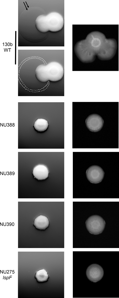
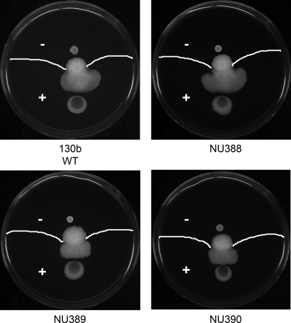
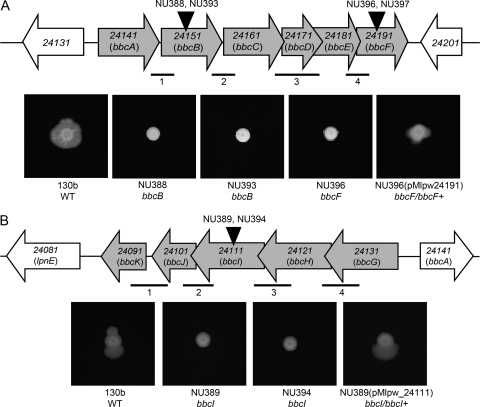
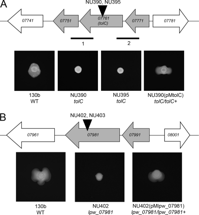
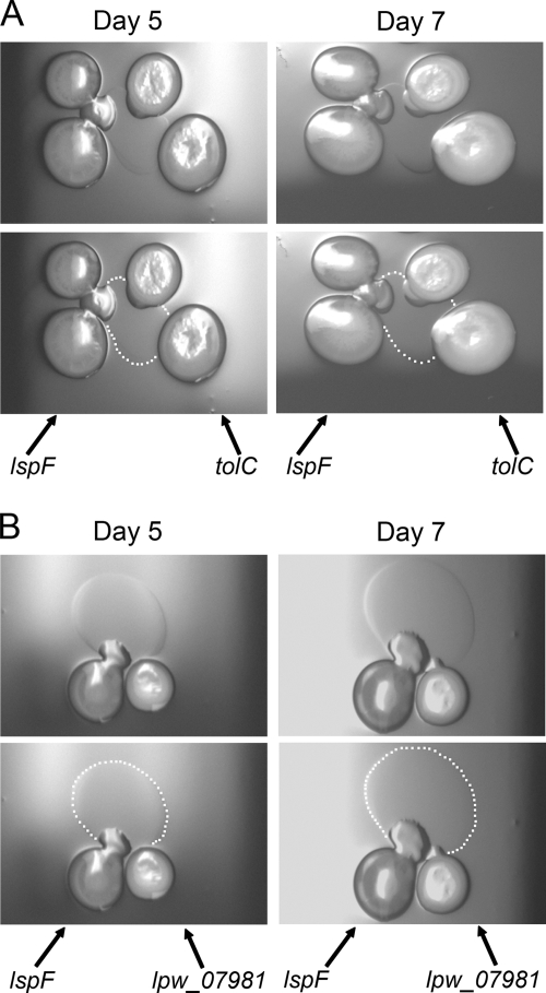
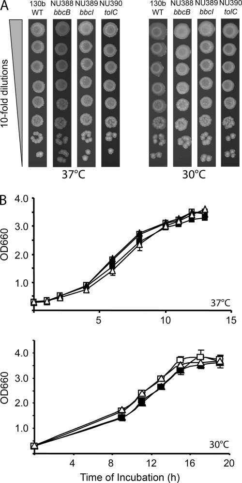
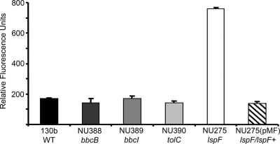
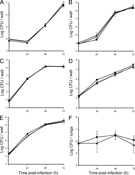
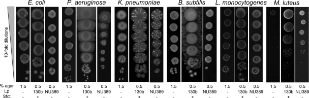
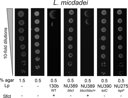
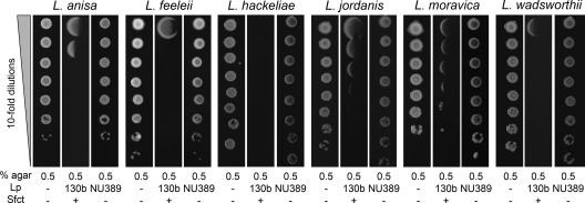
When Legionella pneumophila grows on agar plates, it secretes a surfactant that promotes flagellum- and pilus-independent "sliding" motility. We isolated three mutants that were defective for surfactant. The first two had mutations in genes predicted to encode cytoplasmic enzymes involved in lipid metabolism. These genes mapped to two adjacent operons that we designated bbcABCDEF and bbcGHIJK. Backcrossing and complementation confirmed the importance of the bbc genes and suggested that the Legionella surfactant is lipid containing. The third mutant had an insertion in tolC. TolC is the outer membrane part of various trimolecular complexes involved in multidrug efflux and type I protein secretion. Complementation of the tolC mutant restored sliding motility. Mutants defective for an inner membrane partner of TolC also lacked a surfactant, confirming that TolC promotes surfactant secretion. L. pneumophila (lspF) mutants lacking type II protein secretion (T2S) are also impaired for a surfactant. When the tolC and lspF mutants were grown next to each other, the lsp mutant secreted surfactant, suggesting that TolC and T2S conjoin to mediate surfactant secretion, with one being the conduit for surfactant export and the other the exporter of a molecule that is required for induction or maturation of surfactant synthesis/secretion. Although the surfactant was not required for the extracellular growth, intracellular infection, and intrapulmonary survival of L. pneumophila, it exhibited antimicrobial activity toward seven other species of Legionella but not toward various non-Legionella species. These data suggest that the surfactant provides L. pneumophila with a selective advantage over other legionellae in the natural environment.


Figures

Fig. 1.
Fig. 1.
L. pneumophila mutants that lack surfactant and sliding motility. Wild-type (WT) strain 130b, mini-Tn10-containing mutants NU388, NU389, and NU390, and type II secretion (lspF) mutant NU275 were grown on BCYE plates containing 0.5% agar at 30°C. Imaged after 4 days, the left panel depicts, in the case of the WT but not the mutants, the early stage of surface translocation and a surfactant film front displaying two delineating rings. To aid in visualization of the surfactant film, two images are presented for the WT, with the upper one showing the delineating rings marked by two black arrows and the lower one having white dots mark the boundary of the film. Imaged after 10 days, the right panel shows the progression of surface translocation by the WT and its continued absence for the mutants. The images presented are representative of those observed for at least 12 experiments.
Fig. 2.
Fig. 2.
Surface translocation by L. pneumophila mutants when spotted onto film produced by wild-type legionellae. WT 130b was inoculated onto the center of a BCYE plate containing 0.5% agar at 30°C. After 10 days, when a layer of surface film was clearly evident beyond the spreading legionellae, a 10-μl aliquot of the WT, mutant NU388, mutant NU389, and mutant NU390 was spotted onto the agar surface either in the surfactant-covered area (“+”) or at a point outside of it but still equidistant from the center of WT growth (“−” area). After 3 days of further incubation, images were taken in order to visualize any differences in spreading. The white lines show the film front. The images are representative of three independent experiments.
Fig. 3.
Fig. 3.
L. pneumophila bbcABCDEF and bbcGHIJK operons and their impact on surfactant and surface translocation. (A, top) Depiction of the region of the strain 130b chromosome containing genes lpw_24141 through lpw_24191, i.e., the bbcABCDEF operon. The horizontal gray arrows denote the relative size and orientation of the bbc genes, whereas the horizontal white arrows denote the two flanking genes. The vertical black arrowheads mark the approximate locations of the insertion mutations in mutants NU388, NU393, NU396, and NU397. The thinner horizontal lines below the gene map signify the approximate size and location of the four intergenic transcripts identified by RT-PCR analysis. (A, bottom) Surface translocation and surfactant phenotypes of bbcB and bbcF mutants. WT 130b, bbcB surfactant mutants NU388 and NU393, bbcF surfactant mutant NU396, and complemented mutant NU396(pMlpw24191) were grown on BCYE plates containing 0.5% agar at 30°C for 7 days. The bbcF mutant NU397 also consistently lacked surface translocation and surfactant (data not shown). (B, top) Depiction of the region of the strain 130b chromosome containing genes lpw_24131 through lpw_24091, i.e., the bbcGHIJK operon. The black arrowhead marks the location of the insertion in mutants NU389 and NU394, and the thinner horizontal lines signify the four intergenic transcripts identified by RT-PCR analysis. (B, bottom) Surface translocation and surfactant phenotypes of L. pneumophila bbcI mutants. WT 130b, bbcI surfactant mutants NU389 and NU394, and complemented mutant NU389(pMlpw_24111) were grown on BCYE plates containing 0.5% agar at 30°C for 7 days. The images of bacterial growth presented in panels A and B are representative of those obtained from at least three independent experiments.
Fig. 4.
Fig. 4.
L. pneumophila tolC and lpw_07981 and their impact on surfactant and surface translocation. (A, top) Depiction of the region of the 130b chromosome containing genes lpw_07771, lpw_07761, and lpw_07751; i.e., the tolC operon. The horizontal gray arrows denote the size and orientation of the tolC operon, and the white arrows denote the two flanking genes. The vertical black arrowheads mark the locations of the insertion mutations in mutants NU390 and NU395. The thinner horizontal lines below the gene map signify the size and location of the two intergenic transcripts identified by RT-PCR. (A, bottom) Surface translocation and surfactant phenotypes of tolC mutants. WT 130b, tolC mutants NU390 and NU395, and complemented mutant NU390(pMtolC) were grown on BCYE plates containing 0.5% agar and 0.1 mM IPTG (isopropyl-β-d-thiogalactopyranoside) at 30°C for 7 days. (B, top) Depiction of the region of the 130b chromosome containing genes lpw_07991 and lpw_07981. The black arrowhead marks the locations of the insertions in lpw_07981 mutants NU402 and NU403. In the 130b database, the lpw_07981 ORF is also (erroneously) denoted lpw_07971. (B, bottom) Surface translocation and surfactant phenotypes of lpw_07981 mutants. WT 130b, lpw_07981 mutant NU402, and complemented mutant NU402(pMlpw_07981) were grown on BCYE plates containing 0.5% agar at 30°C for 7 days. The lpw_07981 mutant NU403 also consistently lacked surface translocation and surfactant (data not shown). The images presented in panels A and B are representative of those obtained from at least three independent experiments.
Fig. 5.
Fig. 5.
Surfactant secretion by an L. pneumophila lspF mutant when grown in the presence of an L. pneumophila tolC or lpw_07981 mutant. (A) As indicated by the arrows, two inocula of the lspF mutant NU275 and two inocula of the tolC mutant NU390 were spotted next to each other on a BCYE plate containing 0.5% agar. After incubation at 30°C for 5 days or 7 days, bacterial growth and surface translocation were imaged (top row). To help with visualization of the surfactant that was produced by the sliding lspF mutant, white dots have been added to mark the boundary of the spreading film (bottom row). The surfactant film was also evident when single spots of the two mutants were placed next to each other (data not shown). (B) As indicated, lspF mutant NU275 and lpw_07981 mutant NU402 were spotted next to each other on low-agar BCYE plates, and then after incubation at 30°C for 5 or 7 days, bacterial growth, surface translocation, and surfactant production were imaged. As in panel A, the bottom images depict the boundary of the film produced by the lspF mutant with white dots. The results depicted in panels A and B are representative of those obtained from three independent experiments.
Fig. 6.
Fig. 6.
Extracellular growth of the L. pneumophila wild type and bbcB, bbcI, and tolC mutants. (A) Growth of legionellae on BCYE agar. We spotted dilutions of WT 130b, bbcB mutant NU388, bbcI mutant NU389, and tolC mutant NU390 onto standard BCYE agar and incubated the plates at various temperatures. Bacterial growth was then imaged at 3 days for the 37°C plates and 4 days for the 30°C plates. (B) Growth of legionellae in BYE broth. WT 130b (▪), bbcB mutant NU388 (□), bbcI mutant NU389 (▴), and tolC mutant NU390 (▵) were inoculated into BYE broth and then incubated at 37°C or 30°C. At various times postinoculation, the extent of growth was monitored spectrophotometrically. The data points represent the means and standard deviations for triplicate cultures. The results presented in both panels A and B are representative of three independent experiments.
Fig. 7.
Fig. 7.
PHB in the L. pneumophila wild type and bbcB, bbcI, tolC, and lspF mutants. WT 130b, bbcB mutant NU388, bbcI mutant NU389, tolC mutant NU390, lspF mutant NU275, and complemented NU275(pMF) were grown in BYE to early log phase. The cells were then subcultured into CDM and grown at 37°C. After staining with Nile red, fluorescence was measured using a spectrophotometer blanked with unstained bacteria. Data are the means and standard deviations for three replicates obtained from duplicate cultures. The levels of PHB exhibited by the NU275 mutant were significantly increased compared to those for the other strains (P < 0.05, Student's t test). The results shown here are representative of at least four independent experiments.
Fig. 8.
Fig. 8.
Intracellular and intrapulmonary growth of the L. pneumophila wild type and bbcB and bbcI mutants. (A to E) H. vermiformis (A), A. castellanii (B), U937 macrophages (C), A549 type II epithelial cells (D), and WI-26 type I epithelial cells (E) were infected with WT 130b (▪), bbcB mutant NU388 (□), and bbcI mutant NU389 (▴), and then at the indicated times, the numbers of CFU in the infected monolayers were determined by plating. Data are the means and standard deviations for four infected wells. Each panel is representative of three independent experiments. (F) WT 130b (▪) and bbcI mutant NU389 (▴) were inoculated into the lungs of A/J mice, and then after 24, 48, and 72 h, the numbers of CFU in total lung homogenates were determined by plating. Data are the means and standard deviations for five infected animals. While there might appear to be no growth and reduced recovery of the mutant at 24 h, this result was not statistically significant and was not obtained in the repeat experiment (data not shown).
Fig. 9.
Fig. 9.
Effect of the L. pneumophila surfactant on heterologous bacteria. As indicated, we spotted 10-fold dilutions of E. coli, P. aeruginosa, K. pneumoniae, B. subtilis, L. monocytogenes, and M. luteus onto BCYE agar plates that were free of L. pneumophila (Lp −), contained a nearby row of WT L. pneumophila that had grown and made a surfactant film (Lp 130b), or contained a row of a bbcI mutant that had grown but not produced a surfactant (Lp NU389). After incubation at 37°C for another day, the growth of the heterologous bacteria in the absence (Sfct −) or presence (Sfct +) of a surfactant was imaged. The results depicted here were observed in at least three independent experiments. On BCYE agar not containing L. pneumophila, the efficiencies of plating for these bacteria were the same whether the medium contained 0.5% agar or 1.5% agar, although plating on low-percentage-agar BCYE did result in greater spreading for those bacteria, such as P. aeruginosa, that are known to make a surfactant and/or undergo surface translocation (see Fig. S3 in the supplemental material).
Fig. 10.
Fig. 10.
Effect of the L. pneumophila surfactant on the growth of L. micdadei. For the two leftmost panels, we spotted dilutions of L. micdadei onto BCYE plates containing either 1.5% agar or 0.5% agar but no nearby L. pneumophila (Lp −). For the next five panels, we spotted L. micdadei onto 0.5% agar BCYE containing a nearby row of WT L. pneumophila that had grown and made a surfactant film (Lp 130b; third panel), a row of a bbcI mutant that had grown but not produced a surfactant (Lp NU389; fourth panel), a row of the complemented bbcI mutant that had restored surfactant production [Lp NU389(pMlpw_24111); fifth panel], a row of a tolC mutant that had grown but not made a surfactant (Lp NU390; sixth panel), or a row of an lspF mutant that had grown but lacked a surfactant (Lp NU275; last panel). After incubation at 37°C for another 3 days, growth of L. micdadei in the absence (Sfct −) or presence (Sfct +) of a surfactant was imaged. The results depicted here were observed in at least three independent experiments.
Fig. 11.
Fig. 11.
Effect of the L. pneumophila surfactant on the growth of L. anisa, L. feeleii, L. hackeliae, L. jordanis, L. moravica, and L. wadsworthii. For the leftmost panel, we spotted dilutions of the indicated strains onto BCYE plates containing 0.5% agar but no nearby L. pneumophila (Lp −). For the next two panels, we spotted the bacteria onto 0.5% agar BCYE containing either a nearby row of WT L. pneumophila that had grown and made a surfactant film (Lp 130b; second panel) or a row of a bbcI mutant that had grown but not produced a surfactant (Lp NU389; third panel). After incubation at 37°C for another 3 days, growth of the legionellae in the absence (Sfct −) or presence (Sfct +) of a surfactant was imaged. The results depicted here were observed in at least three independent experiments.

Similar articles

Cited by

References

    1. Abdel-Mawgoud A. M., Lepine F., Deziel E. 2010. Rhamnolipids: diversity of structures, microbial origins and roles. Appl. Microbiol. Biotechnol. 86:1323–1336 - PMC - PubMed
    1. Agusti G., Astola O., Rodriguez-Guell E., Julian E., Luquin M. 2008. Surface spreading motility shown by a group of phylogenetically related, rapidly growing pigmented mycobacteria suggests that motility is a common property of mycobacterial species but is restricted to smooth colonies. J. Bacteriol. 190:6894–6902 - PMC - PubMed
    1. Allard K. A., et al. 2009. Purification of legiobactin and the importance of this siderophore in lung infection by Legionella pneumophila. Infect. Immun. 77:2887–2895 - PMC - PubMed
    1. Allard K. A., Viswanathan V. K., Cianciotto N. P. 2006. lbtA and lbtB are required for production of the Legionella pneumophila siderophore legiobactin. J. Bacteriol. 188:1351–1363 - PMC - PubMed
    1. Aragon V., Kurtz S., Cianciotto N. P. 2001. Legionella pneumophila major acid phosphatase and its role in intracellular infection. Infect. Immun. 69:177–185 - PMC - PubMed

Publication types

MeSH terms

Substances