

Trichoscopy in genetic hair shaft abnormalities
- PMID: 21886705
- PMCID: PMC3157768
- DOI: 10.3315/jdcr.2008.1009
Trichoscopy in genetic hair shaft abnormalities
Abstract
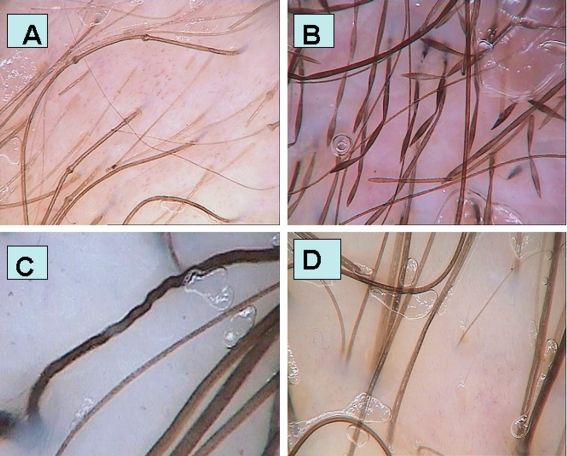
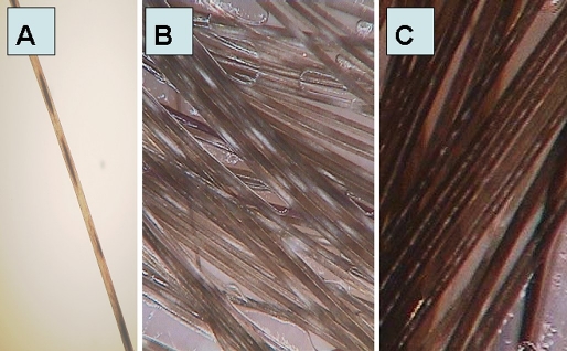
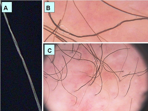
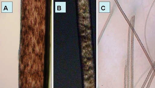
Background: Diagnosis of hair shaft abnormalities is based on light microscopic examination of more than 50 plucked hairs. The aim of this study was to verify whether hair shaft abnormalities may be visualized by trichoscopy (hair and scalp videodermoscopy) and to analyze trichoscopic features of common genetic hair shaft dysplasias.
Method: Patients with known genetic hair shaft disorders were included into the study. Trichoscopy was performed with the use of Fotofinder II videodermoscope. Images performed at 20-fold and 70-fold magnification were analysed. In selected cases 160-fold magnification was used for better visualization of hair shafts.
Results: Our results show that characteristic light microscopy features of Netherton syndrome, monilethrix, woolly hair syndrome, pili torti, pili annulati and trichothiodystrophy may be visualized by trichoscopy.
Conclusion: Genetic hair shaft abnormalities may be diagnosed by trichoscopy in a single diagnostic session without the need of plucking or cutting them for diagnostic purposes.
Keywords: alopecia; dermoscopy; hair; trichoscopy; videodermoscopy.
Figures
Similar articles
-
Trichoscopy in Hair Shaft Disorders.Dermatol Clin. 2018 Oct;36(4):421-430. doi: 10.1016/j.det.2018.05.009. Epub 2018 Aug 16. Dermatol Clin. 2018. PMID: 30201151 Review.
-
Trichoscopy update 2011.J Dermatol Case Rep. 2011 Dec 12;5(4):82-8. doi: 10.3315/jdcr.2011.1083. J Dermatol Case Rep. 2011. PMID: 22408709 Free PMC article.
-
Hair shaft videodermoscopy in netherton syndrome.Pediatr Dermatol. 2009 May-Jun;26(3):320-2. doi: 10.1111/j.1525-1470.2008.00778.x. Pediatr Dermatol. 2009. PMID: 19706096
-
Trichoscopy: a new method for diagnosing hair loss.J Drugs Dermatol. 2008 Jul;7(7):651-4. J Drugs Dermatol. 2008. PMID: 18664157 Review.
-
Hair Shaft Abnormalities as a Dermoscopic Feature of Mycosis Fungoides: Pilot Results.Dermatol Ther (Heidelb). 2024 Aug;14(8):2119-2126. doi: 10.1007/s13555-024-01206-z. Epub 2024 Jul 3. Dermatol Ther (Heidelb). 2024. PMID: 38961037 Free PMC article.
Cited by
-
Pili Torti: A Feature of Numerous Congenital and Acquired Conditions.J Clin Med. 2021 Aug 30;10(17):3901. doi: 10.3390/jcm10173901. J Clin Med. 2021. PMID: 34501349 Free PMC article. Review.
-
Usefulness of Trichoscopy over Hair Light Microscopy in Menkes Disease.Skin Appendage Disord. 2022 Jan;8(1):57-60. doi: 10.1159/000518368. Epub 2021 Sep 2. Skin Appendage Disord. 2022. PMID: 35118132 Free PMC article.
-
Dermoscopy in female androgenic alopecia: method standardization and diagnostic criteria.Int J Trichology. 2009 Jul;1(2):123-30. doi: 10.4103/0974-7753.58555. Int J Trichology. 2009. PMID: 20927234 Free PMC article.
-
A Comment on Trichoscopy.Int J Trichology. 2018 Jul-Aug;10(4):147-149. doi: 10.4103/ijt.ijt_13_18. Int J Trichology. 2018. PMID: 30386072 Free PMC article.
-
Dermoscopic Patterns of Genodermatoses: A Comprehensive Analysis.Biomedicines. 2023 Oct 6;11(10):2717. doi: 10.3390/biomedicines11102717. Biomedicines. 2023. PMID: 37893091 Free PMC article. Review.
References
-
- Silengo M, Valenzise M, Sorasio L, Ferrero GB. Hair as a diagnostic tool in dysmorphology. Clin Genet. 2002;62:270–272. - PubMed
-
- Furdon SA, Clark DA. Scalp hair characteristics in the newborn infant. Adv Neonatal Care. 2003;3:286–296. - PubMed
-
- Itin PH, Fistarol SK. Hair shaft abnormalities - clues to diagnosis and treatment. Dermatology. 2005;211:63–71. - PubMed
-
- Zalaudek I, Argenziano G, Di Stefani A, Ferrara G, Marghoob AA, Hofmann-Wellenhof R, Soyer HP, Braun R, Kerl H. Dermoscopy in general dermatology. Dermatology. 2006;212:7–18. - PubMed
-
- Olszewska M, Rudnicka L, Rakowska A, Kowalska-Oledzka E, Slowinska M. Trichoscopy. Arch Dermatol. 2008;144:1007. - PubMed
LinkOut - more resources
Full Text Sources
Other Literature Sources