. 2012 Mar;19(3):478-87.
doi: 10.1038/cdd.2011.117. Epub 2011 Sep 2.

PI3K/AKT signaling determines a dynamic switch between distinct KSRP functions favoring skeletal myogenesis

Affiliations

PI3K/AKT signaling determines a dynamic switch between distinct KSRP functions favoring skeletal myogenesis

P Briata et al. Cell Death Differ. 2012 Mar.

Abstract

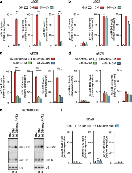
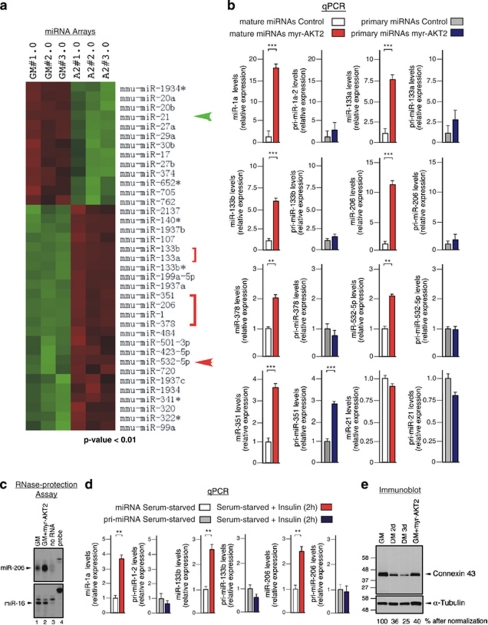
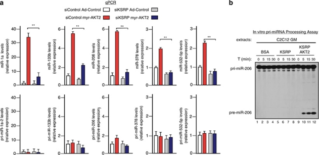
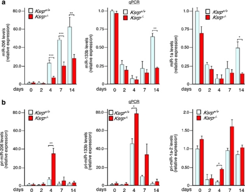
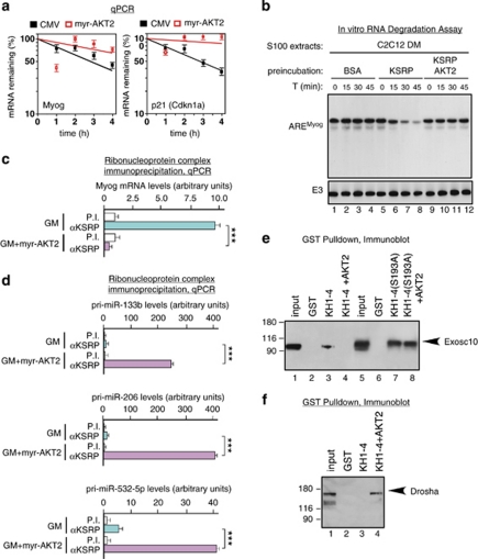
Skeletal myogenesis is orchestrated by distinct regulatory signaling pathways, including PI3K/AKT, that ultimately control muscle gene expression. Recently discovered myogenic micro-RNAs (miRNAs) are deeply implicated in muscle biology. Processing of miRNAs from their primary transcripts is emerging as a major step in the control of miRNA levels and might be well suited to be regulated by extracellular signals. Here we report that the RNA binding protein KSRP is required for the correct processing of primary myogenic miRNAs upon PI3K/AKT activation in myoblasts C2C12 and in the course of injury-induced muscle regeneration, as revealed by Ksrp knock-out mice analysis. PI3K/AKT activation regulates in opposite ways two distinct KSRP functions inhibiting its ability to promote decay of myogenin mRNA and activating its ability to favor maturation of myogenic miRNAs. This dynamic regulatory switch eventually contributes to the activation of the myogenic program.


Figures

Figure 1
Figure 1
PI3K/AKT signaling controls maturation of myogenic miRNAs in differentiating C2C12 myoblasts. (a and b) Quantitative RT-PCR (qPCR) analysis of miR-1a, miR-133b, miR-206 (a), and their respective primary transcripts (b) in C2C12 cells cultured either in growth medium (GM) or differentiation medium (DM) for 2 days in the presence of either DMSO (the solvent of LY294002 (LY), labeled as DM), or LY (25 μM, labeled as DM+LY). The values shown are averages (±S.E.M.) of three independent experiments performed in triplicate. (c and d) qPCR analysis of miR-1a, miR-133b, miR-206 (c), and their respective primary transcripts (d) in C2C12 cells transfected with siControl, siAkt1, or siAkt2 (40 nM), and cultured either in GM or in DM for 2 days. The values shown are averages (±S.E.M.) of three independent experiments performed in triplicate. (e) Northern blot analysis of total RNA purified from C2C12 myoblasts cultured either in GM or DM (1 day) and then infected with either a control empty adenoviral vector (1d DM) or an adenoviral vector expressing constitutively active myristoylated AKT2 (1d DM+myr-AKT2) for further 2 days. Representative blots sequentially hybridized with miR-133, miR-1a, and U6 probes (left panels) or with miR-206, let-7a, and U6 probes (right panels) are displayed. (f) qPCR analysis of pri-miR-1a-2, pri-miR-133b, and pri-miR-206 in the RNA samples from C2C12 cells treated as in panel e. Statistical significance: ***P<0.001 (Student's t-test)
Figure 2
Figure 2
Identification and validation of miRNAs whose expression is induced by PI3K/AKT signaling in C2C12 myoblasts cultured in GM. (a) C2C12 myoblasts cultured in GM were infected with either a control empty adenoviral vector (GM no.;1.0, 2.0, and 3.0 represent three distinct experiments) or an adenoviral vector expressing myr-AKT2 (A2no.;1.0, 2.0, and 3.0 represent three distinct experiments) and harvested after 2 days. Total RNA was extracted and analyzed by miRNA microarrays. Green or red color on the heat map indicates a decrease or increase of miRNA levels, respectively, and color intensities correspond to relative signal levels on a logarithmic scale. The green arrow points to miR-21, whose expression was significantly reduced also by a 2-day treatment of C2C12 myoblasts with DM. The red arrow and the red brackets indicate miRNAs whose expression was significantly enhanced also by a 2-day treatment of C2C12 myoblasts with DM. Statistical analysis was performed using Student's t-test. (b) qPCR analysis of miRNAs and their respective primary transcripts (as indicated) in C2C12 myoblasts cultured in GM and infected with either a control or a myr-AKT2-expressing adenoviral vector. The values shown are averages (±S.E.M.) of three independent experiments performed in triplicate. (c) RNase protection assay performed on the same RNA samples displayed in panel b. Samples were analyzed for the expression of either miR-206 or miR-16 (as control). Lanes corresponding to assays performed in the absence of RNA (no RNA) or in the presence of only RNA probes without digestion (probe) are also displayed. (d) qPCR analysis of miRNAs and their respective primary transcripts (as indicated) in C2C12 myoblasts serum-starved for 16 h and cultured for two additional hours in the same medium in the absence or in the presence of 10−6 M insulin. The values shown are averages (±S.E.M.) of three independent experiments performed in triplicate. (e) Immunoblot analysis of total cell extracts from C2C12 cells cultured either in GM (and infected with control or myr-AKT2 vectors) or in DM (for 2 or 3 days). Blots were sequentially hybridized with anti-connexin 43 and anti-alpha-tubulin antibodies and films subjected to densitometric scanning. Densitometric values (%) of connexin 43 expression after normalization for alpha-tubulin expression are presented. Representative blots are displayed. Statistical significance: **P<0.01 and ***P<0.001 (Student's t-test)
Figure 3
Figure 3
KSRP is required for AKT-induced myomiR maturation and phosphorylation by AKT activated KSRP ability to process pri-miR-206. (a) qPCR analysis of miRNAs (upper) and their respective primary transcripts (lower) in C2C12 myoblasts cultured in GM, transfected with either siControl or siKsrp, and infected with either a control (Ad-Control) or a myr-AKT2-expressing adenoviral vector. The values shown are averages (±S.E.M.) of three independent experiments performed in triplicate. (b) In vitro pri-miR-206 processing assays performed using total extracts from either C2C12 myoblasts cultured in GM pre-incubated with BSA (lanes 1–4), KSRP (30 nM, lanes 5–8), AKT2-phosphorylated KSRP (30 nM, lanes 9–12), respectively. Internally 32P-labeled, pri-miR-206 RNA substrate was added and its processing monitored as described by Briata et al. Statistical significance: **P<0.01 (Student's t-test)
Figure 4
Figure 4
Impaired maturation of myomiRs during muscle regeneration in Ksrp knock-out mice. (a and b) qPCR analysis of miRNAs (a) and their respective primary transcripts (b) in total RNA samples extracted from tibialis anterioris muscles of either wild-type (Ksrp+/+) or Ksrp−/− mice before and at different intervals of time after injection of cardiotoxin as indicated. The values shown are averages (±S.D.) of five independent animals for each experimental group. Assays were performed in triplicate. Statistical significance: *P<0.05, **P<0.01, and ***P<0.001 (Student's t-test)
Figure 5
Figure 5
AKT signaling activation inhibits KSRP ability to participate in a Myogenin mRNA decay complex while favors its association with a primary myomiR processing complex. (a) qPCR analysis of myogenin and p21 (also known as Cdkn1a) expression in C2C12 myoblasts cultured in GM and infected with either a control (GM) or a myr-AKT2-expressing adenoviral vector. Total RNA was isolated at the indicated times after the addition of DRB. The values shown are averages (±S.E.M.) of three independent experiments performed in triplicate. (b) In vitro RNA degradation assays using S100 extracts from C2C12 myoblasts cultured in DM pre-incubated with BSA (lanes 1–4), KSRP (30 nM, lanes 5–8), AKT2-phosphorylated KSRP (30 nM, lanes 9–12), respectively. Internally 32P-labeled and capped RNA substrates were added and their decay monitored as described under Material and Methods. E3 is a stable unrelated transcript. Representative gels are displayed. (c) C2C12 myoblasts were cultured in GM and infected with either control (GM) or myr-AKT2-expressing adenoviral vector. After 2 days cells were lysed, and total cell extracts were immunoprecipitated as indicated. RNA was purified from immunocomplexes and analyzed by qPCR to detect Myogenin mRNA. The values shown are averages (±S.E.M.) of three independent experiments performed in triplicate. (d) C2C12 myoblasts were cultured in GM and infected with either control (GM) or myr-AKT2-expressing adenoviral vector. After 2 days cells were lysed, and total cell extracts were immunoprecipitated as indicated. RNA was purified from immunocomplexes and analyzed by qPCR to detect primary myomiRs as indicated. The values shown are averages (±S.E.M.) of three independent experiments performed in triplicate. (e and f) GST pull-down of endogenous Exosc10 (e) and Drosha (f) present in total extracts from C2C12 cells cultured in GM using the following proteins: GST, GST-KH1-4, GST-KH1-4 in vitro phosphorylated by AKT2, GST-KH1-4(S193A), or GST-KH1-4(S193A) in vitro phosphorylated by AKT2 as indicated. GST-KH1-4(S193A) is mutated in Ser 193 in order to abolish the AKT phosphorylation site as described by Gherzi et al. Proteins were analyzed by immunoblotting using the indicated antibodies. Representative blots are displayed. Statistical significance: ***P<0.001 (Student's t-test)

Similar articles

Cited by

References

    1. Yun K, Wold B. Skeletal muscle determination and differentiation: story of a core regulatory network and its context. Curr Opin Cell Biol. 1996;8:877–889. - PubMed
    1. Charge SB, Rudnicki MA. Cellular and molecular regulation of muscle regeneration. Physiol Rev. 2004;84:209–238. - PubMed
    1. Tedesco FS, Dellavalle A, Diaz-Manera J, Messina G, Cossu G. Repairing skeletal muscle: regenerative potential of skeletal muscle stem cells. J Clin Invest. 2010;120:11–19. - PMC - PubMed
    1. Tajbakhsh S. Skeletal muscle stem cells in developmental versus regenerative myogenesis. J Intern Med. 2009;266:372–389. - PubMed
    1. Guasconi V, Puri PL. Chromatin: the interface between extrinsic cues and the epigenetic regulation of muscle regeneration. Trends Cell Biol. 2009;19:286–294. - PMC - PubMed

Publication types

MeSH terms