

Impaired spatial and contextual memory formation in galectin-1 deficient mice
- PMID: 21884595
- PMCID: PMC3179925
- DOI: 10.1186/1756-6606-4-33
Impaired spatial and contextual memory formation in galectin-1 deficient mice
Abstract
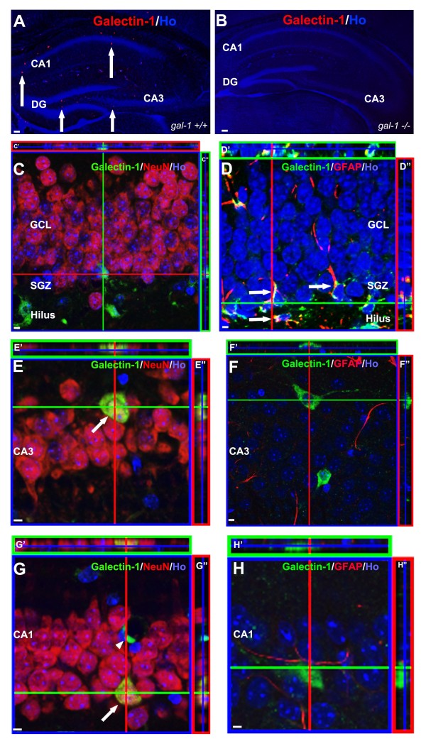
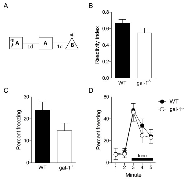
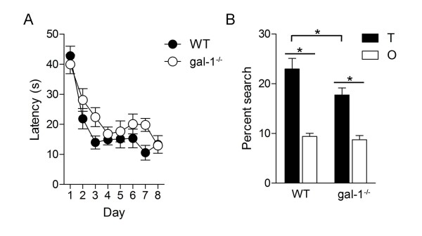
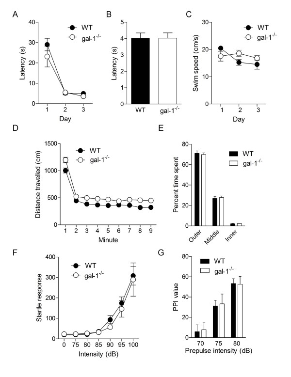
Galectins are a 15 member family of carbohydrate-binding proteins that have been implicated in cancer, immunity, inflammation and development. While galectins are expressed in the central nervous system, little is known about their function in the adult brain. Previously we have shown that galectin-1 (gal-1) is expressed in the adult hippocampus, and, in particular, in putative neural stem cells in the subgranular zone. To evaluate how gal-1 might contribute to hippocampal memory function here we studied galectin-1 null mutant (gal-1-/-) mice. Compared to their wildtype littermate controls, gal-1-/- mice exhibited impaired spatial learning in the water maze and contextual fear learning. Interestingly, tone fear conditioning was normal in gal-1-/- mice suggesting that loss of gal-1 might especially impact hippocampal learning and memory. Furthermore, gal-1-/- mice exhibited normal motor function, emotion and sensory processing in a battery of other behavioral tests, suggesting that non-mnemonic performance deficits are unlikely to account for the spatial and contextual learning deficits. Together, these data reveal a role for galectin-carbohydrate signalling in hippocampal memory function.
Figures

Similar articles
-
Neuroinflammation induced by the peptide amyloid-β (25-35) increase the presence of galectin-3 in astrocytes and microglia and impairs spatial memory.Neuropeptides. 2019 Apr;74:11-23. doi: 10.1016/j.npep.2019.02.001. Epub 2019 Feb 14. Neuropeptides. 2019. PMID: 30795916
-
Learning and memory performance in mice lacking the GAL-R1 subtype of galanin receptor.Eur J Neurosci. 2004 Mar;19(5):1384-96. doi: 10.1111/j.1460-9568.2004.03214.x. Eur J Neurosci. 2004. PMID: 15016096
-
Impaired synaptic clustering of postsynaptic density proteins and altered signal transmission in hippocampal neurons, and disrupted learning behavior in PDZ1 and PDZ2 ligand binding-deficient PSD-95 knockin mice.Mol Brain. 2012 Dec 26;5:43. doi: 10.1186/1756-6606-5-43. Mol Brain. 2012. PMID: 23268962 Free PMC article.
-
Galanin impairs performance on learning and memory tasks: findings from galanin transgenic and GAL-R1 knockout mice.Neuropeptides. 2005 Jun;39(3):239-43. doi: 10.1016/j.npep.2004.12.026. Neuropeptides. 2005. PMID: 15944016 Review.
-
Galectin-3 Involvement in Cognitive Processes for New Therapeutic Considerations.Front Cell Neurosci. 2022 Jul 5;16:923811. doi: 10.3389/fncel.2022.923811. eCollection 2022. Front Cell Neurosci. 2022. PMID: 35875353 Free PMC article. Review.
Cited by
-
Vatairea macrocarpa lectin (VML) induces depressive-like behavior and expression of neuroinflammatory markers in mice.Neurochem Res. 2013 Nov;38(11):2375-84. doi: 10.1007/s11064-013-1150-9. Epub 2013 Sep 12. Neurochem Res. 2013. PMID: 24026569
-
Galectin-1 promotes an M2 macrophage response to polydioxanone scaffolds.J Biomed Mater Res A. 2017 Sep;105(9):2562-2571. doi: 10.1002/jbm.a.36113. Epub 2017 Jun 15. J Biomed Mater Res A. 2017. PMID: 28544348 Free PMC article.
-
Calcineurin/P-ERK/Egr-1 Pathway is Involved in Fear Memory Impairment after Isoflurane Exposure in Mice.Sci Rep. 2017 Oct 24;7(1):13947. doi: 10.1038/s41598-017-13975-z. Sci Rep. 2017. PMID: 29066839 Free PMC article.
-
Metabolic fingerprints of fear memory consolidation during sleep.Mol Brain. 2021 Feb 10;14(1):30. doi: 10.1186/s13041-021-00733-6. Mol Brain. 2021. PMID: 33568175 Free PMC article.
-
Novel Galectin-3 Roles in Neurogenesis, Inflammation and Neurological Diseases.Cells. 2021 Nov 5;10(11):3047. doi: 10.3390/cells10113047. Cells. 2021. PMID: 34831271 Free PMC article. Review.
References
-
- Taylor ME, Drickamer K. Introduction to Glycobiology. 2. Oxford University Press; 2006.
-
- Varki A, Cummings RD, Esko JD, Freeze HH, Stanely P, Bertozzi CR, Hart GW, Etzler ME, Eds. Essentials of Glycobiology. 2. Cold Spring Harbor Laboratory Press; 2008. - PubMed
-
- Yang RY, Rabinovich GA, Liu FT. Galectins: structure, function and therapeutic potential. Expert Rev Mol Med. 2008;10:e17. - PubMed
-
- Leffler H, Carlsson S, Hedlund M, Qian Y, Poirier F. Introduction to galectins. Glycoconj J. 2004;19(7-9):433–440. - PubMed
Publication types
MeSH terms
Substances
Grants and funding
LinkOut - more resources
Full Text Sources
Medical
Research Materials
