. 2011 Oct;138(20):4443-50.
doi: 10.1242/dev.071001. Epub 2011 Aug 31.

Individual axons regulate the myelinating potential of single oligodendrocytes in vivo

Affiliations

Individual axons regulate the myelinating potential of single oligodendrocytes in vivo

Rafael G Almeida et al. Development. 2011 Oct.

Abstract

The majority of axons in the central nervous system (CNS) are eventually myelinated by oligodendrocytes, but whether the timing and extent of myelination in vivo reflect intrinsic properties of oligodendrocytes, or are regulated by axons, remains undetermined. Here, we use zebrafish to study CNS myelination at single-cell resolution in vivo. We show that the large caliber Mauthner axon is the first to be myelinated (shortly before axons of smaller caliber) and that the presence of supernumerary large caliber Mauthner axons can profoundly affect myelination by single oligodendrocytes. Oligodendrocytes that typically myelinate just one Mauthner axon in wild type can myelinate multiple supernumerary Mauthner axons. Furthermore, oligodendrocytes that exclusively myelinate numerous smaller caliber axons in wild type can readily myelinate small caliber axons in addition to the much larger caliber supernumerary Mauthner axons. These data indicate that single oligodendrocytes can myelinate diverse axons and that their myelinating potential is actively regulated by individual axons.


Figures

Fig. 1.
Fig. 1.
Transgenic reporters reveal first axon myelinated in vivo in zebrafish CNS. (A) Lateral view of a stable Tg(mbp:EGFP-CAAX) zebrafish at 8 dpf. Myelinating glia of the CNS and PNS are labelled, as is the heart, which serves as a marker of transgenesis. (A′) Lateral view of the spinal cord (area indicated by box in A). Prominent myelinated tracts myelinated in the dorsal spinal cord and ventral spinal cord are apparent. (B) Lateral views of a stable Tg(mbp:EGFP-CAAX) zebrafish at 60 hpf show that the very first axon to be myelinated is the large Mauthner axon in the ventral spinal cord, which is first myelinated in the anterior spinal cord. (C) Lateral views of a stable Tg(mbp:EGFP-CAAX) zebrafish at 70 hpf. Myelination of the Mauthner axon has now commenced in the more posterior part of the spinal cord. At this stage, oligodendrocytes have started to myelinate axons in the dorsal spinal cord. Dorsal is up and anterior is to the left in all images. Scale bars: 500 μm in A; 20 μm in C.
Fig. 2.
Fig. 2.
Single-cell analysis reveals morphological diversity of individual CNS oligodendrocytes. (A) Lateral views of single mbp:EGFP-expressing oligodendrocytes associating with one Mauthner axon (top) and both Mauthner axons (bottom). (B) Lateral view of a single mbp:EGFP-expressing oligodendrocyte associating with the large Mauthner axon (left) and an axon of much smaller caliber (right). (C) Lateral views of single oligodendrocytes associating with multiple axons in the dorsal spinal cord (top) and ventral spinal cord (bottom). (D) Myelin sheath number per oligodendrocyte (excluding those that myelinate the Mauthner axons) at 4 dpf. (E) Myelin sheath length in four sample oligodendrocytes (that do not associate with the Mauthner axon) at 4 dpf. (F) Average myelin sheath number per cell over time. This does not include oligodendrocytes that myelinate the Mauthner axon. (G) Average myelin sheath length per cell over time. This does not include oligodendrocytes that myelinate the Mauthner axon. Error bars represent s.d. Scale bars: 10 μm.
Fig. 3.
Fig. 3.
Supernumerary Mauthner axons are ensheathed by myelinating glia. (A-C) Ventral views of the hindbrains of wild-type (A), notch1a morphant (B) and hoxb1 mRNA-injected (C) larvae at 5 dpf labelled with the 3A10 antibody, which recognises neurofilament-associated proteins. Mauthner axons are indicated by arrowheads. Scale bar: 20 μm. (A′-C′) Analysis of Tg(sox10:mRFP) (A′-C′) and Tg(mbp:EGFP-CAAX) (A′-C′) shows that wild-type (A′,A′), notch1a morphant (B′,B′) and hoxb1-injected (C′,C′) Mauthner axons are all ensheathed and myelinated. Arrowheads indicate Mauthner axons. Scale bars: 20 μm. (D) RT-PCR analyses show that levels of notch1a mRNA are reduced in morpholino (MO)-injected animals relative to controls at 10 hpf but not at 72 hpf.
Fig. 4.
Fig. 4.
Supernumerary Mauthner axons are robustly myelinated. (A-C′) Transmission electron microscope images of transverse sections through the spinal cord of wild-type (A,A′), notch1a morphant (B,B′) and hoxb1 mRNA-injected (C,C′) zebrafish larvae at 9 dpf shows that all Mauthner axons are robustly myelinated (A-C) and that there is a similar number of axons myelinated in the ventral spinal cord despite the presence of supernumerary Mauthner (M) axons (A′-C′). Scale bars: 2 μm. (D) Total circumference of Mauthner axon(s) as a function of the number of Mauthner axons present per hemi-spinal cord. (E) Number of myelinated axons, excluding Mauthner axon(s), present in the ventral spinal cord as a function of the number of Mauthner axons present per hemi-spinal cord. Error bars represent s.d.
Fig. 5.
Fig. 5.
Presence of supernumerary Mauthner axons does not affect oligodendrocyte number or distribution. (A-C) Lateral views of the spinal cords in Tg(mbp:EGFP)-expressing wild-type (A), notch1a morphant (B) and hoxb1 mRNA-injected (C) animals shows that oligodendrocyte number and distribution are not affected by the presence of supernumerary Mauthner axons. Arrows point to oligodendrocytes in the dorsal spinal cord with projections to the ventral spinal cord. Compare with Fig. 7B. Scale bar: 20 μm. (D). Total oligodendrocyte number per 425 μm length of tissue in the mid-trunk spinal cord of wild-type, notch1a morphant and hoxb1 mRNA-injected animals and, in grey, the number of oligodendrocytes specifically located in the dorsal domain of the spinal cord. Error bars represent s.d.
Fig. 6.
Fig. 6.
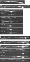
Individual oligodendrocytes myelinate multiple supernumerary Mauthner axons. (A-F) All images are of live single mbp:EGFP-expressing oligodendrocytes in the spinal cord of larvae at 5 dpf (A-C) and 9 dpf (D-F). (A,D) Wild-type oligodendrocytes associated with single Mauthner axons. (B,E) Oligodendrocytes in hoxb1 mRNA-injected animals with myelin sheaths on two Mauthner axons. (C,F) Oligodendrocytes in notch1a morphants that make seven (C) and five (F) myelin sheaths on supernumerary Mauthner axons. Maximum intensity projections and individual confocal z-sections are indicated for clarity. Individual myelin sheaths indicated by brackets. Scale bars: 20 μm.
Fig. 7.
Fig. 7.
Individual oligodendrocytes readily myelinate supernumerary Mauthner axons and smaller caliber axons. (A) In wild type, oligodendrocytes in the dorsal spinal cord myelinate only small caliber axons. (B) Oligodendrocytes in the dorsal spinal cords of animals with supernumerary Mauthner axons can myelinate the large Mauthner axons in the ventral spinal cord in addition to smaller caliber axons (e.g. arrow). Scale bars: 20 μm.

Similar articles

Cited by

References

    1. Bakiri Y., Karadottir R., Cossell L., Attwell D. (2011). Morphological and electrical properties of oligodendrocytes in the white matter of the corpus callosum and cerebellum. J. Physiol. 589, 559-573 - PMC - PubMed
    1. Barres B. A., Raff M. C. (1999). Axonal control of oligodendrocyte development. J. Cell Biol. 147, 1123-1128 - PMC - PubMed
    1. Barres B. A., Lazar M. A., Raff M. C. (1994). A novel role for thyroid hormone, glucocorticoids and retinoic acid in timing oligodendrocyte development. Development 120, 1097-1108 - PubMed
    1. Blakemore W. F. (1974). Pattern of remyelination in the CNS. Nature 249, 577-578 - PubMed
    1. Brinkmann B. G., Agarwal A., Sereda M. W., Garratt A. N., Muller T., Wende H., Stassart R. M., Nawaz S., Humml C., Velanac V., et al. (2008). Neuregulin-1/ErbB signaling serves distinct functions in myelination of the peripheral and central nervous system. Neuron 59, 581-595 - PMC - PubMed

Publication types

LinkOut - more resources