. 2011 Oct;17(10):1922-31.
doi: 10.1261/rna.2855511. Epub 2011 Aug 30.

Interactions of a Pop5/Rpp1 heterodimer with the catalytic domain of RNase MRP

Affiliations

Interactions of a Pop5/Rpp1 heterodimer with the catalytic domain of RNase MRP

Anna Perederina et al. RNA. 2011 Oct.

Abstract

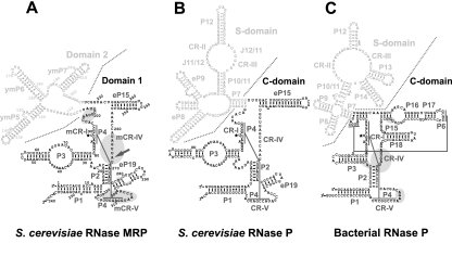
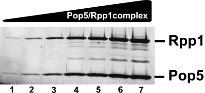
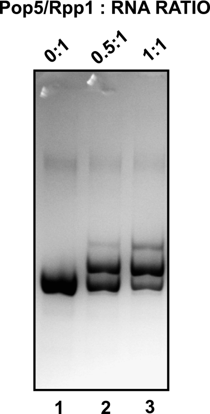
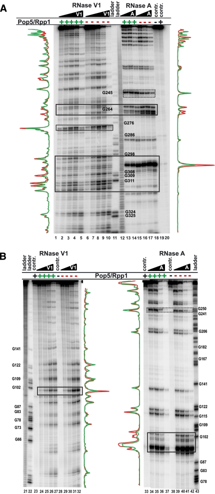
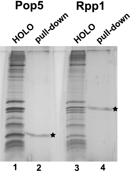
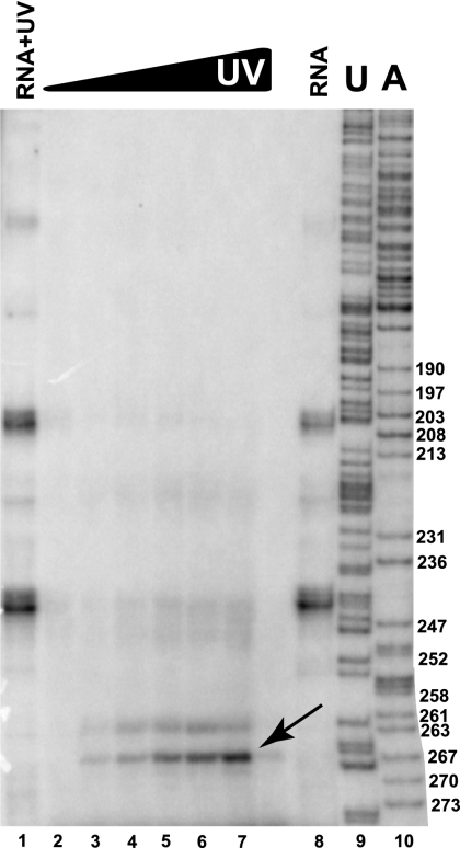
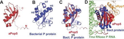
Ribonuclease (RNase) MRP is a multicomponent ribonucleoprotein complex closely related to RNase P. RNase MRP and eukaryotic RNase P share most of their protein components, as well as multiple features of their catalytic RNA moieties, but have distinct substrate specificities. While RNase P is practically universally found in all three domains of life, RNase MRP is essential in eukaryotes. The structural organizations of eukaryotic RNase P and RNase MRP are poorly understood. Here, we show that Pop5 and Rpp1, protein components found in both RNase P and RNase MRP, form a heterodimer that binds directly to the conserved area of the putative catalytic domain of RNase MRP RNA. The Pop5/Rpp1 binding site corresponds to the protein binding site in bacterial RNase P RNA. Structural and evolutionary roles of the Pop5/Rpp1 heterodimer in RNases P and MRP are discussed.


Figures

FIGURE 1.
FIGURE 1.
RNA components of yeast RNase MRP (A), yeast RNase P (B), and bacterial RNase P (Thermotoga maritima) (C). Catalytic (C-) domains of RNase P and Domain 1 of RNase MRP are shown in black; specificity (S-) domains of RNase P and Domain 2 of RNase MRP are shown in gray. (A) Areas of RNase MRP RNA that are protected in the presence of the Pop5/Rpp1 heterodimer are highlighted in gray; the location of the Pop5–RNA UV cross-link in the RNase MRP holoenzyme is indicated by an arrow. (C) Areas of bacterial RNase P RNA that are within 5 Å of the protein component (Reiter et al. 2010) are highlighted in gray. The diagrams are based on Esakova and Krasilnikov (2010).
FIGURE 2.
FIGURE 2.
Varying amounts (75–1600 ng) of the Pop5/Rpp1 complex analyzed on a SYPRO Orange-stained 15% SDS–polyacrylamide gel. Quantification of the intensities of the protein bands shows a 1:1 Pop5:Rpp1 molar ratio. Combined with the Dynamic Light Scattering data (particle size 3.19 nm), this indicates that Pop5 and Rpp1 form a heterodimer.
FIGURE 3.
FIGURE 3.
Binding of the Pop5/Rpp1 heterodimer to RNase MRP RNA. A total of 2 μg (final concentration 1.8 μM) of the full-length RNase MRP RNA was incubated with varying amounts of the Pop5/Rpp1 heterodimer and analyzed on a 5% native polyacrylamide gel stained with Toluidine Blue. (Lane 1) no Pop5/Rpp1; (lane 2) 0.9 μM Pop5/Rpp1; (lane 3) 1.8 μM Pop5/Rpp1. The concentrations of the components of the RNA–protein complex were at least an order of magnitude higher than the dissociation constant for the complex (∼60 nM).
FIGURE 4.
FIGURE 4.
Footprinting analysis of the Pop5/Rpp1–RNase MRP RNA complex. Pop5/Rpp1 was mixed with an equimolar amount of refolded in vitro-transcribed RNA. (A) 3′-end 32P-labeled RNase MRP RNA; (B) 5′-end 32P-labeled RNase MRP RNA. (Lanes 1–5,24–27) increasing concentrations of RNase V1 in the presence of the Pop5/Rpp1 heterodimer; (lanes 6–10,29–32) increasing concentrations of RNase V1 without Pop5/Rpp1; (lanes 11,22,43) RNase T1 digestion of RNase MRP RNA (sequence ladders); (lanes 12,21) alkaline hydrolysis of RNase MRP RNA (ladders); (lanes 13–15,34–37) increasing concentrations of RNase A in the presence of the Pop5/Rpp1 heterodimer; (lanes 16–18,39–42) increasing concentrations of RNase A without Pop5/Rpp1; (lanes 19,28,38) undigested RNase MRP RNA (controls); (lanes 20,23,33) undigested complex of RNase MRP with Pop5/Rpp1 (controls). Regions protected in the presence of Pop5/Rpp1 are traced with solid lines; the region displaying hypersensitivity in the presence of proteins is traced with a dotted line. RNase MRP RNA nucleotide numbering (shown next to sequence ladders) matches that in Figure 1A. Graphs next to the gels represent bands’ intensities in the presence of Pop5/Rpp1 (shown in green; lanes 3,13,27,35 were quantified) and for RNA alone (shown in red; lanes 8,16,32,40 were quantified).
FIGURE 5.
FIGURE 5.
Isolation of His6-tagged proteins from purified RNase MRP/P holoenzymes. (Lane 1) Proteins in the RNase MRP/P holoenzyme mix purified from yeast strain EK-Pop5 (TAP-tagged Pop4 and His6-tagged Pop5); (lane 2) Pop5 isolated from holoenzymes under denaturing conditions; a star indicates the Pop5 protein band; (lane 3) proteins in the RNase MRP/P holoenzyme mix purified from yeast strain EK-Rpp1 (TAP-tagged Pop4 and His6-tagged Rpp1); (lane 4) Rpp1 isolated from holoenzymes under denaturing conditions; a star indicates the Rpp1 protein band. Silver-stained 15% SDS–polyacrylamide gel.
FIGURE 6.
FIGURE 6.
UV cross-linking analysis of interactions of Pop5 with RNA in the RNase MRP holoenzyme purified from yeast. RNase MRP holoenzyme was purified from yeast, subjected to UV cross-linking, and disassembled under denaturing conditions; then, His6-tagged Pop5 with cross-linked RNA was isolated, and RNA cross-linked to Pop5 was extracted and finally subjected to primer extension analysis. (Lane 1) RNase MRP RNA extracted from purified holoenzyme and subjected to UV irradiation at 1.28 J/cm2 (control for RNA–RNA cross-links); (lanes 2–7) UV-irradiated RNase MRP holoenzyme (0, 0.04, 0.08, 0.16, 0.32, and 0.64 J/cm2, respectively); (lane 8) RNase MRP RNA extracted from the purified holoenzyme (control, no irradiation); (lanes 9,10) sequence ladder. 5% denaturing (8 M urea) polyacrylamide gel.
FIGURE 7.
FIGURE 7.
Structural elements in the archaeal homolog of Pop5 (A) and in the protein component of bacterial RNase P (B) have distinct connectivities, but adopt similar overall mutual orientations (C). (A) Crystal structure of an archaeal homolog of Pop5 (PDB ID 2CZV) shown in red; N and C termini are indicated. (B) Crystal structure of T. maritima RNase P protein (PDB ID 3OKB) shown in blue; N and C termini are indicated. Secondary structure elements (H: α-helices, E: β-strands) are marked according to the designation in Figure 8 and Supplemental Figure S1. (C) Archaeal homolog of Pop5 (PDB ID 2CZV, shown in red) superposed on the protein component of bacterial RNase P (PDB ID 3OKB, shown in blue). (D) An archaeal aPop5/aRpp1 heterodimer (PDB ID 2CZV; aPop5 is in red, aRpp1 is in gold) is shown superposed on the crystal structure of T. maritima RNase P (PDB ID 3OKB; the protein component is in blue, the RNA component is in green).
FIGURE 8.
FIGURE 8.
Spatially superposable elements in Pop5 and bacterial RNase P protein are connected differently, but show resembling patterns of phylogenetic conservation in areas corresponding to the protein–RNA interface in bacterial RNase P. Sequences of major secondary structure elements (line 1) in bacterial RNase P proteins (lines 3–10) shown against corresponding (in space) elements (line 11) in archaeal (lines 12,13) and eukaryotic (lines 14–19) Pop5 proteins. The bacterial and archaeal/eukaryotic structure elements are superposed as shown in Figure 7C. The complete alignment is shown in Supplemental Figure S1C. (Line 1) Secondary structure elements in bacterial RNase P protein (PDB ID 3OKB) marked according to Figure 7B and Supplemental Figure S1, A and B. α-helices are shown by cylinders; β-strands are shown by arrows. (Line 2) Amino acids of the bacterial RNase P protein that are involved in interactions with the RNA component of T. maritima RNase P (Reiter et al. 2010) are indicated by red asterisks; amino acids involved in interactions with bacterial RNase P substrate (Reiter et al. 2010) are indicated by red letters (s). (Line 3) T. maritima; (line 4) Bacillus subtilis; (line 5) Staphylococcus aureus; (line 6) Mycobacterium leprae; (line 7) Mycobacterium tuberculosis; (line 8) E. coli; (line 9) Haemophilus influenzae; (line 10) Chlamydophila pneumoniae. (Line 11) Secondary structure elements in the archaeal homolog of Pop5 (PDB ID 2CZV); eukaryotic Pop5 is expected to be similar. (Line 12) Pyrococcus horikoshii OT3; (line 13) Pyrococcus furiosus; (line 14) S. cerevisiae; (line 15) Zygosaccharomyces rouxii; (line 16) Candida glabrata; (line 17) Kluyveromyces lactis; (line 18) Xenopus laevis; (line 19) human. Residues showing similar patterns of conservation in bacterial RNase P protein versus archaeal/eukaryotic Pop5 are highlighted as follows: nonpolar aliphatic (GAPVLIM, yellow), aromatic (FYW, dark blue), polar uncharged (STCNQ, green), positively charged (KHR, light blue), negatively charged (DE, brown).

Similar articles

Cited by

References

    1. Altman S 2010. History of RNase P and overview of its catalytic activity. In Ribonuclease P, protein reviews 10 (ed. Liu F. and Altman S.), pp. 1–15 Springer, New York
    1. Aspinall TV, Gordon JMB, Bennett HJ, Karahalios P, Bukowski J-P, Walker SC, Engelke DR, Avis JM 2007. Interactions between subunits of Saccharomyces cerevisiae RNase MRP support a conserved eukaryotic RNase P/MRP architecture. Nucleic Acids Res 35: 6439–6450 - PMC - PubMed
    1. Brachmann CB, Davis A, Cost GJ, Caputo E, Li J, Hieter P, Boeke JD 1998. Designer deletion strains derived from Saccharomyces cerevisiae S288C: a useful set of strains and plasmids for PCR-mediated gene disruption and other applications. Yeast 14: 115–132 - PubMed
    1. Brown JW 1999. The ribonuclease P database. Nucleic Acids Res 27: 314–314 - PMC - PubMed
    1. Cai T, Reilly TR, Cerio M, Schmitt ME 1999. Mutagenesis of SNM1, which encodes a protein component of the yeast RNase MRP, reveals a role for this ribonucleoprotein endoribonuclease in plasmid segregation. Mol Cell Biol 19: 7857–7869 - PMC - PubMed

Publication types

MeSH terms

LinkOut - more resources