. 2011 Nov 15;20(22):4395-410.
doi: 10.1093/hmg/ddr366. Epub 2011 Aug 24.

Functional and physical interaction between the mismatch repair and FA-BRCA pathways

Affiliations

Functional and physical interaction between the mismatch repair and FA-BRCA pathways

Stacy A Williams et al. Hum Mol Genet. .

Abstract

Fanconi anemia (FA) is a rare genetic disorder characterized by bone marrow failure and an increased risk for leukemia and cancer. Fifteen proteins thought to function in the repair of DNA interstrand crosslinks (ICLs) comprise what is known as the FA-BRCA pathway. Activation of this pathway leads to the monoubiquitylation and chromatin localization of FANCD2 and FANCI. It has previously been shown that FANCJ interacts with the mismatch repair (MMR) complex MutLα. Here we show that FANCD2 interacts with the MMR proteins MSH2 and MLH1. FANCD2 monoubiquitylation, foci formation and chromatin loading are greatly diminished in MSH2-deficient cells. Human or mouse cells lacking MSH2 or MLH1 display increased sensitivity and radial formation in response to treatment with DNA crosslinking agents. Studies in human cell lines and Drosophila mutants suggest an epistatic relationship between FANCD2, MSH2 and MLH1 with regard to ICL repair. Surprisingly, the interaction between MSH2 and MLH1 is compromised in multiple FA cell lines, and FA cell lines exhibit deficient MMR. These results suggest a significant role for MMR proteins in the activation of the FA pathway and repair of ICLs. In addition, we provide the first evidence for a defect in MMR in FA cell lines.


Figures
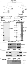
Figure 1.
Figure 1.
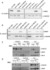
Identification of novel FANCD2-binding partners. (A) Whole-cell extracts from PD20 cells stably expressing FLAG-tagged FANCD2 were subjected to the chromatography scheme represented here. Three distinct FANCD2-containing protein complexes were identified by western blot and named ‘small’, ‘middle’ and ‘large’ complexes. (B) The ‘small’ and ‘middle’ protein complexes were immunoprecipitated using anti-FLAG resin, eluted using FLAG peptide and subjected to SDS–PAGE followed by silver-staining. The ‘small’ complex appeared as a single band, whereas silver-staining of the ‘middle’ complex revealed many protein bands, suggesting the presence of several different FANCD2-containing protein complexes. Bands were cut out, trypsin-digested and analyzed by mass spectrometry to reveal the novel FANCD2-binding partners, MSH2 and MLH1. (CE) Immunoprecipitation of endogenous FANCD2 and MSH2 from HeLa whole-cell extracts confirms that FANCD2, MSH2 and MLH1 co-precipitate and this interaction is induced upon damage with MMC.
Figure 2.
Figure 2.
Characterization of the FANCD2–MSH2 and FANCD2–MLH1 interactions. (A) Endogenous FANCD2 was immunoprecipitated from whole-cell extracts of human cell lines deficient in FA core complex proteins or stably expressing wild-type FANCD2 or FANCD-K561R after an 18 h treatment with 50 nm MMC. MSH2 co-precipitates with FANCD2-L only. (B) Endogenous FANCD2 was immunoprecipitated from whole-cell extracts of cell lines deficient in ATR, ATM and BRCA1 after an 18 h treatment with 50 nm MMC. ATR is required for the interaction of FANCD2 and MSH2, but not ATM or BRCA1. (C) Endogenous FANCD2 was immunoprecipitated from extracts of MSH2-deficient and corrected cells that are untreated or treated with MMC for 24 h. MLH1 does not co-precipitate with FANCD2 in MSH2-deficient cells. (D) Endogenous FANCD2 was immunoprecipitated from MLH1-deficient cell extracts that are untreated or treated with MMC for 24 h. MSH2 co-precipitates with FANCD2 to a greater extent in MLH1-corrected cells, indicating that MLH1 enhances the interaction between FANCD2 and MSH2.
Figure 3.
Figure 3.
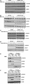
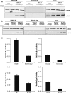
MSH2 is required for normal kinetics of monoubiquitylation and chromatin loading of FANCD2 and FANCI. (A) MSH2-deficient and corrected cells or (B) MLH1-deficient and corrected cells were treated with 500 nm MMC over several time points for 24 h. Immunoblotting shows FANCD2 and FANCI monoubiquitylation is greatly diminished in MSH2-deficient cells, but not MLH1-deficient cells. (C) MSH2-deficient and corrected cells or (D) MLH1-deficient and corrected cells were treated with 75 μm CDDP over several time points for 24 h. Immunoblotting reveals a similar deficiency in FANCD2 monoubiquitylation in MSH2-deficient cells, but not MLH1-deficient cells. (E) MSH2-deficient and corrected cells or (F) MLH1-deficient and corrected cells were treated with 500 nm MMC for 24 h and then subjected to cellular fractionation. Immunoblotting reveals that although FANCD2 and FANCI chromatin loading is normal in MLH1-deficient cells, chromatin loading of both proteins is nearly undetectable in MSH2-deficient cells.
Figure 4.
Figure 4.
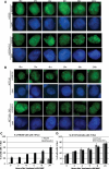
FANCD2 foci formation is impaired in MSH2-deficient cells. (A) MSH2-deficient and corrected cells or (B) MLH1-deficient and corrected cells were treated with 500 nm MMC over several time points for 24 h. Immunostaining for FANCD2 shows that FANCD2 foci formation is greatly diminished in MSH2-deficient cells through all time points up to 24 h, but not in MLH1-deficient cells. At least 100 cells were counted to determine the percentage of cells with greater than five foci in both (C) MSH2-deficient and corrected cells and (D) MLH1-deficient and corrected cells. Graphs clearly show a significant delay and reduction in FANCD2 foci formation in MSH2-deficient cells.
Figure 5.
Figure 5.
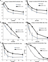
MSH2 and MLH1 play a key role in ICL repair. (A) Human MSH2-deficient and corrected cells, (B) MSH2-deficient MEFs and their wild-type counterparts and (C) human MLH1-deficient and corrected cells were treated with increasing concentrations of MMC and CDDP and assessed for survival using crystal violet staining and extraction. Both MSH2- and MLH1-deficient cells are hypersensitive to DNA interstrand crosslinking agents. (D) Depletion of MSH2, MLH1, FANCD2, MSH2 and FANCD2, or MLH1 and FANCD2 by siRNA transfection followed by crystal violet survival assay shows that MSH2, MLH1 and FANCD2 are epistatic with regard to ICL repair. (E) MLH1-deficient and corrected cells and (F) MSH2-deficient and corrected cells were treated with MMC and dropped onto slides for chromosome breakage analysis. Increased radial formation is evident in both MSH2- and MLH1-deficient cells (marked by arrows). (G) Analysis of metaphase spreads shows a >3-fold increase in radial formation in MSH2- and MLH1-deficient cells versus their corrected counterparts.
Figure 5.
Figure 5.
MSH2 and MLH1 play a key role in ICL repair. (A) Human MSH2-deficient and corrected cells, (B) MSH2-deficient MEFs and their wild-type counterparts and (C) human MLH1-deficient and corrected cells were treated with increasing concentrations of MMC and CDDP and assessed for survival using crystal violet staining and extraction. Both MSH2- and MLH1-deficient cells are hypersensitive to DNA interstrand crosslinking agents. (D) Depletion of MSH2, MLH1, FANCD2, MSH2 and FANCD2, or MLH1 and FANCD2 by siRNA transfection followed by crystal violet survival assay shows that MSH2, MLH1 and FANCD2 are epistatic with regard to ICL repair. (E) MLH1-deficient and corrected cells and (F) MSH2-deficient and corrected cells were treated with MMC and dropped onto slides for chromosome breakage analysis. Increased radial formation is evident in both MSH2- and MLH1-deficient cells (marked by arrows). (G) Analysis of metaphase spreads shows a >3-fold increase in radial formation in MSH2- and MLH1-deficient cells versus their corrected counterparts.
Figure 6.
Figure 6.
Mutation of MSH2 in Drosophila results in DEB hypersensitivity and increased mutagenesis. (A) spel1 mutant flies (MSH2-deficient) were assessed for sensitivity to DEB as percent of surviving progeny. The expected percentage of each genotype is 33% based on Mendelian ratios. Spel1 mutant flies are hypersensitive to DNA crosslinking agents. (B) Flies with heterozygous mutations in the tumor-suppressor gene lats were treated with 0.25 mm DEB and tumors were counted in resulting progeny to assess mutation frequency. Examples of tumors from Fanconi RNAi flies are shown in (C). Fanconi RNAi, spel1/− and Fanconi RNAi spel1/− flies had significantly more tumors than control flies, but Fanconi RNAi spel1/− flies did not have significantly more tumors than Fanconi RNAi alone or spel1 mutation alone, indicating an epistatic relationship between Drosophila FANCD2 and MSH2 in ICL repair.
Figure 7.
Figure 7.
MMR is defective in FA cell lines. (A) Endogenous MSH2 was immunoprecipitated from extracts of FANCA-deficient and corrected cells. MLH1 does not co-precipitate with MSH2 in FANCA-deficient cells. (B) Endogenous MSH2 was immunoprecipitated from extracts of FANCD2-deficient and corrected cells. The interaction between MSH2 and MLH1 is reduced in FANCD2-deficient cells. (C) Several FA mutant and corrected cell lines in addition to the MSH2-deficient cell lines HEC59 and its corrected counterpart were transfected with the pCAR-OF reporter vector to assess MMR activity. Both the MSH2-deficient cells and several different FA mutant cells displayed at least a 4-fold increase in β-gal expression compared with their corrected counterparts, indicating a defect in MMR in all FA cell lines tested on par with the defect observed in MSH2-deficient cells.
Figure 8.
Figure 8.
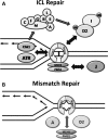
A model showing the overlapping functions of MMR and FA proteins in ICL and MMR. MSH2 is likely involved in the detection of ICLs and early signaling events leading to the monoubiquitylation and chromatin loading of FANCD2 and FANCI, such as recruitment of ATR. MLH1 may play a role in ICL repair downstream of FANCD2 and FANCI monoubiquitylation, dependent on its interaction with both FANCD2 and FANCJ. Conversely, FANCA and FANCD2, along with other members of the FA pathway, may be required for efficient binding between MSH2 and MLH1. This role in the MSH2–MLH1 interaction renders all FA cell lines tested defective in MMR.

Similar articles

Cited by

References

    1. Kennedy R.D., D'Andrea A.D. The Fanconi anemia/BRCA pathway: new faces in the crowd. Genes Dev. 2005;19:2925–2940. doi:10.1101/gad.1370505. - DOI - PubMed
    1. Wang W. Emergence of a DNA-damage response network consisting of Fanconi anaemia and BRCA proteins. Nat. Rev. Genet. 2007;8:735–748. doi:10.1038/nrg2159. - DOI - PubMed
    1. Collins N., Kupfer G.M. Molecular pathogenesis of Fanconi anemia. Int. J. Hematol. 2005;82:176–183. doi:10.1532/IJH97.05108. - DOI - PubMed
    1. McCabe K.M., Olson S.B., Moses R.E. DNA interstrand crosslink repair in mammalian cells. J. Cell. Physiol. 2009;220:569–573. doi:10.1002/jcp.21811. - DOI - PMC - PubMed
    1. Williams S.A., Kupfer G.M. Ubiquitin and FANC stress responses. In: Bradshaw R.A., Dennis E.A., editors. Handbook of Cell Signaling. 2nd edn. Oxford: Oxford University Press; 2009. pp. 2265–2272.

Publication types

MeSH terms