. 2011 Aug;7(8):e1002209.
doi: 10.1371/journal.pgen.1002209. Epub 2011 Aug 11.

Gamma-tubulin is required for bipolar spindle assembly and for proper kinetochore microtubule attachments during prometaphase I in Drosophila oocytes

Affiliations

Gamma-tubulin is required for bipolar spindle assembly and for proper kinetochore microtubule attachments during prometaphase I in Drosophila oocytes

Stacie E Hughes et al. PLoS Genet. 2011 Aug.

Abstract

In many animal species the meiosis I spindle in oocytes is anastral and lacks centrosomes. Previous studies of Drosophila oocytes failed to detect the native form of the germline-specific γ-tubulin (γTub37C) in meiosis I spindles, and genetic studies have yielded conflicting data regarding the role of γTub37C in the formation of bipolar spindles at meiosis I. Our examination of living and fixed oocytes carrying either a null allele or strong missense mutation in the γtub37C gene demonstrates a role for γTub37C in the positioning of the oocyte nucleus during late prophase, as well as in the formation and maintenance of bipolar spindles in Drosophila oocytes. Prometaphase I spindles in γtub37C mutant oocytes showed wide, non-tapered spindle poles and disrupted positioning. Additionally, chromosomes failed to align properly on the spindle and showed morphological defects. The kinetochores failed to properly co-orient and often lacked proper attachments to the microtubule bundles, suggesting that γTub37C is required to stabilize kinetochore microtubule attachments in anastral spindles. Although spindle bipolarity was sometimes achieved by metaphase I in both γtub37C mutants, the resulting chromosome masses displayed highly disrupted chromosome alignment. Therefore, our data conclusively demonstrate a role for γTub37C in both the formation of the anastral meiosis I spindle and in the proper attachment of kinetochore microtubules. Finally, multispectral imaging demonstrates the presences of native γTub37C along the length of wild-type meiosis I spindles.


Conflict of interest statement

The authors have declared that no competing interests exist.

Figures
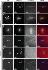
Figure 1
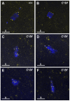
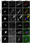
Figure 1. γtub37C mutations cause spindle and chromosome defects in oocytes during prometaphase I.
Fixed oocytes were treated with antibodies against α-tubulin and histone H3 phosphorylated at serine 10 (phH3S10), as well as the DNA dye DAPI. All oocytes were from prometaphase I-enriched preparations of oocytes. (A) Shows a wild-type oocyte with symmetrical-looking chromosomes and a bipolar spindle. Arrowheads highlight the phH3S10 positive thread projecting from the achiasmate 4th chromosomes. (B) Shows autosomal slippage (arrowheads) on a monopolar spindle in a γtub37CP162L mutant oocyte. (C) Shows a barrel-like spindle and chromosomes with an abnormal and round morphology in a γtub37CP162L mutant oocyte. (D) Shows a γtub37C 3 /Df mutant oocyte with a barrel-like spindle with abnormal microtubule density and chromosome misalignment. (E) Shows a γtub37C 3 /Df mutant oocyte with chromosomes and microtubules that lack clear orientation. Additionally, the phH3S10 antibody highlights threads that project away from the chromosomes rather than connecting achiasmate homologs (arrowheads). (F) Shows a γtub37C 3 /Df mutant oocyte with a spindle that is more similar to wild type with clear microtubule directionality and nearly tapered bipolar morphology. The chromosomes are associated and aligned along the spindle axis. (A) and (E) are single Z sections to highlight the phH3S10-labeled threads while (B–D, F) are maximum intensity projections from Z stacks. The scale bars are in microns.
Figure 2
Figure 2. Kinetochores fail to properly biorient during prometaphase I in γtub37C 3 /Df mutant oocytes.
Fixed oocytes were treated with antibodies against CID (yellow), as well as the DNA dye DAPI (blue). All oocytes were from prometaphase I-enriched preparations of oocytes. (A) In a wild-type oocyte the eight CID foci are properly bioriented. (B) A γtub37C 3 /Df mutant oocyte with bioriented kinetochores as based on CID foci. (C) A γtub37C 3 /Df mutant oocyte with CID foci that failed to properly biorient at opposite ends of the chromosome mass. (D) A γtub37C 3 /Df mutant oocyte with kinetochores that appear to be mono-oriented based on CID foci. (E) A γtub37C 3 /Df mutant oocyte with clustered CID foci. (F) More than eight CID foci are observed in a γtub37C 3 /Df mutant oocyte, suggesting loss of sister chromatid cohesion. Images are maximum intensity projections from partial Z stacks. The scale bars are in microns.
Figure 3
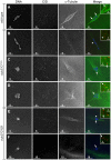
Figure 3. Kinetochores fail to make direct contacts to kinetochore microtubules in γtub37C mutant oocytes.
Fixed prometaphase I oocytes were treated with antibodies against α-tubulin and CID, as well as the DNA dye DAPI. Selected regions are magnified in the insets. Insets show only CID and α-tubulin for better visualization of microtubule interactions with CID foci. (A) CID foci directly interact with kinetochore microtubules (arrows) in a wild-type oocyte. (B–D) CID foci show aberrant kinetochore microtubule attachments in γtub37CP162L mutant oocytes. Arrows point to examples of CID foci with no apparent direct microtubule interactions and the arrowhead points to an example of a CID focus with potentially a lateral microtubule interaction. (E–F) Shows spindles from γtub37C 3 /Df mutant oocytes. The arrows point to examples of CID foci lacking clear microtubule interactions and the arrowhead points to an example of a CID focus showing only a potential lateral interaction to microtubules. Images are single Z sections to highlight the interaction of CID foci with the microtubules. Black levels were adjusted to allow better visualization of CID foci. Scale bars are in microns.
Figure 4
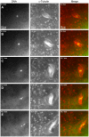
Figure 4. Defects in spindle morphology and chromosome positioning are observed in a living γtub37CP162L mutant oocyte.
Shown is a time-lapse of a spindle from a living prometaphase I γtub37CP162L mutant oocyte. OliGreen labels the DNA (green) and rhodamine-conjugated-tubulin labels the microtubules (red). The time-lapse is shown in Video S3. (A) At the start of the time-lapse the chromosomes are associated, but the spindle is wide and lacks tapered poles. (B) At 8.8 minutes the spindle has changed shape due to the movement of the microtubules. (C) At 22.7 minutes the microtubules continue to move and the whole spindle has rotated clockwise. The chromosomes also appear to have changed position. (D) At 37.8 minutes the chromosomes have moved apart, the spindle further broadens, and microtubules are shed into the cytoplasm. (E) By the end of Video S3 at 45.7 minutes the chromosomes have formed two masses. The spindle has narrowed and rotated an additional 30° clockwise. Images are maximum intensity projections from Z stack and scale bars are in microns.
Figure 5
Figure 5. Oocytes with a missense mutation in γtub37C mostly recover by metaphase I arrest while a null mutation causes splitting of the chromosomes.
Fixed oocytes were treated with antibodies against α-tubulin and histone H3 phosphorylated at serine 10 (phH3S10), as well as the DNA dye DAPI. Oocytes were from virgin mothers (to enrich for metaphase I arrested oocytes). (A) A wild-type metaphase I-arrested oocyte with chromosomes that have congressed to the metaphase plate to form a lemon-shaped morphology. The bipolar spindle has shortened. (B) An oocyte from a γtub37CP162L mutant oocyte displays properly congressed chromosomes, but the spindle is extremely narrow. (C) A γtub37C3/Df mutant oocyte with a DNA mass that has split into 2 parts (arrowheads) which have moved far apart in the cytoplasm. Microtubules do not appear to be associated with the chromosome masses. (D) A γtub37C3/Df mutant oocyte with a chromosome mass split into two parts (arrowheads) that are relatively close together. Each piece is associated with its own set of microtubules. (E) A γtub37C3/Df mutant oocyte that has achieved a wild-type metaphase I chromosome morphology and a bipolar spindle. All images are maximum intensity projections from Z stacks. Scale bars are in microns.
Figure 6
Figure 6. At metaphase I chromosomes congress randomly in γtub37CP162L mutant oocytes.
Shown is localization of FISH probes to the 359 base pair (bp) heterochromatic repeat on the X (green) and to the AATAT heterochromatic repeats on a large region of the 4th chromosomes and a small region of the X chromosomes (red), as well as DAPI (blue) in oocytes from metaphase I-enriched preparations. (A) A wild-type oocyte with an X and 4th chromosome on each end of the chromosome mass indicating proper alignment of chromosomes in preparation for segregation at anaphase I. (B) A γtub37CP162L mutant oocyte with X and 4th chromosomes properly positioned on each end of the chromosome mass. (C) A γtub37CP162L mutant oocyte with both 4th chromosomes on the same side of the chromosome mass. (D) A γtub37CP162L mutant oocyte with both X chromosomes on the same side of the chromosome mass. (E) A γtub37CP162L mutant oocyte with the 4th and X chromosomes on opposite sides of the chromosome mass. Numbers indicate the number of oocytes displaying each chromosome configuration over the total oocytes scored. For the probe recognizing the AATAT heterochromatic repeat, assignment of signal to the 4th or X chromosomes was based on the size of the signal and proximity to the 359 bp probe signal. Images are projections from Z stacks and scale bars are in microns.
Figure 7
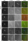
Figure 7. The spindle pole component D-TACC is mislocalized in γtub37CP162L mutant oocytes.
Oocytes were immuno-stained to D-TACC (green), α-tubulin (red) and DAPI (blue). (A) A wild-type prometaphase I spindle with polar and diffuse D-TACC localization. (B) A wild-type metaphase I spindle with D-TACC staining at the spindle poles and diffuse staining along the microtubules. (C) Large, bright patches of D-TACC cover part of the spindle while D-TACC is absent in other regions in a prometaphase I γtub37CP162L mutant oocyte. (D) Small puncta of D-TACC are present near the DNA, but D-TACC is extremely weak or absent near the poles in a prometaphase I γtub37CP162L mutant oocyte. (E) D-TACC is absent or extremely weak from the spindle in a prometaphase I γtub37CP162L mutant oocyte despite strong localization of D-TACC to cytoplasmic structures. (F) A metaphase I spindle from a γtub37CP162L mutant oocyte lacking clear D-TACC staining. Shown are single Z slices since projection of the entire Z stack results in images with high background in the D-TACC channel. For the examples of γtub37CP162L mutant oocytes, spindles that displayed directionality were analyzed to rule out D-TACC mislocalization being caused by lack of a clear spindle. Scale bars are in microns.
Figure 8
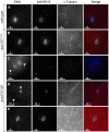
Figure 8. Endogenous γTub37C is present on the meiosis I spindle in Drosophila oocytes.
The DrosC γTub37C-specific and α-tubulin antibodies in A–F were acquired simultaneously using 488 excitation, while Hoescht (A–E) or DAPI (F) was acquired separately using the exact same Z-stack parameters. (A and B) γTub37C localizes to wild-type prometaphase I spindles in a pattern similar to α-tubulin (N = 20). (C) γTub37C continues to localize to the spindle microtubules in a wild-type metaphase I oocyte (N = 10). (D–E) The γTub37C-specific antibody fails to recognize the spindle in γtub37C3/Df mutant oocytes (N = 18). (F) γTub37C is not detected on a spindle from a γtub37CP162L mutant oocyte (N = 20). Images are projections of a few Z slices. Scale bars are in microns.
Figure 9
Figure 9. A speculative hypothesis for the functions of γTub37C in meiosis I in Drosophila oocytes.
In wild-type oocytes (top) microtubules are recruited to the chromosomes after nuclear envelope breakdown (NEB). γTub37C complexes nucleate and stabilize additional microtubules, especially the kinetochore microtubules that attach kinetochores to the developing spindle poles. Ncd bundles the microtubules together to further develop the tapered, bipolar spindle with the help of additional factors, such as the spindle pole component, D-TACC, and the chromosome passenger complex (not shown). Nod utilizes the microtubules to push the chromosomes toward the spindle midzone. In the absence of functional γTub37C (bottom) microtubules can still be recruited to the chromosomes after NEB but nucleation of additional microtubules, particularly kinetochore microtubules, is delayed and existing microtubules are unstable. In the absence of robust microtubules the ability of Ncd to bundle microtubules into a tapered, bipolar spindle is inhibited. Nod function would also be inhibited which, in combination with disrupted kinetochore microtubule attachments, would lead to chromosome orientation and alignment defects.

Similar articles

Cited by

References

    1. Raynaud-Messina B, Merdes A. Gamma-tubulin complexes and microtubule organization. Curr Opin Cell Biol. 2007;19:24–30. - PubMed
    1. Schuh M, Ellenberg J. Self-organization of MTOCs replaces centrosome function during acentrosomal spindle assembly in live mouse oocytes. Cell. 2007;130:484–498. - PubMed
    1. Barrett SL, Albertini DF. Allocation of gamma-tubulin between oocyte cortex and meiotic spindle influences asymmetric cytokinesis in the mouse oocyte. Biol Reprod. 2007;76:949–957. - PubMed
    1. Burbank KS, Groen AC, Perlman ZE, Fisher DS, Mitchison TJ. A new method reveals microtubule minus ends throughout the meiotic spindle. J Cell Biol. 2006;175:369–375. - PMC - PubMed
    1. Combelles CM, Albertini DF. Microtubule patterning during meiotic maturation in mouse oocytes is determined by cell cycle-specific sorting and redistribution of gamma-tubulin. Dev Biol. 2001;239:281–294. - PubMed

Publication types

MeSH terms

LinkOut - more resources